.webp&w=384&q=75)

Loading, please wait...
Loading, please wait...
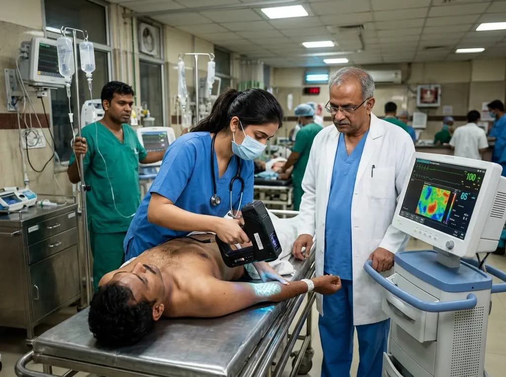
Trauma care rapidly evolves as non-invasive tools emerge to assess tissue perfusion at the microvascular level. Recent clinical investigations have evaluated hyperspectral imaging in trauma to determine whether microcirculatory alterations can guide early resuscitation. Specifically, severe physical injury triggers significant physiological stress, often leading to microcirculatory impairment long before macrohemodynamic parameters collapse. Consequently, clinicians require fast, objective measurements to detect subtle microvascular dysfunction during initial patient intake. Hyperspectral imaging provides non-contact, rapid spectroscopic evaluation of tissue oxygenation, water content, and hemoglobin concentration without requiring contrast media or radiation. Therefore, researchers conducted a prospective observational study at a German level I trauma center to evaluate its diagnostic accuracy during acute trauma resuscitation. The prospective trial included adult trauma patients admitted to the resuscitation room. Measurements occurred across three specific time points: during primary survey, after resuscitation room care, and twenty-four hours post-admission. By capturing spectral reflections, the camera system provides quantitative color-coded maps of microcirculatory parameters. However, researchers needed to establish whether these non-invasive metrics directly reflect systemic hemodynamic instability or predict critical outcomes. Understanding these physiological relationships is vital for emergency physicians and trauma surgeons seeking to refine patient triage.
To perform comprehensive non-invasive assessment, the study utilized the TIVITA 2.0 system, which calculates four primary microcirculatory metrics. Specifically, these metrics include tissue oxygenation saturation, tissue water index, tissue hemoglobin index, and near-infrared perfusion index. These parameters offer detailed insights into tissue oxygen availability, capillary volume, superficial perfusion, and localized edema. During trial execution, investigators evaluated one hundred sixty-three trauma patients. Among this cohort, eighteen patients presented with clinical hemodynamic instability, while one hundred forty-five patients remained stable. The authors defined hemodynamic instability using clinical criteria, including prolonged systolic blood pressure below ninety millimeters of mercury, sustained catecholamine administration, or obvious signs of systemic hypoperfusion lasting over ten minutes. The primary study endpoint focused on detecting significant differences in initial tissue oxygenation saturation between stable and unstable cohorts. Additionally, secondary outcomes examined correlations between imaging parameters, Injury Severity Score, essential biomarkers, and overall in-hospital mortality. Statistical evaluations included standard t-tests, receiver operating characteristic curve analyses, diagnostic accuracy measurements, and DeLong's test for comparing area under the curve values. Consequently, this methodology created a robust framework for evaluating non-invasive optical microvascular monitoring.
Surprisingly, primary endpoint analysis demonstrated no statistically significant difference in initial tissue oxygenation saturation between hemodynamically unstable and stable trauma cohorts. Specifically, patients presenting with hemodynamic instability recorded a mean initial oxygen saturation of fifty-six point five percent, whereas stable patients recorded fifty-nine point forty-eight percent. Likewise, secondary microcirculatory parameters—including tissue water index, tissue hemoglobin index, and near-infrared perfusion index—showed no significant variations between groups. These findings indicate that initial macroscopic hemodynamic collapse does not uniformly correlate with superficial microcirculatory values measured via hyperspectral imaging. However, a significant physiological correlation emerged when comparing imaging metrics against metabolic biomarkers. Notably, patients presenting with elevated initial blood lactate levels of two millimoles per liter or higher displayed significantly lower tissue oxygenation saturation values. Blood lactate serves as an established surrogate marker for cellular hypoxia and anaerobic metabolism during shock. Consequently, the strong correlation between reduced optical oxygenation and elevated lactate confirms that hyperspectral imaging accurately reflects localized metabolic stress. Therefore, while microvascular imaging may not replace standard blood pressure monitoring, it effectively identifies subclinical tissue hypoperfusion associated with hyperlactatemia during acute trauma resuscitation.
Predicting mortality during initial trauma resuscitation remains a fundamental clinical objective for emergency care providers. In this study, receiver operating characteristic curve analysis evaluating initial tissue oxygenation saturation for predicting in-hospital mortality demonstrated an area under the curve of zero point six seven seven. Furthermore, comparative analysis showed that the predictive accuracy of tissue oxygenation saturation overlapped with established laboratory biomarkers, including serum lactate, bicarbonate, and base excess. Crucially, pairwise comparisons using DeLong's test revealed no significant statistical differences between these diagnostic markers. Through detailed diagnostic accuracy analysis, researchers identified an optimal tissue oxygenation threshold of fifty-four percent or lower for predicting mortality. At this specific threshold, the technique demonstrated a sensitivity of zero point seven zero and a specificity of zero point six six. Although the positive predictive value remained low at zero point two two, the negative predictive value reached an impressive zero point nine four. Consequently, a tissue oxygenation reading above fifty-four percent strongly indicates that a trauma patient maintains a low risk of short-term mortality. Therefore, while low oxygenation values require cautious interpretation, normal microcirculatory values offer robust reassurance regarding patient survival.
The high negative predictive value of hyperspectral imaging in trauma assessment carries substantial operational implications for acute trauma management. Modern emergency departments and level I trauma centers frequently face resource constraints during high-volume periods. Consequently, trauma leaders constantly face decisions regarding resource allocation, clinical downgrading, and stepping down intensive monitoring. Because a normal tissue oxygenation value reliably rules out imminent mortality risk, clinicians can confidently identify low-risk patients who do not require prolonged resuscitation room occupation. Thus, non-invasive microcirculatory monitoring can function as an objective triage tool to support safe, rapid trauma team downgrading. Furthermore, combining optical imaging with point-of-care lactate measurements creates a powerful multi-modal assessment strategy during primary survey procedures. By integrating non-invasive microvascular metrics into standard trauma protocols, clinical teams can improve diagnostic efficiency while minimizing unnecessary invasive monitoring. Additionally, future research should explore sequential imaging to monitor resuscitation efficacy in real time, tracking microvascular recovery following fluid administration or vasopressor therapy. Ultimately, hyperspectral imaging represents a promising non-invasive innovation that enhances objective clinical decision-making in high-stakes emergency environments.
Hyperspectral imaging is a non-invasive, contact-free optical technology that analyzes broad-spectrum light reflected from microvascular tissue. In trauma care, the system rapidly measures key physiological parameters, including tissue oxygenation saturation, water content, and hemoglobin concentration. Consequently, emergency clinicians obtain quantitative, color-coded perfusion maps within seconds, allowing immediate objective evaluation of microcirculatory health during primary trauma survey procedures without requiring contrast media or ionizing radiation.
In the study, initial microcirculatory parameters did not significantly differ between hemodynamically stable and unstable patients because macrovascular blood pressure changes do not always directly parallel localized capillary perfusion. Systemic compensatory mechanisms, vasopressor administration, and localized autoregulation can maintain or alter skin microcirculation independently of systemic blood pressure. Consequently, microcirculatory assessments provide complementary physiological data regarding tissue-level oxygenation rather than merely duplicating systemic macrohemodynamic parameters.
Clinicians can leverage the high negative predictive value of tissue oxygenation saturation to guide trauma team downgrading decisions safely. In the prospective study, a tissue oxygenation saturation above fifty-four percent yielded a negative predictive value of ninety-four percent for in-hospital mortality. Therefore, finding normal microvascular oxygenation during initial assessment gives trauma leaders reliable objective reassurance that the patient carries a very low mortality risk, facilitating safe de-escalation of trauma team resources.
Disclaimer: This content is for informational and educational purposes only and does not constitute medical advice, diagnosis, or treatment. Always seek the advice of a qualified healthcare provider with any questions regarding a medical condition or clinical intervention. Refer to the latest local and national guidelines for clinical practice.
References

Read summarized clinical updates, watch expert medical content, and earn CME certifications right from your smartphone.
Open your smartphone camera to scan and install.





A prospective study evaluated hyperspectral imaging (HSI) in trauma resuscitation. While HSI did not differentiate hemodynamic stability, StO2 correlated with lactate and achieved a 94% NPV for mortality, supporting its role in early trauma team downgrading.
4 weeks back

Dendritic cells bridge innate and adaptive immunity in myocardial infarction. This review explores their pathological roles, circulating dynamics, novel tolerogenic interventions, and how standard cardiovascular medications modulate dendritic cells to improve post-infarction myocardial repair and patient outcomes.
Today

A premature neonate developed upper limb compartment syndrome after uterine rupture extruded the arm through a scar defect. Conservative management with continuous monitoring yielded complete functional recovery and normal limb growth at 10-year follow-up, highlighting non-operative safety in selected cases.
Today
Atherosclerosis involves extensive glycometabolic reprogramming across immune and vascular cells. This review examines how glycolysis, the pentose phosphate pathway, and lactate-driven epigenetic shifts fuel plaque vulnerability, while highlighting novel therapeutic targets like PFKFB3 and LDHA.
Today
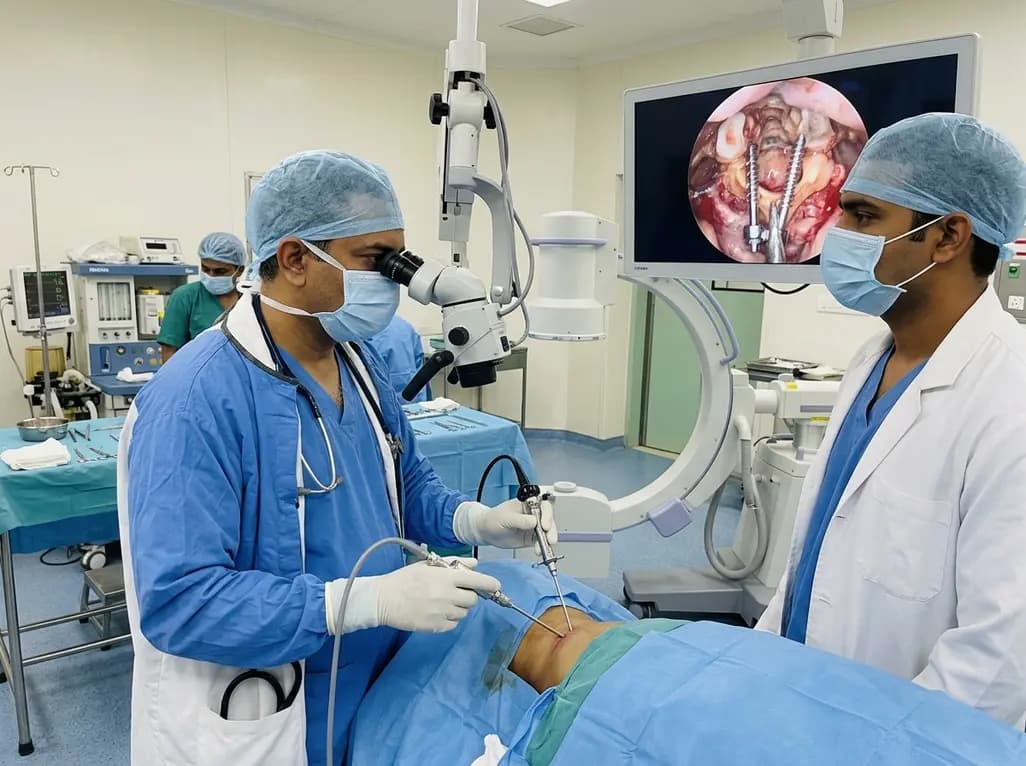
Endoscopic posterior cervical fusion combines minimally invasive decompression, joint preparation, and rigid screw-rod fixation for atlantoaxial pathologies. Early clinical findings demonstrate solid bony union, excellent symptom relief, and minimal soft-tissue morbidity without significant vascular compromise.
Yesterday

The All-India Food Processors' Association has approached the Supreme Court to oppose FSSAI's proposed per-100g benchmark for front-of-pack warning labels, advocating instead for a per-serving threshold. We explore the regulatory showdown, nutritional evidence, and implications for clinical lifestyle counseling.
Today
