.webp&w=384&q=75)

Loading, please wait...
Loading, please wait...

Classic Hodgkin lymphoma typically demonstrates excellent cure rates with frontline ABVD chemotherapy regimens. However, approximately 15% to 30% of patients with advanced-stage disease experience relapse or primary refractory disease. In recent years, programmed death-1 (PD-1) pathway inhibitors such as pembrolizumab have significantly reshaped salvage management paradigms for these challenging cases. Despite their impressive antineoplastic efficacy, clinicians must remain vigilant regarding immune-related adverse events. Among these rare complications, hypereosinophilic syndrome represents an uncommon but potentially life-threatening entity that warrants immediate diagnostic precision. Hypereosinophilic syndrome involves marked peripheral eosinophilia paired with direct eosinophil-mediated organ damage. Although mild, transient eosinophilia occasionally occurs during immune checkpoint blockade as an incidental biomarker of immune activation, severe multi-organ hypereosinophilic presentations remain exceptionally rare. Therefore, oncology teams must understand the clinical trajectory, pathogenic overlap, and therapeutic priorities when immune checkpoint inhibitors trigger profound eosinophilic proliferation.
A recent clinical report details the complex presentation of a 56-year-old male diagnosed with stage IV nodular sclerosis classic Hodgkin lymphoma. The patient had previously exhausted multiple lines of standard cytotoxic and targeted salvage treatments. Specifically, his therapeutic history included ABVD, DHAP, brentuximab vedotin, and ICE protocols. Following disease recurrence, oncologists initiated combination therapy utilizing pembrolizumab alongside GVD chemotherapy (gemcitabine, vinorelbine, and liposomal doxorubicin). Consequently, the patient achieved a complete metabolic response on functional imaging. However, shortly after this remarkable oncologic response, routine laboratory surveillance revealed dramatic hematologic derangements. The patient developed marked leukocytosis with a total white blood cell count of 63,000/μL. Most notably, his absolute eosinophil count surged to 53,000/μL. In addition to these laboratory findings, the patient exhibited significant clinical deterioration characterized by profound dehydration, worsening acute renal dysfunction, and widespread anasarca requiring urgent inpatient hospitalization.
Evaluating severe eosinophilia in patients receiving immunotherapy requires systematic exclusion of secondary reactive etiologies alongside comprehensive clonal assessment. In this patient, the diagnostic evaluation fulfilled formal criteria for hypereosinophilic syndrome due to persistent extreme eosinophilia coupled with overt end-organ injury. Furthermore, advanced molecular testing demonstrated an unexpected clonal abnormality. Specifically, fluorescence in situ hybridization identified a FIP1L1-PDGFRA gene rearrangement in approximately 2% of analyzed cells. This finding raised strong suspicion for an underlying or therapy-related myeloid neoplasm with eosinophilia. Prior exposure to multiple lines of alkylating agents and topoisomerase inhibitors may have created genomic instability that favored clonal emergence. Alternatively, profound PD-1 blockade might have released immunologic constraints on a pre-existing occult clonal hematopoietic population. Therefore, this case highlights that clinicians must pursue comprehensive cytogenetic and molecular panels whenever severe hypereosinophilia emerges during immune checkpoint inhibitor regimens.
The pathophysiology connecting immune checkpoint inhibition to hypereosinophilic syndrome involves complex immune dysregulation. Under physiological conditions, PD-1 signaling restrains T-cell activation and balances cytokine synthesis. Blocking this checkpoint pathway promotes robust T-helper-2 (Th2) polarization in susceptible individuals. Consequently, activated T cells release substantial quantities of eosinophilopoietic cytokines, most notably interleukin-5 (IL-5), interleukin-3, and granulocyte-macrophage colony-stimulating factor. These signaling molecules accelerate bone marrow eosinophil differentiation, prolong peripheral eosinophil survival, and promote systemic tissue infiltration. Moreover, extensive prior chemotherapy regimens often induce enduring alterations in the bone marrow microenvironment. When immunotherapy superimposes profound T-cell disinhibition onto a vulnerable marrow architecture, uncontrolled eosinophil expansion can rapidly manifest. Additionally, the activated eosinophils release toxic cationic proteins, major basic protein, and reactive oxygen species into tissues. This localized degranulation drives endothelial damage, vascular permeability, tissue edema, and severe multi-organ dysfunction.
Managing severe hypereosinophilia during immune checkpoint therapy demands rapid intervention to prevent irreversible organ damage. When acute organ failure or extreme leukocytosis occurs, stabilizing the patient takes absolute clinical precedence over continuing cancer-directed immunotherapy. Clinicians initially attempted cytoreduction with hydroxyurea in this reported case; however, patient intolerance prompted a rapid transition to systemic corticosteroids. Corticosteroid therapy serves as the standard first-line intervention for severe immune-related toxicities and hypereosinophilic syndromes. Glucocorticoids suppress cytokine production, induce eosinophil apoptosis, and rapidly reduce circulating eosinophil burdens. Furthermore, identifying the FIP1L1-PDGFRA fusion provides a crucial targeted therapeutic alternative. Low-dose imatinib mesylate represents a highly effective, definitive treatment for PDGFRA-rearranged myeloid disorders. Additionally, biologic agents targeting the IL-5 pathway, such as mepolizumab or benralizumab, offer valuable corticosteroid-sparing options in refractory immune-mediated eosinophilic disorders, ensuring comprehensive symptom control and biochemical stabilization.
This remarkable case provides pivotal clinical lessons for hematologists and oncologists managing complex lymphoma cohorts. First, clinicians must incorporate routine absolute eosinophil count monitoring into standard safety protocols for patients undergoing immune checkpoint blockade. Second, when extreme eosinophil elevations arise, teams must promptly differentiate reactive immune-mediated processes from primary clonal myeloid neoplasms. Third, early multidisciplinary collaboration between oncology, hematology, and nephrology specialists ensures timely initiation of organ-preserving immunosuppression. In addition, oncologists must weigh the risks and benefits of re-challenging patients with immunotherapy after severe adverse events resolve. Molecular characterization remains indispensable, as actionable fusions such as PDGFRA rearrangements directly alter therapeutic trajectories. Ultimately, maintaining high clinical vigilance facilitates early diagnosis, guides targeted pharmacotherapy, and mitigates life-threatening complications while preserving durable oncologic responses.
Hypereosinophilic syndrome is defined by peripheral blood eosinophil counts exceeding 1,500/μL on repeated assessments, accompanied by documented eosinophil-induced end-organ damage or dysfunction, after excluding parasitic, allergic, and infectious causes. In immunotherapy recipients, this condition manifests through cytokine-mediated eosinophil activation or the unmasking of clonal myeloid processes, causing complications such as renal injury, skin changes, pulmonary infiltrates, or widespread anasarca.
Clinicians must immediately interrupt immune checkpoint inhibitor therapy when hypereosinophilia is accompanied by organ dysfunction. Initial management centers on prompt high-dose systemic corticosteroid administration to induce eosinophil apoptosis and reduce tissue inflammation. Supportive care for organ failure, thorough secondary workups, and multidisciplinary specialist consultation are critical before determining if safe immunotherapy resumption is feasible.
Molecular testing identifies specific clonal mutations and gene fusions, such as FIP1L1-PDGFRA, that define distinct myeloid neoplasms with eosinophilia. Detecting this rearrangement carries critical therapeutic implications because it predicts exceptional clinical sensitivity to low-dose tyrosine kinase inhibitors like imatinib, providing a definitive targeted therapeutic pathway beyond non-specific immunosuppressive therapies.
Disclaimer: This content is for informational and educational purposes only, and should not be taken as medical advice. It is not intended to substitute for professional medical advice, diagnosis, or treatment. Always seek the advice of your physician or other qualified health provider with any questions you may have regarding a medical condition. Refer to the latest local and national guidelines for clinical practice.
References

Read summarized clinical updates, watch expert medical content, and earn CME certifications right from your smartphone.
Open your smartphone camera to scan and install.





A rare case report highlights hypereosinophilic syndrome and suspected myeloid neoplasia with FIP1L1-PDGFRA rearrangement in a patient receiving pembrolizumab and GVD for relapsed classic Hodgkin lymphoma, emphasizing diagnostic vigilant monitoring and prompt corticosteroid therapy.
4 weeks back
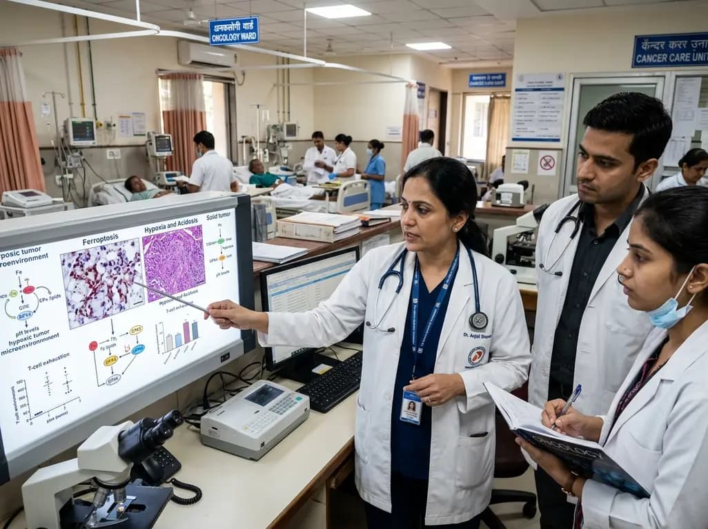
Targeting ferroptosis provides a compelling strategy to dismantle hypoxia- and acidosis-driven immunosuppression in solid tumors, enhancing response to modern immunotherapy.
Today

Findings from Project Viva indicate that sustained postpartum weight retention, rather than excessive gestational weight gain alone, directly worsens women's mid-life cardiovascular health, degrading blood pressure and glycemic parameters decades after delivery.
Today

A large UK Biobank study reveals that plasma proteomic signatures capture preclinical organ damage and improve multiorgan risk prediction across early CKM syndrome stages 0 to 2 beyond traditional PREVENT clinical models.
Today

A retrospective CBCT study reveals that superior TMJ space asymmetry correlates moderately with occipital condyle-atlas-axis asymmetry in unilateral TMJ osteoarthritis, highlighting an imaging correlate between jaw biomechanics and upper cervical alignment rather than a standalone diagnostic marker.
Today

A Bayesian multilevel meta-analysis reveals that aerobic training combined with moderate carbohydrate restriction modestly lowers HbA1c in type 2 diabetes. However, sparse data and very low certainty leave incremental benefits over exercise or diet alone unproven, highlighting the need for individualized care.
Today
