

Loading, please wait...
Loading, please wait...

Hurler syndrome, or mucopolysaccharidosis type IH, represents the most severe manifestation of alpha-L-iduronidase deficiency. Consequently, systemic glycosaminoglycan accumulation disrupts normal connective tissue architecture, neurodevelopment, and cerebrospinal fluid dynamics. Hurler syndrome hydrocephalus frequently emerges during infancy due to multifactorial hydrodynamics. Specifically, progressive glycosaminoglycan infiltration thickens leptomeningeal membranes, impairing cerebrospinal fluid resorption through arachnoid granulations. Furthermore, cranial base dysostosis often compresses major dural venous outflow pathways, which increases intracranial venous pressure. Left untreated, escalating intracranial pressure causes irreversible cognitive regression, optic atrophy, and early mortality. Hematopoietic stem cell transplantation remains the established standard of care for halting central nervous system deterioration. However, clinicians historically lacked definitive longitudinal evidence regarding ventricular size trajectories after successful donor engraftment. Consequently, clinical guidelines remained ambiguous regarding optimal neuroimaging surveillance schedules before and after cell therapy. Medical teams frequently ordered repetitive brain magnetic resonance imaging scans under general anesthesia to monitor ventricular anatomy. Because pediatric anesthesia carries established neurodevelopmental and airway risks, clarifying true hydrocephalus progression rates after transplantation became an urgent priority for pediatric specialists and neurosurgeons worldwide. Therefore, researchers launched a comprehensive investigation to define post-transplant ventricular changes and assess routine imaging necessity.
To resolve clinical monitoring dilemmas, researchers at the University of Minnesota conducted a rigorous single-center retrospective cohort study. The investigators analyzed clinical records from fifty-seven consecutive pediatric patients with Hurler syndrome who received allogeneic hematopoietic stem cell transplantation. Furthermore, the investigative team evaluated 291 longitudinal brain magnetic resonance imaging examinations performed across multiple post-transplant years. Rather than relying on qualitative interpretations, researchers measured ventricular size using the fronto-occipital horn width ratio. This objective linear radiographic index divides the maximal width of the frontal horns and occipital horns by the biparietal skull diameter. Consequently, this standardized measurement allowed investigators to track lateral ventricular dimensions over time with exceptional consistency. In addition, the authors stratified participants based on the presence of prior ventriculoperitoneal shunting to separate natural disease resolution from hardware-mediated drainage. They meticulously recorded every surgical intervention, intracranial pressure measurement, and neurologic exam finding. By tracking patients over extended follow-up periods, the team generated the most extensive longitudinal dataset on ventricular dynamics in this population. Thus, this methodology provided an unprecedented look at anatomical ventricular remodeling after curative cellular therapy.
The investigative findings provide remarkable clarity and reassurance regarding long-term ventricular anatomy following stem cell transplantation. Among the fifty-seven participants, fifty-one children did not have a ventriculoperitoneal shunt at baseline. Notably, these non-shunted patients demonstrated a gradual, consistent decrease in ventricular size over time. Furthermore, their ventricular dimensions reached definitive anatomical stability approximately two years following stem cell infusion. Most importantly, zero patients without preexisting shunts developed progressive hydrocephalus requiring surgical diversion as part of their natural disease course post-transplant. This translates to an incidence rate of zero percent, with a tight upper ninety-five percent confidence limit of 5.8 percent. Therefore, successful donor cell engraftment halted the pathological accumulation of glycosaminoglycans within meningeal tissues. Although some patients displayed persistent, mild ventriculomegaly, their ventricles remained entirely quiescent without signs of intracranial hypertension. Thus, mild ventriculomegaly reflects stable, preexisting structural changes rather than active hydrodynamic failure. Donor-derived microglial and macrophage populations provide sufficient localized enzymatic activity to maintain patent cerebrospinal fluid pathways. Consequently, patients who undergo successful transplantation without preexisting hydrocephalus remain remarkably protected from late-onset ventricular decompensation.
Given that ventricular dimensions reliably stabilize within two years, the study authors strongly questioned the utility of serial surveillance neuroimaging. Historically, institutional protocols recommended obtaining annual or biennial brain magnetic resonance imaging scans to track ventricular changes indefinitely. However, repetitive neuroimaging in young children requires deep sedation or general anesthesia. Pediatric anesthesia poses significant airway challenges, especially in mucopolysaccharidosis due to upper airway narrowing and cervical spine instability. Furthermore, routine neuroimaging creates immense logistical strain, emotional distress, and substantial financial costs for families. Because asymptomatic patients without shunts did not progress to surgical hydrocephalus, scheduled surveillance imaging provided negligible clinical benefit. Instead, clinicians should transition from indiscriminate calendar-based neuroimaging to symptom-triggered assessments. Medical teams must educate parents to monitor for specific red-flag symptoms of elevated intracranial pressure. For instance, morning vomiting, unremitting headaches, sudden visual disturbances, or accelerated head circumference crossing percentiles warrant immediate magnetic resonance imaging. In contrast, stable, thriving children do not require repeated scans once two years have elapsed following successful stem cell transplantation. Thus, discontinuing mandatory routine scans spares children unnecessary anesthesia while preserving high standards of patient safety and neurosurgical oversight.
While non-shunted children experienced structural stability, patients with preexisting ventriculoperitoneal shunts presented unique management requirements. In this cohort, six individuals required surgical shunting before or around their transplant procedure. Notably, allogeneic transplantation does not eliminate hardware-related complications such as catheter blockage, bacterial colonization, or mechanical valve malfunction. Therefore, neurosurgeons and pediatricians must remain vigilant whenever a shunted patient displays acute neurologic deterioration. However, physicians should clearly distinguish mechanical shunt failure from progressive primary metabolic disease. Additionally, timely diagnosis and prompt hematopoietic stem cell transplantation remain crucial for preventing initial shunt dependence. Initiating donor therapy before extensive meningeal fibrosis develops protects normal arachnoid architecture and preserves natural cerebrospinal fluid drainage pathways. For clinical centers managing inborn errors of metabolism, these findings underscore the value of coordinated care among pediatric hematologists, neurosurgeons, and metabolic specialists. Routine physical evaluations, regular head circumference tracking, and fundoscopic examinations to exclude papilledema offer sensitive, non-invasive surveillance without requiring sedation. By embracing targeted, evidence-based neuroimaging strategies, clinicians reduce intervention risks, lower healthcare costs, and optimize long-term neurodevelopmental outcomes for children with Hurler syndrome. Consequently, personalized follow-up protocols deliver superior, compassionate pediatric care.
Hurler syndrome causes hydrocephalus through widespread accumulation of glycosaminoglycans within the central nervous system. This metabolic buildup thickens leptomeningeal membranes, obstructs arachnoid granulations, and impedes normal cerebrospinal fluid reabsorption. Furthermore, skeletal dysplasias often compress major dural venous outflow channels at the skull base, elevating venous pressure and triggering communicating hydrocephalus.
Recent clinical evidence shows routine brain MRIs offer minimal utility in asymptomatic patients without preexisting shunts. Ventricular size stabilizes within two years post-transplant, with no new surgical hydrocephalus cases emerging naturally. Therefore, clinicians should avoid repetitive scans requiring general anesthesia, reserving neuroimaging specifically for patients exhibiting signs of elevated intracranial pressure.
Hematopoietic stem cell transplantation introduces donor-derived mononuclear cells that cross into recipient tissues, including the brain. These engrafted cells continuously secrete the missing enzyme, alpha-L-iduronidase. Consequently, cellular enzymatic activity progressively degrades accumulated glycosaminoglycans within arachnoid granulations and dural structures, restoring balanced cerebrospinal fluid dynamics and preventing progressive ventricular enlargement.
Disclaimer: This content is for informational and educational purposes only. It does not constitute medical advice and should not be used as a substitute for professional healthcare guidance, diagnosis, or treatment. Refer to the latest local and national guidelines for clinical practice.
References

Read summarized clinical updates, watch expert medical content, and earn CME certifications right from your smartphone.
Open your smartphone camera to scan and install.





A longitudinal study of 57 patients with Hurler syndrome shows that ventricular size stabilizes within 2 years after hematopoietic stem cell transplantation. With no new hydrocephalus cases requiring shunts in non-shunted patients, researchers question the clinical necessity of routine post-transplant surveillance brain MRIs.
Today

A large retrospective cohort study demonstrates that explainable machine learning models, notably XGBoost, accurately predict hospital admission at emergency triage. By analyzing routine triage parameters, hospitals can streamline bed allocation, decrease boarding times, and alleviate acute care overcrowding.
Today

A new quality-improvement initiative reveals that universal maternal mental health screening in the NICU increased screening rates from 29.5% to 95.0%, with nearly 60% of screened mothers requiring psychosocial referrals, highlighting the urgent need for embedded mental health resources.
Today
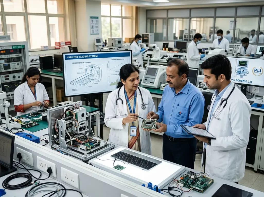
The Department of Pharmaceuticals has extended the medical devices PLI scheme to 2035-36 with a Rs 2,700 crore outlay. Featuring relaxed investment barriers and eliminated sales thresholds, the revised framework targets deep manufacturing, high-end diagnostics, critical MedTech components, and supply resilience.
Today
A metabolomic study reveals that aging delays glutathione synthesis and gamma-glutamylcysteine accumulation following muscle injury. Although precursor supplementation targets this enzymatic bottleneck, in vitro tests show no immediate effect on myogenesis, highlighting the complexity of muscle repair in older adults.
Today

A systematic review evaluates psychological interventions for smoking cessation in diabetes, showing short-term success but highlighting challenges in long-term abstinence.
Today
