.webp&w=384&q=75)

Loading, please wait...
Loading, please wait...
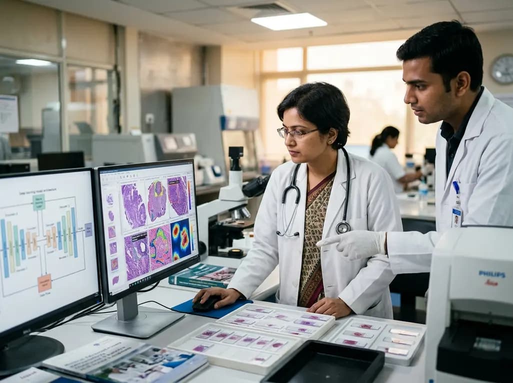
Pathological evaluation serves as the cornerstone of definitive clinical diagnosis and translational biomedical research. Automated histological image classification powered by advanced artificial intelligence has emerged as a transformative tool to support clinical pathologists in routine diagnostic workflows. By processing tissue morphology rapidly and systematically, deep neural networks can augment diagnostic precision, identify subtle microstructural changes, and alleviate administrative burdens on laboratory staff. However, deploying machine learning algorithms in clinical and experimental laboratories introduces substantial technical challenges. Primary among these hurdles is out-of-distribution generalization, where algorithms falter when encountering unseen staining variations, tissue preparation artifacts, or inter-species biological differences. Traditional deep learning models frequently achieve near-perfect metrics during internal cross-validation. Yet, they often suffer significant performance degradation when tested on independent external cohorts. Consequently, assessing how neural network encoders generalize across distinct biological domains has become a critical research priority. Understanding these performance discrepancies allows investigators and healthcare providers to develop robust decision support systems that withstand real-world diagnostic variability across diverse healthcare institutions.
To evaluate out-of-distribution robustness systematically, researchers conducted a comprehensive benchmark using frozen feature extraction paired with a linear probe across nine distinct neural network architectures. The experimental design utilized an internal dataset of 4,307 hematoxylin and eosin stained images representing male rat lung, cerebellum, and adipose tissues. To test genuine generalization capabilities, the models were subsequently evaluated on an external cohort consisting of 600 mixed human and animal tissue images. The study analyzed diverse architecture families, including lightweight convolutional models like MobileNetV3-Small and MobileNetV3-Large, established residual architectures including DenseNet121 and DenseNet201, modern convolutional designs such as ConvNeXt-Small and ConvNeXt-Base, self-supervised vision transformers like DINOv3 ViT-Small and DINOv3 ViT-Huge+, and the dedicated pathology foundation model UNI2-h. By rigorously measuring classification accuracy, sensitivity, specificity, F1 score, Cohen's kappa, and receiver operating characteristic curves alongside per-image inference speed, this benchmark provides objective clarity regarding true cross-domain transferability rather than simple model underfitting during the initial training phase.
The experimental findings revealed significant performance divergence across different tissue types and network architectures during external validation. All competitive deep learning models demonstrated exceptional proficiency when classifying relatively homogenous and structurally distinct specimens, such as adipose and cerebellar tissues. Specifically, these architectures achieved F1 scores exceeding 95% for both tissue categories during cross-domain testing. However, distinguishing lung tissue presented a far greater diagnostic challenge due to its highly variable alveolar morphology, irregular air spaces, and intricate cellular arrangements. In this complex category, the pathology foundation model UNI2-h clearly demonstrated superior capability, achieving a remarkable F1 score of 97.4% and a diagnostic recall of 95.0%. Self-supervised foundation models pretrained on vast computational pathology repositories demonstrated an innate capacity to extract robust cellular representations. In contrast, standard vision models without specialized pathological pretraining suffered notable performance drops, underscoring the necessity of domain-specific feature learning for interpreting heterogeneous anatomical microenvironments.
Achieving consistent diagnostic accuracy in histological image classification requires feature encoders capable of capturing subtle microenvironmental details while ignoring non-diagnostic domain shifts. The exceptional resilience of the UNI2-h model highlights the tremendous value of massive pretraining on domain-specific histological data. Because foundation models encounter millions of varied whole-slide patches during self-supervised pretraining, they learn universal histological representations that generalize effectively across diverse species and laboratory preparation methods. Conversely, general-purpose vision encoders trained exclusively on natural images often struggle when confronted with fine chromatin textures, delicate basement membranes, and variable hematoxylin dye intensities. Nevertheless, modern convolutional architectures like ConvNeXt-Small demonstrated impressive structural feature preservation without requiring massive computational footprints. By incorporating large kernel convolutions and modern design principles, ConvNeXt-Small captured essential spatial hierarchies effectively, making it highly competitive against much larger vision transformer backbones across external validation datasets.
Although diagnostic precision remains paramount, computational speed and resource consumption are equally decisive factors for real-world laboratory adoption. Digital pathology workflows frequently generate gigapixel whole-slide images containing thousands of individual patches that require rapid processing. In high-volume clinical settings, inference latency directly impacts diagnostic turnaround time and operational infrastructure costs. In the comparative benchmark, MobileNetV3-Small demonstrated the lowest processing latency across all testing scenarios, achieving an inference time of 9.4 milliseconds on central processing units and 12.5 milliseconds on graphics processing units. However, its classification performance on structurally complex lung tissue lagged behind larger architectures. In contrast, ConvNeXt-Small established an optimal operational equilibrium between classification accuracy and computational throughput. It delivered diagnostic metrics approaching those of large foundation models while maintaining minimal hardware requirements, establishing itself as an exceptional option for resource-constrained clinical settings and edge-device diagnostic applications.
The rapid evolution of computational pathology offers significant promise for modern healthcare systems, promising enhanced diagnostic consistency, reduced inter-observer variability, and accelerated turnaround times for critical pathology reports. However, successful clinical translation requires selecting artificial intelligence architectures tailored to specific institutional workflows and technical resources. Centralized tertiary referral centers handling complex oncological biopsies and high-complexity consultations may benefit most from deploying advanced foundation models like UNI2-h to maximize diagnostic sensitivity. Meanwhile, peripheral community hospitals, diagnostic laboratories in resource-constrained regions, and point-of-care facilities can effectively implement optimized convolutional networks like ConvNeXt-Small to achieve rapid and dependable automated screening. Moving forward, establishing standardized benchmarking protocols and cross-species validation frameworks will be essential to ensure that automated diagnostic decision support tools deliver safe, equitable, and reproducible clinical benefits across diverse patient populations.
Cross-species generalization evaluates an artificial intelligence model on tissue specimens from species different from its training data. Because staining protocols, cellular architecture, and histological textures vary across species, this testing method exposes whether a network relies on superficial shortcuts or learns true biological features. Consequently, successful cross-species performance confirms robust out-of-distribution reliability, ensuring the algorithm can handle real-world laboratory variations and preparation differences effectively.
The UNI2-h architecture excelled because it is a dedicated pathology foundation model pretrained on hundreds of millions of diverse histology image patches. This extensive domain-specific pretraining allows the model to learn intricate spatial representations and subtle morphological variations. Therefore, when encountering heterogeneous and structurally complex pulmonary tissue, UNI2-h captured delicate alveolar architectures and cellular nuances that general-purpose vision models frequently overlooked.
Laboratories should choose ConvNeXt-Small when operating under computational hardware constraints or requiring rapid, high-throughput inference for large volumes of whole-slide images. While giant foundation models provide slightly higher precision on challenging tissues, ConvNeXt-Small delivers competitive accuracy with significantly lower processing latency and memory consumption. This makes it an ideal, cost-effective solution for routine diagnostic workflows and edge-computing pathology environments.
Disclaimer: This content is for informational and educational purposes only and should not be used as a substitute for professional medical advice, diagnosis, or treatment. Refer to the latest local and national guidelines for clinical practice.
References

Read summarized clinical updates, watch expert medical content, and earn CME certifications right from your smartphone.
Open your smartphone camera to scan and install.





A comparative analysis of deep neural network architectures reveals key insights into out-of-distribution generalization, speed, and accuracy for histological image classification in digital pathology.
last month
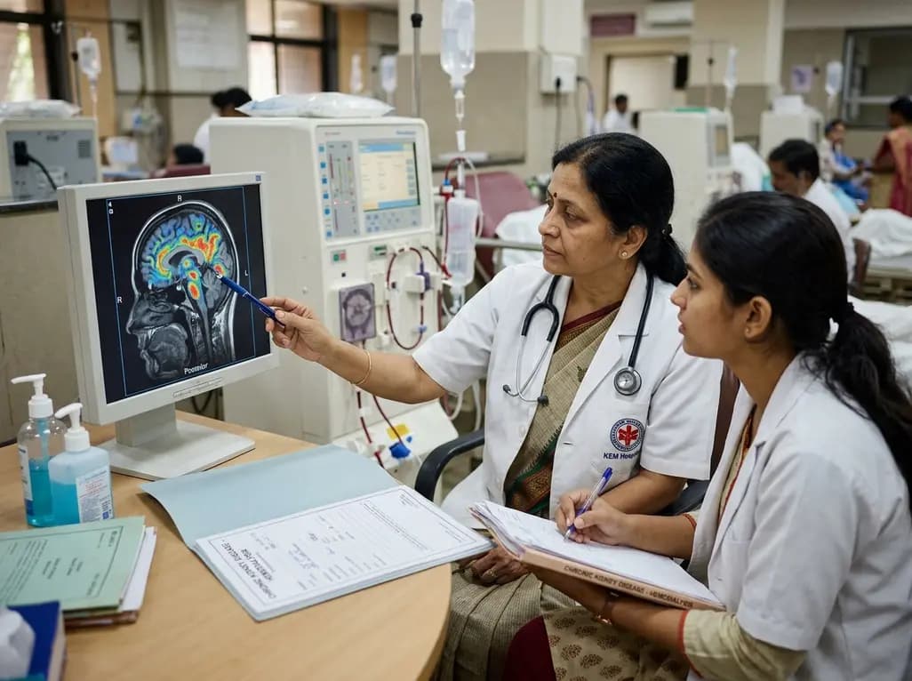
A retrospective clinical analysis reveals that hemodialysis-associated cognitive dysfunction arises from synergistic pathological mechanisms, including severe chronic anemia, worsening renal clearance, abnormal regional brain iron deposition, and an imbalance between neuroinflammatory and neurotrophic factors.
Today

A multiparametric MRI approach combining quantitative T2 shading, apparent diffusion coefficient (ADC) measurements, and morphological signs provides superior diagnostic accuracy over subjective evaluation to differentiate ovarian endometriomas from benign hemorrhagic cysts.
Today

Discover a novel laparoscopic approach for iatrogenic diaphragmatic hernia repair following pedicled omentoplasty. This modified Sugarbaker technique integrates dorsal pedicle lateralization and round ligament reinforcement to secure the diaphragmatic defect while preserving vital omental vascularity.
Yesterday
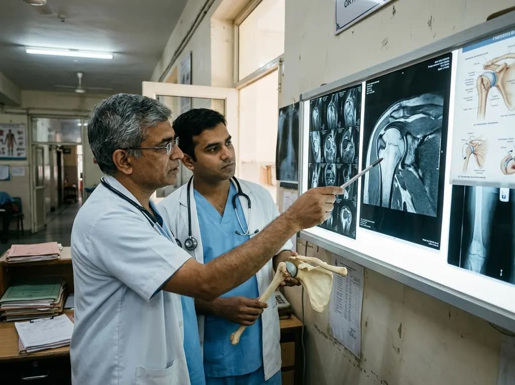
Evaluating Hill-Sachs lesion size and the distance to dislocation is critical in recurrent shoulder instability. As bone loss concepts shift from binary on-track classifications to continuous risk assessment, clinicians must identify when isolated Bankart repair is insufficient and supplementary procedures are needed.
Today

New research reveals that M1 macrophage-derived exosomes aggravate diabetic nephropathy by transferring WTAP to stabilize S1PR2 mRNA. Silencing WTAP in these vesicles attenuates endothelial injury and renal fibrosis, pointing toward innovative nanomedicine therapies.
Today
