.webp&w=384&q=75)

Loading, please wait...
Loading, please wait...

Despite extensive characterisation of systemic immune responses across peripheral organs, the immunological landscape of the central nervous system remains incompletely defined. Among the resident immune cell populations residing in brain tissue, mast cells and microglia have emerged as key modulators of neuroinflammatory processes. Additionally, these specialized cells control fundamental homeostatic functions, including sleep regulation, affective states, and energy balance. These cellular populations engage in complex bidirectional communication that is mediated significantly by the biogenic amine histamine. Although histamine was discovered over a century ago, clinicians and neuroscientists still face gaps in understanding its multifaceted roles in central nervous system homeostasis and surveillance. Investigating the link between histamine and neuroinflammation provides vital insights into neuropathophysiology and clinical neurology. Consequently, emerging research aims to clarify how histaminergic signals modulate microglial dynamics under physiological and pathological conditions.
Neuroinflammation involves complex cellular interactions where microglia continuously monitor their local microenvironment. Furthermore, mast cells located near brain blood vessels act as early responders to physiological stress, releasing prestored mediators into extracellular spaces. Therefore, understanding histamine signaling bridges immunology and neuroscience, opening new therapeutic avenues for acute and chronic neurological conditions across various clinical disciplines.
Histamine biosynthesis in the central nervous system occurs through the enzymatic decarboxylation of L-histidine by histidine decarboxylase. Histaminergic neurons originate primarily within the tuberomammillary nucleus of the posterior hypothalamus, sending widespread projections throughout the brain. However, non-neuronal sources, including mast cells and microglial populations, also synthesize and store histamine for rapid release during immunological activation. Once released into the synaptic or extracellular space, histamine exerts its actions through four distinct G protein-coupled receptors, designated as H1, H2, H3, and H4 receptors. Microglia express all four receptor subtypes, each driving unique intracellular cascades that regulate cell migration, cytokine secretion, and phagocytic activity.
Specifically, activation of H1 and H4 receptors typically promotes pro-inflammatory signaling pathways, enhancing microglial activation and the secretion of inflammatory mediators like interleukin-6 and tumor necrosis factor-alpha. In contrast, H2 receptor activation often increases intracellular cyclic adenosine monophosphate, which can suppress excessive immune reactivity. Meanwhile, presynaptic H3 receptors act as autoreceptors and heteroreceptors, modulating histamine release along with other neurotransmitters. Consequently, the selective activation or inhibition of these specific receptor subtypes allows precise tuning of neuroinflammatory responses. Understanding this intricate signaling network enables clinicians to better appreciate how pharmacological agents targeting histamine receptors influence overall central nervous system reactivity.
The functional interactions between brain mast cells and microglial populations represent a crucial axis in central immune surveillance. Mast cells strategically reside at neurovascular interfaces, such as the meninges, choroid plexus, and parenchyma, positioning them to act as sentinels. Upon degranulation, mast cells release a spectrum of bioactive substances, including histamine, tryptase, and preformed cytokines. Histamine acts directly on adjacent microglial cells, stimulating their transition from a homeostatic state to an activated phenotype. Microglia, in turn, synthesize and release inflammatory cytokines and chemokines that further stimulate mast cell degranulation, establishing a robust positive feedback loop.
This bidirectional crosstalk significantly amplifies neuroinflammatory cascades within localized brain regions. Furthermore, mast cell-derived histamine alters blood-brain barrier permeability by disrupting endothelial tight junctions. As a result, peripheral immune cells can infiltrate brain tissue, exacerbating existing inflammatory cascades. Consequently, suppressing aberrant mast cell-microglia communication offers a promising therapeutic target for limiting unchecked neuroinflammation. Modern experimental models demonstrate that disrupting this cellular dialogue attenuates microglial hyperactivation, thereby preserving neuronal integrity and protecting synaptic architecture during neuroinflammatory stress. Indian medical specialists in neurology and psychiatry must recognize these cellular dynamics when evaluating neuroinflammatory conditions.
Understanding the impact of histamine and neuroinflammation on neurodevelopmental disorders represents a rapidly evolving frontier in pediatric neurology and psychiatry. During early brain development, microglia execute indispensable functions, including synaptic pruning, axonal guidance, and neuronal survival. Disruption of these homeostatic microglial operations by aberrant histaminergic signaling can lead to altered neural circuit formation. Mast cells, which accumulate in the developing brain, release histamine in response to environmental stressors, maternal immune activation, or localized inflammation. Consequently, elevated histaminergic tone can drive microglia toward dysregulated, inflammatory states during sensitive developmental windows.
Recent clinical and preclinical studies link dysregulated histamine pathways to conditions such as autism spectrum disorder, Tourette syndrome, and attention-deficit/hyperactivity disorder. In these neurodevelopmental conditions, persistent low-grade neuroinflammation disrupts critical developmental trajectories. Furthermore, microglial activation alters neurotransmitter metabolism and synaptic plasticities, culminating in cognitive and behavioral changes. Understanding how histamine and neuroinflammation drive these aberrant microglial phenotypes allows clinicians to explore novel biomarker candidates and early intervention strategies. Thus, targeted modulation of histaminergic signaling during early development may offer new therapeutic possibilities to mitigate long-term neurodevelopmental deficits.
Pharmacological intervention targeting central histaminergic pathways relies heavily on the ability of drugs to cross the blood-brain barrier. Centrally acting antihistamines, particularly first-generation H1 receptor antagonists and selective H3/H4 receptor modulators, demonstrate significant capacity to alter microglial activity. Recent experimental evidence reveals that specific antihistamines can shift activated microglia from a pro-inflammatory phenotype toward a neuroprotective, repair-promoting state. By blocking H1 or H4 receptors on microglial membranes, these agents reduce the expression of pro-inflammatory cytokines, inducible nitric oxide synthase, and reactive oxygen species.
Additionally, novel H3 receptor antagonists and inverse agonists enhance endogenous histamine release while paradoxically regulating neuroinflammatory responses through complex feedback mechanisms. These compounds enhance cognitive performance and promote neuroprotection in preclinical models of neurodegenerative and neurodevelopmental diseases. However, classical first-generation H1 antihistamines often induce sedation and anticholinergic side effects, limiting their utility in long-term therapy. Therefore, neuropharmacologists are actively developing brain-penetrable compounds with high receptor selectivity to maximize anti-inflammatory efficacy while minimizing sedating adverse effects. These therapeutic developments hold substantial promise for treating complex neuroinflammatory disorders in clinical settings.
The integration of histaminergic pharmacology into clinical neurology promises to redefine current approaches toward neuroinflammatory disorders. Historically, antihistamines were viewed primarily as symptomatic treatments for peripheral allergic conditions. However, accumulating evidence highlights their profound capacity to modulate neuroimmune responses within the central nervous system. Translating these mechanistic insights into effective clinical therapies requires rigorous clinical trials to establish safety, optimal dosing, and brain permeability profiles. Furthermore, researchers must identify reliable biomarkers to monitor microglial activation and treatment responses in real time.
For medical practitioners, including neurologists, psychiatrists, and general physicians, understanding histamine's role in neuroinflammation expands therapeutic horizons. Combining selective histamine receptor modulators with existing anti-inflammatory or disease-modifying agents could produce synergistic therapeutic outcomes. Moreover, addressing mast cell-microglia crosstalk may prevent disease progression in conditions characterized by chronic neuroinflammation. As ongoing research clarifies these pathways, central histaminergic modulation will likely play a prominent role in personalized medicine, providing tailored interventions for patients suffering from complex neurological and psychiatric disorders.
Histamine binds to specific G protein-coupled receptors expressed on microglial surfaces, including H1, H2, H3, and H4 receptors. Activation of H1 and H4 receptors generally promotes a pro-inflammatory phenotype, increasing cytokine production and cell motility. Conversely, H2 receptor stimulation increases intracellular cyclic AMP, suppressing inflammatory mediator release. Therefore, histamine dynamic signaling directly dictates whether microglia adopt a pro-inflammatory or neuroprotective phenotype during central nervous system inflammatory challenges.
During brain development, mast cells release histamine and cytokines near neural tissue, signaling adjacent microglial cells. Microglia respond by regulating synaptic pruning, neuronal migration, and circuit maturation. However, excessive mast cell degranulation due to maternal stress or immune activation causes microglial hyperactivation. This dysregulated crosstalk alters neurodevelopmental processes, potentially contributing to conditions such as autism spectrum disorder, Tourette syndrome, and other neurodevelopmental conditions.
Yes, centrally acting antihistamines that cross the blood-brain barrier show significant neuroprotective potential. Selective H1 and H4 receptor antagonists reduce microglial pro-inflammatory cytokine release and oxidative stress. Furthermore, selective H3 receptor modulators regulate neurotransmitter release and enhance cognitive function. By shifting microglial activation from a harmful pro-inflammatory phenotype toward a reparative state, these agents offer potential therapeutic benefits for managing neuroinflammatory and neurodegenerative disorders.
Disclaimer: This content is for informational and educational purposes only. It is not intended as medical advice or a substitute for professional clinical judgment. Refer to the latest local and national guidelines for clinical practice.
References

Read summarized clinical updates, watch expert medical content, and earn CME certifications right from your smartphone.
Open your smartphone camera to scan and install.





This review highlights how histamine and centrally acting antihistamines influence microglial regulation, mast cell crosstalk, and neuroinflammation. It discusses biosynthesis, GPCR signaling pathways, and translational potential for treating neurodevelopmental and central nervous system disorders.
last month

Dendritic cells bridge innate and adaptive immunity in myocardial infarction. This review explores their pathological roles, circulating dynamics, novel tolerogenic interventions, and how standard cardiovascular medications modulate dendritic cells to improve post-infarction myocardial repair and patient outcomes.
Today

A premature neonate developed upper limb compartment syndrome after uterine rupture extruded the arm through a scar defect. Conservative management with continuous monitoring yielded complete functional recovery and normal limb growth at 10-year follow-up, highlighting non-operative safety in selected cases.
Today
Atherosclerosis involves extensive glycometabolic reprogramming across immune and vascular cells. This review examines how glycolysis, the pentose phosphate pathway, and lactate-driven epigenetic shifts fuel plaque vulnerability, while highlighting novel therapeutic targets like PFKFB3 and LDHA.
Today
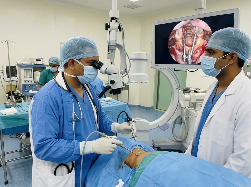
Endoscopic posterior cervical fusion combines minimally invasive decompression, joint preparation, and rigid screw-rod fixation for atlantoaxial pathologies. Early clinical findings demonstrate solid bony union, excellent symptom relief, and minimal soft-tissue morbidity without significant vascular compromise.
Yesterday

The All-India Food Processors' Association has approached the Supreme Court to oppose FSSAI's proposed per-100g benchmark for front-of-pack warning labels, advocating instead for a per-serving threshold. We explore the regulatory showdown, nutritional evidence, and implications for clinical lifestyle counseling.
Today
