

Loading, please wait...
Loading, please wait...

Endovascular intervention provides a vital therapeutic pathway for patients diagnosed with complex cerebral vascular lesions. However, achieving curative occlusion through a single procedure remains a formidable challenge for neurointerventional teams. Clinicians frequently encounter unexpected residual shunting that forces repeated catheterizations. Therefore, selecting candidates likely to succeed with a single intervention requires objective predictive markers. In modern neurovascular practice, brain AVM embolization demands nuanced evaluation that transcends traditional morphological visual assessments. Angiographic suites generate extensive dynamic data during digital subtraction angiography (DSA), yet interventionalists rarely exploit these quantitative signals to their full potential. Recent breakthrough clinical research demonstrates that quantitative hemodynamic parameters and radiomic texture features extracted from the AVM nidus accurately forecast whether patients require staged endovascular procedures.
Endovascular embolization aims to obliterate abnormal vascular connections while preserving normal brain perfusion. Nevertheless, interventionalists often face unpredictable treatment trajectories during curative brain AVM embolization. Standard clinical practice primarily relies on morphological grading systems, including the Spetzler-Martin and Buffalo scores. While these anatomical metrics evaluate lesion size, venous drainage patterns, and eloquence, they completely overlook microscopic flow dynamics. Consequently, operators cannot reliably determine whether a patient will achieve complete cure in one intervention or require multiple sequential sessions. Staged embolization minimizes abrupt intranidal pressure fluctuations and mitigates normal perfusion pressure breakthrough edema. However, multiple interventions increase cumulative anesthetic exposure, arterial access complications, radiation dose, and interim hemorrhagic risk. Therefore, clinicians urgently require objective biomarkers to anticipate procedural complexity before navigating microcatheters into fragile feeding pedicles. By identifying patients who inherently require staged interventions, multidisciplinary teams can tailor embolization strategies, optimize staged hospitalizations, or select alternative modalities like microsurgical resection or stereotactic radiosurgery.
To overcome qualitative limitations, investigators analyzed dynamic contrast flow through the AVM nidus and draining veins on DSA sequences. Specifically, quantitative DSA software calculates hemodynamic metrics from time-density curves generated within manually placed regions of interest. These objective parameters include time to peak (TTP), inflow gradients, outflow gradients, stasis index, and full width at half maximum (FWHM). Notably, lesions requiring multiple-stage embolizations demonstrated significantly lower FWHM values (1.788 ± 1.524 seconds) compared to lesions occluded in a single session (2.637 ± 1.471 seconds; p = 0.037). A shortened FWHM duration directly reflects accelerated contrast transit and high-velocity fistulous shunting through the nidus. In contrast, longer FWHM indicates slower, more tortuous intranidal circulation. Furthermore, rapid transnidal blood velocity limits the dwell time of liquid embolic agents such as Onyx or Squid during initial injection. As a result, interventionalists must exercise extreme caution to prevent non-target venous migration and premature draining vein occlusion. Hence, identifying a shortened FWHM warns operators that rapid shunting will likely impede complete single-session casting and dictate a multi-stage approach.
Beyond macroscopic hemodynamics, computational radiomics reveals intricate texture details invisible to the naked human eye. Interventionalists extract first-order and higher-order radiomic descriptors, including energy, entropy, homogeneity, and contrast, from DSA pixel intensity matrices. In the study, AVMs necessitating multiple-stage interventions exhibited strikingly higher energy values compared to single-stage lesions (11.796 ± 1.757 versus 5.763 ± 1.893; p = 0.006). Mathematically, radiomic energy quantifies textural uniformity and repetitive pixel concentration across the segmented region of interest. In vascular biology, this elevated energy reflects a densely compacted, highly organized microvascular network rather than loose, dispersed channels. Consequently, high-energy nidal lesions possess immense capillary bed density and abundant collateral micro-feeders. Moreover, these compacted microvascular tangles resist single-stage penetration because liquid embolic agents cannot effortlessly permeate every micro-channel without causing unacceptable retrograde reflux. Therefore, elevated radiomic energy serves as an objective marker for profound nidal microstructural complexity that inevitably demands repetitive, sequential embolization procedures.
Univariate associations offer valuable preliminary insights, but multivariable regression proves essential to establish independent prognostic power. When researchers performed multivariate logistic regression, nidal energy remained independently associated with incomplete occlusion after a single-stage embolization (Odds Ratio: 1.524; 95% Confidence Interval: 1.106–2.744; p = 0.039). Thus, each incremental unit increase in radiomic energy increases the odds of requiring staged endovascular therapy by over 52%. Furthermore, this predictive relationship persisted even after adjusting for confounding vascular anatomy and patient baseline characteristics. Interestingly, conventional morphological features failed to reach statistical significance in the adjusted model, underscoring the superior sensitivity of computational texture analysis. In addition, the independent predictive value of nidal energy highlights that microscopic vessel packaging dictates embolic diffusion far more than overall lesion diameter. As a result, neurointerventionalists can now quantify procedural difficulty mathematically prior to the initial puncture. This quantitative stratification substantially enhances pre-procedural risk assessment and sets realistic clinical expectations for both clinical teams and anxious families.
Accurate pre-procedural prediction of multiple-stage embolizations transforms clinical decision-making across neurovascular teams. For example, neurosurgeons and interventional neuroradiologists can proactively design safer multi-stage strategies rather than making reactive decisions mid-procedure. Staged embolization aims to incrementally reduce nidal volume while allowing adjacent normal cerebral parenchyma to adapt to altered perfusion pressures. When computational imaging identifies high nidal energy and reduced FWHM, the care team can anticipate staged embolizations from the outset. Consequently, the team avoids aggressive over-embolization in the initial session, markedly lowering the risk of intraoperative nidus rupture or acute venous hypertension. Additionally, this predictive insight guides alternative modality selection. If an unruptured lesion exhibits extreme nidal energy alongside hostile vascular anatomy, clinicians might favor primary stereotactic radiosurgery or planned surgical resection over high-risk endovascular therapy. Furthermore, accurate prediction empowers interventionalists to counsel patients transparently regarding prolonged treatment timelines, potential cumulative risks, and required follow-up imaging.
The integration of hemodynamic and radiomic processing into standard angiographic workstations represents the future of interventional neuroradiology. Modern biplane angiography units already capture high-resolution temporal and spatial datasets during routine diagnostic workups. Therefore, applying automated machine learning algorithms to segmented regions of interest can deliver real-time quantitative metrics without adding procedural time or contrast exposure. Furthermore, standardized open-source radiomic pipelines will allow validation across diverse patient populations and distinct hardware platforms. As software tools mature, interventionalists will receive automated dashboards displaying FWHM, flow gradients, and nidal energy before placing microcatheters. Subsequently, neurovascular specialists will utilize these computational biomarkers to design personalized embolization roadmaps, optimize embolic agent selection, and standardize endovascular trials globally.
Higher energy indicates uniform, densely packed vascular channels within the arteriovenous malformation nidus. Consequently, embolic penetration remains restricted during an initial intervention. Interventional teams must therefore schedule staged procedures to deposit liquid embolic agents safely without precipitating dangerous intranidal pressure shifts, vessel rupture, or catastrophic venous occlusion.
Full width at half maximum quantifies contrast transit duration across the vascular lesion. Specifically, a narrower value indicates rapid shunting and high-velocity transit through direct arteriovenous fistulous connections. Consequently, liquid embolic agents wash out more quickly, which prevents complete single-stage occlusion and necessitates staged procedures to achieve stable devascularization.
Quantitative angiography complements rather than replaces conventional scales like Spetzler-Martin or Buffalo scores. While traditional systems classify macroscopic anatomical geometry, hemodynamic and radiomic metrics capture functional microvascular kinetics. Therefore, integrating computational imaging with established anatomical grading scales substantially improves risk assessment, patient selection, and tailored neurointerventional procedural planning.
Disclaimer: This content is for informational and educational purposes only and does not constitute medical advice. Treating clinicians should exercise their independent professional judgment. Refer to the latest local and national guidelines for clinical practice.
References

Read summarized clinical updates, watch expert medical content, and earn CME certifications right from your smartphone.
Open your smartphone camera to scan and install.





A study identifies hemodynamic and radiomic markers—specifically lower FWHM and higher nidal energy on DSA—that independently predict the need for multiple-stage brain AVM embolization, aiding patient selection and treatment planning.
Today

A complex clinical case reveals how adipsic hypernatremia after traumatic brain injury, combined with hyperosmolar hyperglycemic state, triggers life-threatening dehydration and extensive venous thromboembolism, emphasizing the vital role of meticulous fluid management and vigilance.
Today

Large genomic databases like gnomAD and All of Us offer crucial insights into the population prevalence of partial RHD genotypes. Identifying these variants helps clinicians mitigate anti-D alloimmunization risks in transfusion medicine and obstetric care across diverse ancestral populations.
Yesterday
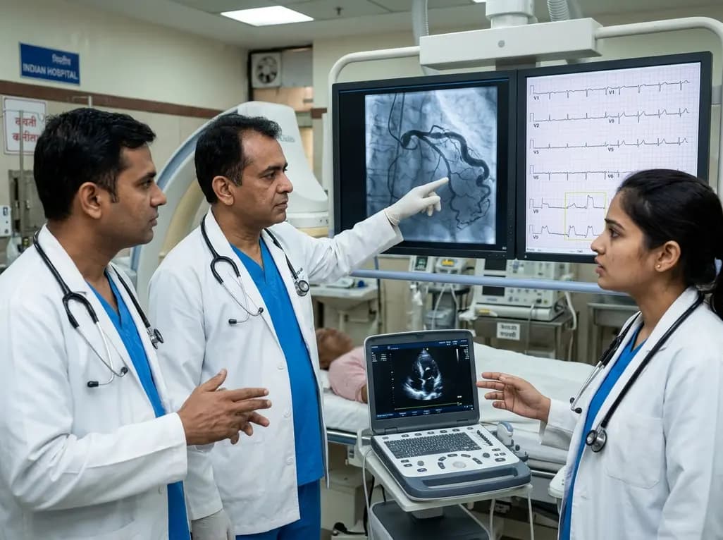
A hyperdominant left circumflex artery supplies extensive myocardial regions, creating a perilous diagnostic blind spot during acute occlusion. Standard ECGs often conceal posterior infarction, requiring clinicians to employ posterior leads and rapid echocardiography for early invasive catheterization.
Today
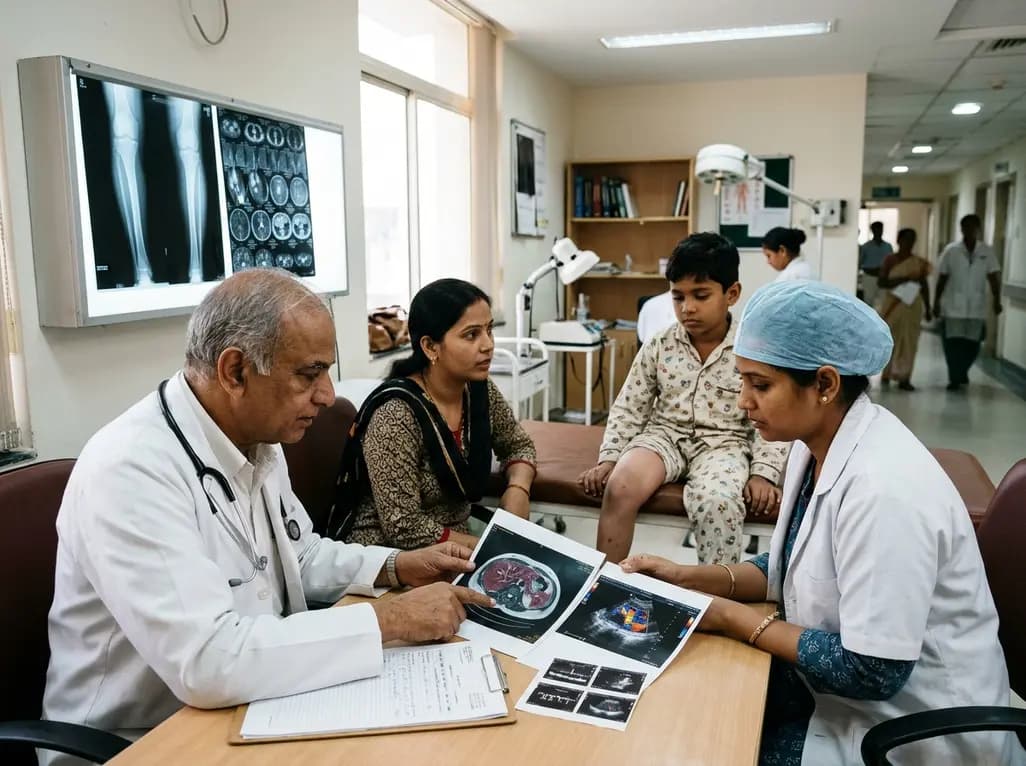
Deep intramuscular vascular malformations in pediatric Klippel-Trenaunay syndrome can mimic Maffucci syndrome. Multimodality imaging with Doppler ultrasound and MRI, along with histopathology, ensures prompt differential diagnosis, targeted surgical debulking, and optimized conservative surveillance.
Today

Discover essential clinical insights into AGLP1-type amyloidosis, a rare localized complication of glucagon-like peptide-1 analog injections. Learn how histopathology and mass spectrometry differentiate benign iatrogenic amyloidomas from systemic disease, preventing misdiagnosis and unwarranted therapies.
Yesterday
