.webp&w=384&q=75)

Loading, please wait...
Loading, please wait...
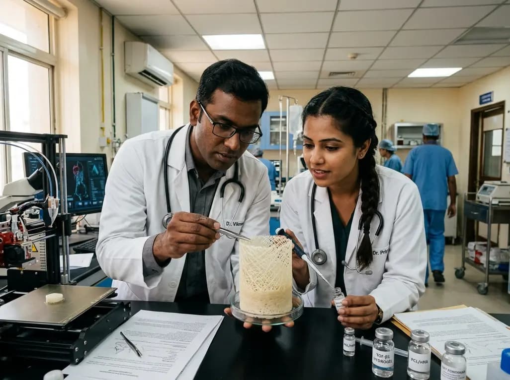
Articular cartilage lesions and deep subchondral defects present major therapeutic dilemmas in modern orthopedic practice. Because native articular cartilage lacks vascularity, spontaneous healing rarely occurs after acute trauma or chronic joint degeneration. Consequently, clinicians frequently observe progressive joint deterioration leading to debilitating osteoarthritis. Traditional surgical techniques, such as microfracture and osteochondral autografts, often yield biomechanically inferior fibrocartilage or suffer from donor-site morbidity. To overcome these critical barriers, bioengineers are developing advanced biomaterial constructs. In particular, achieving seamless osteochondral regeneration demands complex scaffolds that replicate the natural transition between mineralized bone and unmineralized cartilage. Recent breakthroughs in high-resolution manufacturing offer new hope for tissue engineering. A newly published study introduces a one-step melt electrowriting technique that encodes continuous compositional and structural gradients directly into poly(ε-caprolactone) constructs. By mimicking native tissue architecture, this approach enhances cellular differentiation and structural integration. Consequently, this innovation may redefine how orthopedic surgeons address complex joint injuries in the future.
The native osteochondral unit is a sophisticated interfacial tissue that smoothly bridges soft articular cartilage with stiff subchondral bone. Structurally, this interface features a continuous gradient of mineral distribution, collagen fiber orientation, and cellular phenotypes. Furthermore, mechanical stiffness shifts across several orders of magnitude over a span of merely a few hundred micrometers. When joint trauma disrupts this delicate zone, restoring the mechanical gradient becomes essential to prevent shear failure at the boundary. Unfortunately, conventional multi-layered scaffolds often suffer from interfacial delamination due to sharp boundaries between distinct layers. Moreover, standard manufacturing strategies struggle to balance microscale fiber resolution with macroscopic pore interconnectivity. As a result, implanted constructs often fail to withstand physiologic mechanical loads during joint movement. Therefore, orthopedic specialists require next-generation scaffolds that mimic natural gradient architecture without abrupt physical boundaries. Creating such seamless transitions remains a fundamental goal for durable joint preservation and long-term functional recovery.
To address interfacial weakness, researchers employed melt electrowriting (MEW) to establish a generalizable platform for osteochondral regeneration. Melt electrowriting combines high-voltage electric fields with thermal extrusion to deposit microscale polymer filaments with sub-micron positional accuracy. In this pioneering protocol, investigators sequentially loaded nanohydroxyapatite-laden poly(ε-caprolactone) melts into a single printing barrel. Consequently, the predetermined mineral gradient remained stable throughout the additive manufacturing run. This continuous extrusion generated a steady decline in mineral concentration across the total scaffold thickness. Furthermore, the varying inorganic content intrinsically modulated melt viscosity and fiber diameter during printing. Thus, the process achieved simultaneous compositional and architectural gradients in a single automated step. This single-step approach completely eliminates the need for gluing or stitching separate material layers. As a result, the fabricated constructs exhibit exceptional mechanical durability, pore interconnectivity, and structural integrity under dynamic loading conditions.
The newly engineered scaffold features a specialized biomimetic gradient that closely mirrors native osteochondral anatomy. At the bottom layer, high concentrations of nanohydroxyapatite mimic the mineralized microenvironment of subchondral bone. Moving upward toward the superficial articular zone, the mineral content gradually declines to zero. Interestingly, this chemical transition directly influences fiber deposition dynamics. The fibers become progressively thinner toward the chondral surface, creating fine architectural networks suited for cartilage matrix deposition. In addition, researchers incorporated a porous hydrogel coating loaded with transforming growth factor-beta 1 across the upper compartment. This bioactive coating supplies essential biochemical cues to induce chondrogenesis while preserving open pore networks. Therefore, nutrient transport, cell migration, and fluid permeation remain unhindered throughout the three-dimensional construct. By harmonizing chemical composition, physical geometry, and localized growth factors, the scaffold creates distinct biological microenvironments within a unified physical framework.
To evaluate biological performance, investigators seeded human bone marrow mesenchymal stem cells onto the gradient scaffolds. The experimental findings demonstrated coordinated spatial differentiation of stem cells across different depths of the construct. Specifically, cells residing in the mineral-rich lower compartment exhibited robust osteogenic differentiation, marked by elevated alkaline phosphatase activity and calcium deposition. Simultaneously, cells in the upper growth-factor-enriched zone differentiated along the chondrogenic lineage, synthesizing abundant type II collagen and glycosaminoglycans. Furthermore, in vivo evaluations confirmed that these gradient scaffolds promote integrated osteochondral repair without structural failure. The newly formed tissue exhibited smooth integration with host subchondral bone and surrounding articular cartilage. Consequently, the healed defect showed superior compressive strength and histological organization compared to homogenous control scaffolds. These results validate the capacity of graded scaffolds to direct site-specific tissue regeneration in complex joint environments.
These technological advancements hold substantial translational relevance for clinicians managing challenging joint disorders. Current clinical modalities for large osteochondral defects, such as autologous chondrocyte implantation or allograft transplantation, face significant logistical and biological limitations. In contrast, off-the-shelf gradient scaffolds fabricated through melt electrowriting could provide reproducible, customized implants tailored to specific patient defect profiles. Moreover, the robust interface reduces the long-term risk of construct displacement or graft failure under weight-bearing conditions. As regenerative medicine continues to evolve in India and worldwide, integrating biomimetic scaffolds with point-of-care stem cell therapies could improve outcomes for active individuals and athletes. However, extensive pre-clinical validation in large animal models and standardized clinical trials will be necessary before widespread clinical implementation. Nevertheless, this one-step gradient encoding platform represents a vital milestone toward predictable, functional joint restoration.
Melt electrowriting offers exceptional spatial control by producing precisely ordered fibers with diameters ranging from single micrometers to tens of micrometers. Unlike standard extrusion 3D printing, it creates highly porous, compliant networks that replicate native extracellular matrix architecture. Furthermore, melt electrowriting completely avoids toxic organic solvents, thereby supporting robust cellular viability and rapid tissue infiltration during joint repair.
Gradient scaffolds present spatially tailored biophysical and biochemical signals across their depth. The mineralized subchondral zone contains nanohydroxyapatite that actively directs mesenchymal stem cells toward osteogenic differentiation. Meanwhile, the superficial chondral zone features finer fibers and transforming growth factor-beta 1, which stimulates robust chondrogenic extracellular matrix synthesis to regenerate durable articular cartilage.
Key translational hurdles include confirming long-term mechanical durability in large animal weight-bearing joint models and obtaining regulatory approval for advanced combination biomaterials. Additionally, commercial manufacturing must consistently achieve sterile Good Manufacturing Practice compliance at scale. Orthopedic surgeons also require standardized surgical instruments and delivery techniques to ensure precise, secure scaffold fixation during minimally invasive joint repair procedures.
Disclaimer: This content is for informational and educational purposes only and does not constitute medical advice, diagnosis, or treatment. Healthcare professionals should exercise their independent clinical judgment when evaluating new research and therapeutic strategies. Refer to the latest local and national guidelines for clinical practice.
References

Read summarized clinical updates, watch expert medical content, and earn CME certifications right from your smartphone.
Open your smartphone camera to scan and install.





Researchers have engineered a one-step melt electrowriting technique to fabricate graded PCL/nHA scaffolds with TGF-β1 hydrogel coating, advancing osteochondral regeneration and integrated tissue repair.
last month
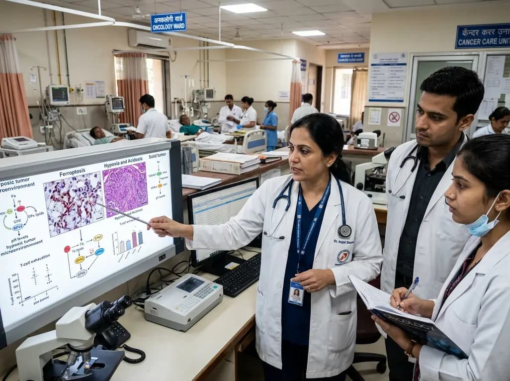
Targeting ferroptosis provides a compelling strategy to dismantle hypoxia- and acidosis-driven immunosuppression in solid tumors, enhancing response to modern immunotherapy.
Today

Findings from Project Viva indicate that sustained postpartum weight retention, rather than excessive gestational weight gain alone, directly worsens women's mid-life cardiovascular health, degrading blood pressure and glycemic parameters decades after delivery.
Today

A large UK Biobank study reveals that plasma proteomic signatures capture preclinical organ damage and improve multiorgan risk prediction across early CKM syndrome stages 0 to 2 beyond traditional PREVENT clinical models.
Today

A retrospective CBCT study reveals that superior TMJ space asymmetry correlates moderately with occipital condyle-atlas-axis asymmetry in unilateral TMJ osteoarthritis, highlighting an imaging correlate between jaw biomechanics and upper cervical alignment rather than a standalone diagnostic marker.
Today

A Bayesian multilevel meta-analysis reveals that aerobic training combined with moderate carbohydrate restriction modestly lowers HbA1c in type 2 diabetes. However, sparse data and very low certainty leave incremental benefits over exercise or diet alone unproven, highlighting the need for individualized care.
Today
