.webp&w=384&q=75)

Loading, please wait...
Loading, please wait...

Total hip arthroplasty provides excellent pain relief for end-stage osteoarthritis. However, persistent lateral hip pain remains a vexing challenge for orthopedic surgeons. Degenerative abductor tendon tears often drive this persistent post-surgical morbidity. When conservative measures fail, open gluteus medius repair offers a viable salvage option to restore stability and function. While clinicians frequently address concurrent abductor tears during primary arthroplasty, isolated repairs performed months or years after arthroplasty represent an understudied clinical scenario. Recent registry investigations now clarify long-term functional recovery after secondary abductor repair.
Lateral hip pain following total hip arthroplasty often stems from greater trochanteric pain syndrome or frank abductor detachment. Consequently, orthopedic surgeons must differentiate benign trochanteric bursitis from progressive tendon tears. Degenerative gluteus medius and minimus tears disrupt the pelvic force couple, leading to severe Trendelenburg lurch and chronic pain. Furthermore, surgical dissection and tissue devascularization during primary surgery can compromise soft-tissue integrity. Patients present with gait instability, sleep disturbance, and abductor weakness.
Recent multicenter evidence demonstrates that tear patterns vary widely among symptomatic patients. Specifically, high-grade partial tears account for over half of presentations, while full-thickness ruptures comprise more than one-third. Additionally, roughly one-third of cases involve concomitant gluteus minimus disruption. Without timely surgical intervention, retracted abductor tendons undergo progressive fatty degeneration. Therefore, clinicians must recognize failing abductors before irreversible muscle atrophy occurs.
Surgeons consider open gluteus medius repair when non-operative treatments fail to resolve chronic lateral hip pain and limp. Standard conservative management includes physical therapy, eccentric loading, nonsteroidal anti-inflammatory drugs, and trochanteric injections. However, refractory abductor insufficiency requires direct anatomical restoration. Open transosseous suture anchor fixation remains the gold standard for robust tendon reattachment over prosthetic hardware.
During surgical exposure, surgeons carefully mobilize retracted tendon edges while protecting nearby neurovascular structures. Subsequently, clinicians prepare the greater trochanteric footprint to bleeding cancellous bone to foster biological healing. High-strength suture anchors secure the tendon in a tension-free configuration. Furthermore, surgeons often repair concurrent gluteus minimus tears to re-establish dynamic hip abductor mechanics. In cases involving severe tissue loss, biological augmentation provides additional mechanical stability. Thus, meticulous operative technique ensures secure fixation and promotes durable long-term healing.
Recent clinical registry findings demonstrate remarkable improvements following open abductor repair after prior primary arthroplasty. At an average follow-up exceeding four and a half years, patients achieved clinically meaningful recovery across standard hip metrics. Specifically, mean modified Harris Hip Scores improved dramatically from baseline values around 44 to post-surgical values exceeding 71. Therefore, surgical reconstruction reliably restores functional independence and ambulatory endurance.
In addition to objective scores, patients experience profound subjective pain relief and improved quality of life. International Hip Outcome Tool scores and visual analogue scale ratings show substantial post-surgical gains. Consequently, most patients successfully achieve established minimal clinically important difference thresholds. Furthermore, patient satisfaction remains consistently high across multi-year evaluations. Despite the technical complexity of secondary abductor surgery, complication rates remain low. These durable results confirm that secondary abductor reconstruction provides lasting relief for recalcitrant trochanteric pain.
Postoperative rehabilitation directly dictates the final outcome of abductor tendon reconstruction. Tendon-to-bone integration requires sustained mechanical protection during the initial biological healing phase. Therefore, physical therapists enforce strict protected weight-bearing protocols with assistive devices for six to eight weeks postoperatively. Moreover, clinicians must strictly restrict active hip abduction, passive adduction, and excessive external rotation to protect suture anchor integrity.
Following the initial healing period, patients transition to progressive passive and active-assisted motion. Subsequently, gentle isometric abductor strengthening begins under close physiotherapeutic supervision. Closed-chain kinetic exercises and gait training help eliminate compensatory pelvic tilting. In addition, core stabilization exercises reinforce lumbo-pelvic balance and decrease shearing forces across the greater trochanter. Patient compliance plays a decisive role during recovery. Ultimately, structured rehabilitation protocols maximize functional recovery and prevent catastrophic re-tears during daily tasks.
Accurate diagnosis of secondary abductor tears requires high clinical suspicion and targeted imaging. Persistent pain over the greater trochanter after successful arthroplasty warrants thorough clinical evaluation. Physical examination typically demonstrates marked local tenderness, weak resisted hip abduction, and an asymmetric single-leg stance. However, clinicians must first exclude prosthetic joint infection, component loosening, and referred lumbar radiculopathy before attributing symptoms to soft tissue failure.
Metal artifact reduction magnetic resonance imaging provides exceptional visualization of peritrochanteric soft tissues around metallic components. Furthermore, dynamic high-resolution musculoskeletal ultrasound serves as an accessible, cost-effective diagnostic alternative for identifying tendon detachment. During primary arthroplasty, surgeons can prevent iatrogenic abductor damage through gentle tissue handling and anatomical repair of detached tendons. When post-arthroplasty tears occur, timely recognition prevents extensive tendon retraction and muscular fatty infiltration. Thus, early multidisciplinary evaluation ensures optimal candidate selection and superior clinical outcomes.
Several prognostic variables influence long-term outcomes following abductor reconstruction in arthroplasty patients. Tissue quality remains the single most critical determinant of structural healing. Severe preoperative muscle fatty degeneration correlates with diminished functional recovery and higher failure risk. In contrast, patients with preserved muscle bulk achieve substantially greater strength gains and walking endurance.
Moreover, patient age, general medical comorbidities, and body mass index influence recovery. Elevated body mass index increases mechanical strain across the repair site. Consequently, surgeons must optimize metabolic health and provide comprehensive pre-surgical counseling. Multicenter registry data confirm that high-grade partial and full-thickness tears both benefit markedly from prompt surgical reconstruction. Therefore, surgeons should offer timely repair before irreversible structural degradation supervenes. Ultimately, robust biomechanical fixation delivers durable functional gains.
Gluteus medius and minimus pathology affects up to twenty percent of patients presenting with degenerative hip osteoarthritis. Following total hip arthroplasty, persistent lateral hip pain occurs in approximately five to ten percent of cases. While bursitis causes many symptoms, significant abductor tears represent a major structural etiology. High-grade partial tears occur most frequently, followed by full-thickness tendon ruptures, necessitating thorough diagnostic evaluation when pain persists despite standard conservative therapy.
Magnetic resonance imaging with metal artifact reduction sequences serves as the gold standard imaging modality around hip prostheses. It clearly delineates tear thickness, tendon retraction, and muscle fatty infiltration. In addition, high-resolution musculoskeletal ultrasound provides an excellent, cost-effective alternative. Ultrasound allows dynamic real-time assessment of tendon continuity without metal distortion. Clinicians routinely combine these imaging modalities with standard radiographs to rule out component loosening, periprosthetic fractures, and hardware malposition.
Protecting the repaired abductor tendon requires strict adherence to physical therapy restrictions during early tissue healing. Patients must maintain protected partial weight-bearing with assistive devices for six to eight weeks. Furthermore, patients must strictly avoid active hip abduction, passive hip adduction across midline, and excessive internal or external rotation. Controlled passive motion begins early, while active abductor strengthening starts only after structural healing occurs, minimizing the risk of suture anchor failure.
Disclaimer: This content is for informational and educational purposes only... Refer to the latest local and national guidelines for clinical practice.
References

Read summarized clinical updates, watch expert medical content, and earn CME certifications right from your smartphone.
Open your smartphone camera to scan and install.





Secondary open gluteus medius repair after total hip arthroplasty yields significant, durable improvements in hip function, pain relief, and patient satisfaction at a minimum two-year follow-up.
Today
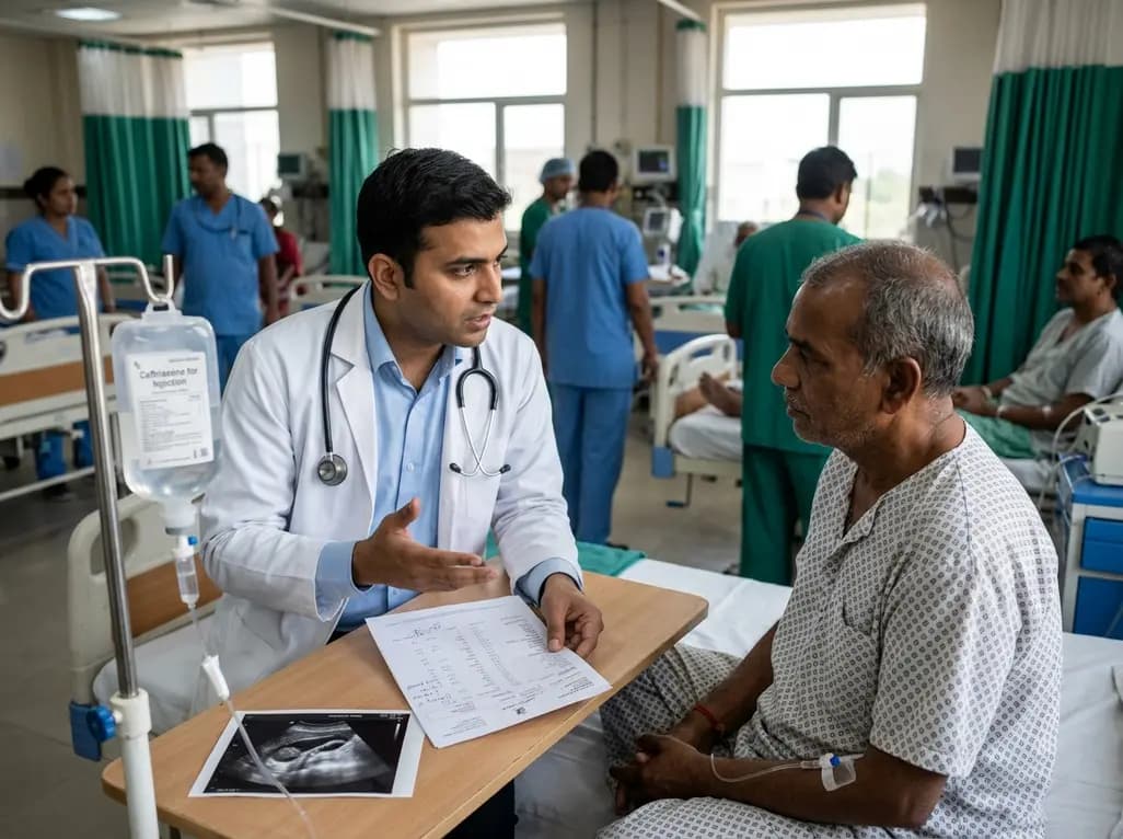
Ceftriaxone therapy can lead to biliary sludge and gallstone formation due to calcium-ceftriaxone precipitation. A recent cohort study shows that the noninvasive Fibrosis-4 (FIB-4) index, alongside treatment duration, independently predicts lithogenesis, enabling proactive risk stratification.
Today

A multiparametric MRI approach combining quantitative T2 shading, apparent diffusion coefficient (ADC) measurements, and morphological signs provides superior diagnostic accuracy over subjective evaluation to differentiate ovarian endometriomas from benign hemorrhagic cysts.
Today

A landmark case report reveals a donor heart beating for a century in a 63-year-old recipient, challenging conventional age limits in cardiac transplantation and offering critical lessons on organ longevity, donor selection, and long-term graft survival.
Today

New research reveals that M1 macrophage-derived exosomes aggravate diabetic nephropathy by transferring WTAP to stabilize S1PR2 mRNA. Silencing WTAP in these vesicles attenuates endothelial injury and renal fibrosis, pointing toward innovative nanomedicine therapies.
Yesterday
