.webp&w=384&q=75)

Loading, please wait...
Loading, please wait...

Major depressive disorder imposes an immense public health burden worldwide, and clinicians continually seek objective biomarkers to understand therapeutic responses. Emerging neuroimaging modalities now allow direct evaluation of central neurochemical architecture in psychiatric patients. In particular, the clinical application of GluCEST MRI in depression provides an unprecedented window into regional neurotransmitter dynamics. Glutamate chemical exchange saturation transfer imaging visualizes proton exchange between solute amine groups and bulk water molecules. Consequently, this technique maps in vivo glutamate distributions at high spatial resolution without requiring radioactive tracers or invasive interventions.
Historically, proton magnetic resonance spectroscopy offered limited anatomical granularity when measuring cerebral amino acids. In contrast, GluCEST technology quantifies magnetization transfer ratio asymmetry values across distinct subcortical and cortical structures with superior spatial definition. A landmark prospective investigation recently examined unmedicated individuals suffering from major depression alongside matched healthy controls. Investigators sought to determine whether baseline glutamatergic deficits characterize acute depressive episodes. Furthermore, they evaluated whether standard pharmacotherapy reverses these neurometabolic abnormalities over eight weeks. Ultimately, this novel molecular imaging approach bridges cellular neuropharmacology and macroscopic neurocircuitry in clinical psychiatry.
The corticolimbic network governs emotional processing, executive function, and stress resilience across human populations. At baseline evaluation, investigators identified significant neurochemical differences between unmedicated depressed patients and healthy volunteers. Specifically, patients with major depressive disorder demonstrated significantly lower magnetization transfer ratio asymmetry values within the left hippocampus. Moreover, bilateral prefrontal cortical regions exhibited comparable glutamatergic suppression. These quantitative reductions reflect impaired synaptic glutamate pooling, diminished cellular storage, or altered glial-neuronal metabolic cycling.
Glutamate serves as the primary excitatory neurotransmitter throughout mammalian neural networks, driving cognitive and affective equilibrium. Therefore, localized neurochemical depletion disrupts dendritic arborization, synaptic plasticity, and long-term potentiation within critical mood circuits. Chronic psychological distress frequently accelerates excitotoxic wear, astrocytic dysfunction, and impaired amino acid clearance. Consequently, subcortical circuits struggle to sustain adaptive functional connectivity during severe depressive episodes. These baseline imaging findings confirm that clinical depression entails profound disturbances beyond classical monoaminergic neurotransmitter depletion. Clinicians must recognize that corticolimbic hypoglutamatergia represents a robust neuropathological hallmark of untreated illness.
Selective serotonin reuptake inhibitors represent the most common first-line pharmacotherapy for managing moderate-to-severe depressive illness. However, the precise downstream mechanisms linking serotonin transporter blockade to cellular remodeling have long remained contentious. In this prospective longitudinal cohort, depressed participants completed eight weeks of standardized antidepressant treatment. Following this therapeutic intervention, repeat GluCEST imaging revealed dramatic physiological changes across targeted cerebral structures. Specifically, patients exhibited significant increases in magnetization transfer ratio asymmetry values compared to their unmedicated baseline scans.
Remarkably, this restorative surge encompassed the bilateral hippocampus and bilateral prefrontal cortex. Antidepressant therapy effectively normalized regional glutamate concentrations toward levels typically observed in healthy individuals. Thus, sustained serotonergic stimulation promotes downstream glutamatergic restoration throughout forebrain networks. Mechanistically, increased synaptic serotonin availability stimulates postsynaptic receptors that trigger essential intracellular neurotrophic signaling pathways. Furthermore, these molecular cascades upregulate astrocytic glutamate transporters and enhance metabolic cycling. As a result, pharmacotherapy successfully re-establishes metabolic equilibrium across vulnerable corticolimbic structures over time.
To establish meaningful clinical relevance, researchers evaluated whether regional neurochemical shifts correlated directly with psychometric improvements. Clinicians utilized the 17-item Hamilton Depression Rating Scale to track symptom severity before and after pharmacotherapy. Notably, longitudinal increases in left hippocampal glutamate signals correlated positively with total score reductions across the cohort. Patients who achieved greater neurochemical recovery experienced superior overall clinical remission.
Furthermore, detailed domain analyses revealed striking associations with distinct vegetative and cognitive symptom clusters. Treatment-induced glutamate replenishment within the left prefrontal cortex and left hippocampus mirrored notable improvements in sleep disturbance scores. Similarly, patients demonstrated concurrent relief from psychomotor retardation as regional amino acid concentrations rose toward normal values. These correlation analyses suggest that glutamatergic modulation directly drives symptomatic relief in responders. Consequently, neurochemical restoration within the dominant hemisphere appears integral for recovering affective vigor, psychomotor speed, and normal circadian function.
For several decades, classical biological psychiatry focused almost exclusively on monoaminergic neurotransmission when explaining antidepressant efficacy. Nonetheless, contemporary neuroscience demonstrates intricate crosstalk between monoamine pathways and amino acid neurotransmitter systems. Serotonin receptor activation directly modulates presynaptic glutamate release and postsynaptic AMPA receptor trafficking across central synapses. In addition, chronic antidepressant exposure enhances brain-derived neurotrophic factor expression within the hippocampus and neocortex.
This neurotrophic surge stimulates synaptogenesis, repairs dendritic branching, and restores astrocyte metabolic viability. Because astrocytes actively clear extracellular glutamate and synthesize glutamine, their functional renewal normalizes parenchymal glutamate storage. Therefore, selective serotonin reuptake inhibitors exert their long-term therapeutic effects by reorganizing excitatory neurotransmission rather than altering serotonin alone. These neuroimaging discoveries align closely with rapid-acting glutamatergic interventions, highlighting a unifying biological pathway. Ultimately, successful antidepressant therapy hinges upon restoring cortical excitability and dynamic neural network plasticity.
These clinical findings provide valuable diagnostic and prognostic perspectives for daily psychiatric and primary care practice. Understanding the central role of glutamate helps physicians explain antidepressant latency and therapeutic mechanisms to patients effectively. Pharmacotherapy requires several weeks because downstream neuroplastic reorganization and metabolic restoration occur gradually. Furthermore, persistent residual symptoms may reflect incomplete normalization of regional excitatory neurochemistry within corticolimbic structures.
In the future, non-invasive molecular imaging could guide personalized treatment selection and therapeutic monitoring. Clinicians might identify specific patient subgroups exhibiting pronounced glutamatergic deficits who require targeted neuromodulatory approaches. Moreover, monitoring regional chemical exchange saturation transfer signals could evaluate early treatment responsiveness before overt behavioral changes manifest clinically. While widespread clinical adoption requires accessible ultra-high-field MRI infrastructure, these insights enrich our understanding of affective neuroscience. Clinicians should incorporate these neurochemical concepts when discussing comprehensive, biologically informed management strategies with patients.
GluCEST MRI measures chemical exchange between glutamate amine protons and bulk water protons after selective radiofrequency saturation. This non-invasive method generates magnetization transfer asymmetry values, providing high-resolution anatomical maps of in vivo glutamate concentrations. Consequently, it surpasses conventional magnetic resonance spectroscopy in spatial resolution without using ionizing radiation.
Chronic stress and depressive episodes impair glial-neuronal metabolic coupling and downregulate astrocytic glutamate recycling. Furthermore, dendritic atrophy and decreased synaptic density diminish the total intracortical glutamate pool. These pathophysiological alterations collectively reduce regional glutamate-weighted imaging signals in vulnerable prefrontal and hippocampal circuits during untreated illness.
Although SSRIs primarily inhibit serotonin reuptake, sustained serotonergic stimulation activates neurotrophic cascades like BDNF and enhances astrocytic function. Consequently, chronic pharmacotherapy promotes synaptic plasticity, improves glial amino acid transport, and elevates glutamate-sensitive signals. This restorative mechanism normalizes corticolimbic neurotransmitter concentrations, facilitating clinical recovery from depressive symptoms.
Disclaimer: This content is for informational and educational purposes only. It is not intended to be a substitute for professional medical advice, diagnosis, or treatment. Always seek the advice of your physician or other qualified health provider with any questions you may have regarding a medical condition. Refer to the latest local and national guidelines for clinical practice.
References

Read summarized clinical updates, watch expert medical content, and earn CME certifications right from your smartphone.
Open your smartphone camera to scan and install.





A prospective GluCEST MRI study demonstrates that unmedicated patients with major depressive disorder exhibit prefrontal and hippocampal glutamate deficits, which significantly increase after eight weeks of SSRI treatment in correlation with clinical symptom improvement.
Today

Survival analyses often report hazard ratios, but mathematical non-collapsibility creates crucial differences between conditional and marginal estimates. Clinicians must account for unmeasured confounding and model selection when interpreting trial endpoints and real-world cardiovascular outcomes.
Today

A cross-sectional study of 700 college students highlights significant contraceptive knowledge disparities between sexually active and inactive young adults. The findings demonstrate a critical need for proactive, comprehensive sexual health counseling before sexual debut.
Yesterday
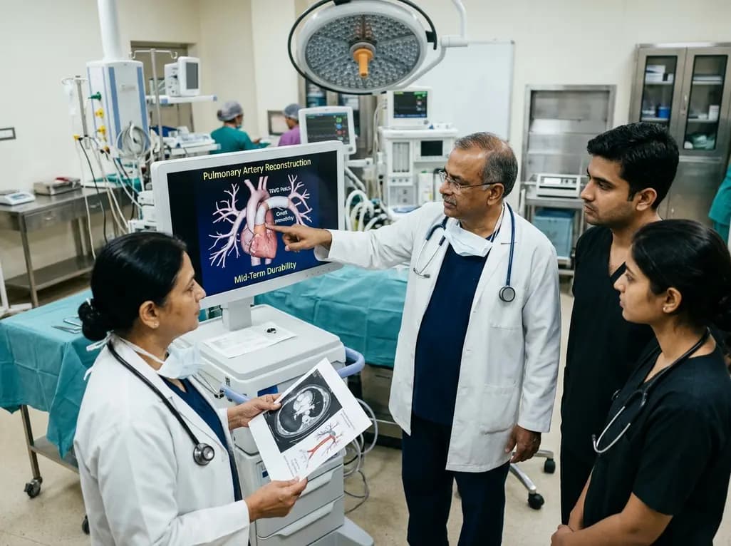
Autologous tissue-engineered vascular graft patches created via in-body tissue architecture show excellent mid-term durability and safety for pulmonary artery reconstruction in congenital heart surgery, particularly for peripheral branch augmentation.
Today

A novel cluster analysis of real-world datasets reveals four distinct CPPD disease phenotypes, validating existing EULAR categories while uncovering previously unrecognized monoarticular and axial presentations to refine patient management.
Today

Recent pharmacokinetic research demonstrates that trace dermal exposure to microgram quantities of trenbolone and metenolone produces detectable urinary metabolites for days to over a week, providing critical objective data for evaluating unintentional contamination claims in sports drug testing.
Today
