.webp&w=384&q=75)

Loading, please wait...
Loading, please wait...
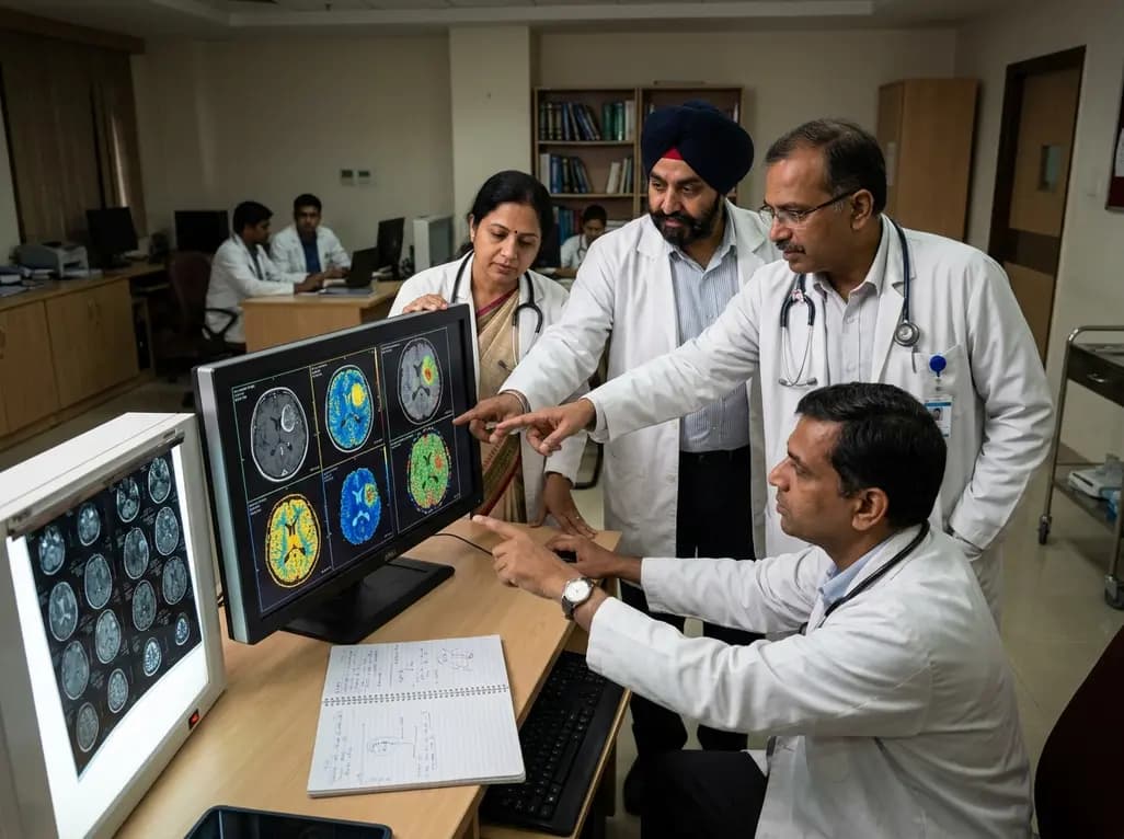
Diffuse gliomas present profound clinical challenges due to their marked spatial heterogeneity and infiltrative biology. Clinicians increasingly rely on advanced neuroimaging modalities to evaluate tumor microenvironments noninvasively. Recent breakthroughs utilizing glioma radio-pathomic maps allow physicians to translate standard MRI signals into microscopic tissue features, including cellular density and interstitial fluid. This digital pathology paradigm offers deep insights into tumor cellularity and infiltrative margins without requiring invasive surgical biopsies.
Conventional magnetic resonance imaging provides structural visualization but fails to capture the true histological complexity of diffuse gliomas. Consequently, neuro-oncologists frequently face uncertainty when delineating active tumor cores from infiltrative margins. To overcome this diagnostic hurdle, investigators developed radio-pathomic mapping by training machine learning algorithms on direct autopsy tissue specimens. These sophisticated models directly correlate multiparametric MRI signal intensities with quantitative histological metrics, such as cell density and extracellular fluid distribution.
Furthermore, these predictive computational tools bridge the gap between microscopic pathology and macroscopic radiology. Rather than relying on non-specific contrast enhancement, clinicians can now visualize tumor cellularity across distinct anatomical compartments. This spatial precision enables teams to identify hidden malignant foci that traditional scans overlook. In addition, the noninvasive nature of these computational algorithms facilitates serial longitudinal monitoring throughout therapy. Ultimately, bridging neuroimaging with computational histopathology provides unprecedented diagnostic accuracy for aggressive intracranial neoplasms.
Validating imaging biomarkers across independent medical institutions remains critical for broad clinical translation. Accordingly, investigators conducted a rigorous retrospective analysis leveraging two prominent open-access datasets, UPenn-GBM and UCSF-PDGM. The team integrated diverse perfusion techniques, analyzing dynamic susceptibility contrast derived relative cerebral blood volume alongside arterial spin labeling derived cerebral blood flow. Additionally, they incorporated diffusion tensor imaging metrics, specifically fractional anisotropy, across all enrolled subjects.
Researchers applied the autopsy-validated radio-pathomic algorithm to standard structural sequences to compute voxel-wise cellularity and extracellular fluid density maps. Subsequently, they performed statistical evaluations across contrast-enhancing tumor regions and non-enhancing peritumoral FLAIR hyperintensity zones. This multi-institutional design directly mitigates site-specific acquisition biases and scanner variations. Furthermore, validating algorithmic robustness across multiple magnetic field strengths ensures exceptional generalizability. Consequently, this multi-center framework establishes a definitive methodological foundation for deploying radio-pathomic pipelines in everyday clinical oncology.
The study established substantial quantitative relationships between perfusion parameters and tumor microstructural density within contrast-enhancing regions. Specifically, both relative cerebral blood volume and arterial spin labeling measurements exhibited robust positive correlations with radio-pathomic cell density. These observations confirm that hypervascular tumor regions correspond closely with heightened neoplastic proliferation and dense cellular clustering.
Moreover, dynamic susceptibility contrast perfusion displayed consistent physiological alignment with calculated extracellular fluid levels. High-grade malignant gliomas exhibit rapid angiogenesis and disordered vessel architecture, which induces elevated vascular permeability and interstitial leakage. Therefore, elevated perfusion metrics directly reflect active tumor neoangiogenesis alongside dense cellular packing. These findings provide compelling biological validation for perfusion-weighted imaging. Clinicians can confidently utilize these cross-validated parameters to assess tumor aggressiveness prior to surgical intervention. Furthermore, this quantitative concordance reinforces the reliability of radio-pathomic computational models in characterizing tumor biology.
Diffusion tensor imaging provides critical complementary information regarding microstructural integrity and cellular organization. In this multi-site cohort, fractional anisotropy demonstrated distinct associations with radio-pathomic parameters across distinct tumor subregions. In contrast-enhancing cores, fractional anisotropy correlated positively with cell density, reflecting directional water diffusion constraints within tightly packed malignant cells.
However, peritumoral FLAIR hyperintensity regions exhibited distinct physiological dynamics. Within these non-enhancing margins, extracellular fluid density significantly influenced diffusion characteristics, demonstrating the complex interplay between infiltrative tumor cells and vasogenic edema. Furthermore, the presence of disrupted white matter tracts in peritumoral zones further altered fractional anisotropy readings. By capturing these subtle microstructural variations, diffusion metrics help neuro-oncologists differentiate purely edematous brain parenchyma from occult tumor infiltration. Consequently, combining diffusion and perfusion metrics with radio-pathomic models yields a comprehensive portrait of the intracranial tumor microenvironment.
Applying glioma radio-pathomic maps into clinical neuro-oncology holds transformative potential for patient management and precision care. Standard imaging often struggles to differentiate true progression from radiation-induced pseudoprogression, creating substantial therapeutic dilemmas. Because radio-pathomic algorithms quantify cellularity directly, clinicians can accurately distinguish hypercellular tumor recurrence from hypocellular treatment-related necrosis.
Furthermore, these predictive maps assist neuro-oncologists in monitoring real-time treatment response during systemic chemotherapy and anti-angiogenic therapy. When patients receive anti-VEGF agents like bevacizumab, vascular normalization can mask tumor growth on routine post-contrast scans. Radio-pathomic analysis circumvents this pseudoresponse artifact by tracking cellular density independently of blood-brain barrier permeability. Additionally, integrating these automated quantitative biomarkers into multidisciplinary tumor boards enhances objective prognostic stratification. As a result, neuro-oncologists can tailor personalized therapeutic regimens, preventing premature treatment cessation and improving patient outcomes.
Surgical resection and adjuvant radiotherapy rely heavily on precise target delineation to maximize efficacy while sparing functional brain tissue. However, infiltrative glioma cells frequently extend far beyond visible contrast-enhancing boundaries into surrounding FLAIR hyperintense zones. Radio-pathomic maps successfully uncover these stealth invasive cellular pockets, offering neurosurgeons clear guidance during supramarginal resection planning.
Similarly, radiation oncologists can harness these quantitative spatial maps to optimize target volume contouring. Rather than applying uniform radiation doses across broad peritumoral areas, clinicians can deliver focused dose escalation to regions demonstrating elevated cell density. Consequently, this targeted approach minimizes radiation-induced neurotoxicity to healthy eloquent cortex while maximizing tumor control. Moreover, post-surgical monitoring gains substantial reliability when baseline radio-pathomic contours are available for serial comparison. In summary, integrating multi-site validated imaging signatures into neurosurgical and radiotherapeutic workflows represents a vital paradigm shift toward tailored glioma management.
Standard MRI protocols primarily display macroscopic anatomical structures and gross vascular permeability through non-specific contrast enhancement. In contrast, radio-pathomic maps translate conventional imaging voxel signals into quantitative microscopic tissue properties. Specifically, they accurately estimate cellular density and extracellular fluid distribution, revealing occult tumor infiltration beyond traditional radiological boundaries.
Multi-site validation confirms that computational imaging biomarkers remain reliable across different hospital settings, scanner manufacturers, and acquisition protocols. By validating algorithms using diverse datasets from institutions like UPenn and UCSF, researchers ensure that radio-pathomic metrics maintain physiological accuracy and statistical robustness, enabling safe clinical translation into everyday neuro-oncology workflows.
Evaluating peritumoral FLAIR hyperintensity enables clinicians to differentiate benign vasogenic edema from active, infiltrative glioma cells. Identifying high cell density within these non-enhancing margins guides neurosurgeons during supramarginal resection and helps radiation oncologists target dose escalation accurately, ultimately reducing local recurrence rates and sparing healthy functional brain tissue.
Disclaimer: This content is for informational and educational purposes only. It is not intended to be a substitute for professional medical advice, diagnosis, or treatment. Always seek the advice of your physician or another qualified health provider with any questions you may have regarding a medical condition. Refer to the latest local and national guidelines for clinical practice.
References

Read summarized clinical updates, watch expert medical content, and earn CME certifications right from your smartphone.
Open your smartphone camera to scan and install.





A multi-site study demonstrates that advanced diffusion and perfusion MRI significantly correlate with glioma radio-pathomic maps of cell density and extracellular fluid, enhancing noninvasive tumor characterization in contrast-enhancing and peritumoral brain regions.
last month

The CARE study protocol introduces a multilingual, RAG-powered conversational interface designed by IIT Bombay to support tobacco cessation in India. Evaluating feasibility and behavioral outcomes across three cessation centers, this digital health initiative aims to enhance quitting support for diverse users.
Today

A large UK cohort study of 1.4 million women shows that pregnancy complications, including gestational diabetes, hypertension, and preterm birth, substantially raise long-term risks for hypertension, chronic kidney disease, type 2 diabetes, and cardiovascular events, highlighting early postpartum intervention needs.
Today
A cross-sectional study reveals that hypertension knowledge, illness perceptions, depression, and substance use significantly predict self-care behaviors. Addressing mental health and cognitive beliefs in primary care is crucial for optimal blood pressure control and cardiovascular disease prevention.
Today
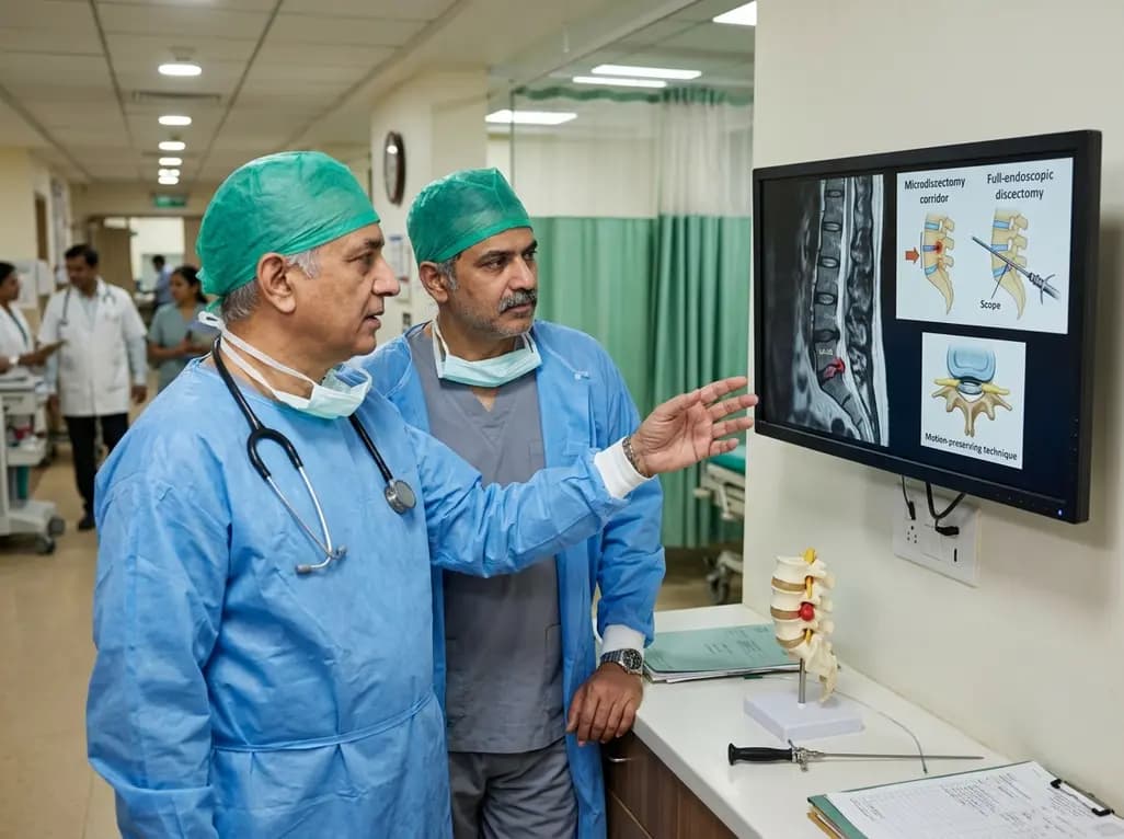
This clinical overview examines recent advancements in lumbar disc herniation surgery, comparing microscopic and full-endoscopic discectomy while highlighting biportal techniques and emerging motion-preserving innovations for spine specialists.
Today
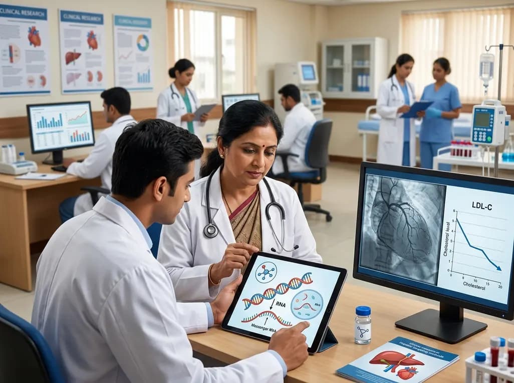
Small interfering RNA (siRNA) therapeutics harness RNA interference to degrade disease-causing messenger RNA. By delivering hepatocyte-targeted, durable gene silencing, agents targeting PCSK9, LPA, and angiotensinogen offer potent cardiovascular risk reduction with infrequent, biannual dosing schedules.
Today
