

Loading, please wait...
Loading, please wait...

Glioblastoma multiforme remains one of the most fatal adult central nervous system malignancies. Clinicians frequently encounter profound therapeutic resistance due to marked intratumoral heterogeneity. Positron emission tomography imaging often reveals distinct regional variations across the tumor mass. Specifically, glioblastoma metabolic lesions display contrasting areas of high and low metabolic activity. However, standard therapeutic protocols fail to account for these spatial distinctions. A groundbreaking study in Neuro-Oncology now reveals that spatial genomic evolution directly shapes these metabolic boundaries. Consequently, these findings offer novel diagnostic markers and therapeutic avenues for modern neuro-oncology practice.
Glioblastoma tumors do not represent homogeneous cellular masses. Instead, they harbor genetically diverse subclonal populations that thrive under distinct physiological stresses. Clinicians evaluate these tumors using amino acid positron emission tomography tracers such as fluoroethyl-L-tyrosine. In clinical practice, these scans consistently distinguish hypermetabolic hotspots from surrounding hypometabolic tumor tissues. Until recently, oncologists struggled to explain how these metabolic differences arose at the genomic level. Furthermore, clinicians questioned whether hypermetabolic zones simply reflected higher cell density or distinct evolutionary trajectories. To address this mystery, researchers designed an elegant multi-regional sampling strategy. Surgical teams performed image-guided stereotactic biopsies to isolate autologous hypometabolic and hypermetabolic tumor fragments from individual patients. Subsequently, investigators interrogated these distinct regions using comprehensive whole-genome, whole-exome, and transcriptome profiling. The resulting data established that glioblastoma metabolic lesions reflect dynamic spatial evolution rather than simple vascular variance. Hypometabolic zones serve as the foundational evolutionary soil. From these quiet territories, aggressive subclonal populations emerge and build intense metabolic lesions. Therefore, spatial metabolic mapping provides a direct window into subclonal divergence. Moreover, understanding these spatial dynamics helps surgeons identify target regions for maximal safe resection. Clinicians can thereby correlate specific imaging phenotypes with localized molecular vulnerability.
Multiomic sequencing revealed that hypermetabolic lesions evolve directly from hypometabolic ancestral regions. Rather than arising independently, hypermetabolic lesions inherit primary driver mutations while acquiring secondary genomic disruptions. Moreover, hypermetabolic lesions demonstrate strategic focal copy number alterations, including selective oncogene amplifications and key tumor suppressor deletions. Hypometabolic regions preserve an earlier evolutionary snapshot of the disease. In contrast, hypermetabolic niches harbor severe structural variants and extensive chromosomal instability. Consequently, these hyperactive clones display heightened proliferative capacity and profound metabolic reprogramming. The investigators also evaluated transcriptomic signatures across both tumor compartments. Importantly, hypermetabolic niches showed elevated expression of genes that mediate cellular survival, nutrient uptake, and biosynthetic flux. Simultaneously, these regions downregulated classical tumor suppressors that restrain uncontrolled cell growth. Thus, the transition from low to high metabolic activity represents a fundamental genomic shift. This shift equips tumor cells to withstand microenvironmental deprivation and immune assault. Neuro-oncologists must recognize that single biopsies fail to capture this complex spatial landscape. Ultimately, multi-regional molecular profiling explains why targeted therapies frequently trigger incomplete clinical responses. Additionally, these genomic divergence patterns indicate that distinct areas of the same mass follow branching evolutionary trees during disease progression.
A pivotal discovery within hypermetabolic glioblastoma lesions involves the dramatic activation of the APOBEC3 cytidine deaminase family. These enzymes induce specific cytosine-to-thymine mutations across single-stranded DNA during replication. Although APOBEC3 enzymes normally defend host cells against viral retroelements, aberrant expression generates pervasive genomic instability in cancers. In this study, hypermetabolic tissues exhibited intense APOBEC3 expression alongside classic mutational signatures. Furthermore, this enzymatic hyperactivity coincided with increased DNA damage response signaling. As a direct result, hypermetabolic clones acquired diverse subclonal passenger mutations at an accelerated rate. This rapid mutagenesis generates extensive genetic variety, providing the raw material for rapid drug resistance. When chemotherapy or radiation challenges the tumor, pre-existing subclonal variants easily escape eradication. Additionally, hypoxic signaling pathways synergize with APOBEC3 activity in these hypermetabolic areas. Because oxygen deprivation stresses replicating cells, it accelerates mutagenesis and drives clonal divergence. Therefore, heightened APOBEC3 expression serves as a critical catalyst for evolutionary progression in aggressive brain tumors. Clinicians must view hypermetabolic lesions not merely as energy-hungry tissues, but as hyper-mutating evolutionary hubs. Consequently, therapeutic strategies designed to inhibit APOBEC enzymes could potentially retard clonal diversification and stabilize treatment sensitivity over time.
Glioblastoma progression relies heavily on pathological angiogenesis to supply burgeoning energetic demands. In this multiomic investigation, researchers identified dipeptidase 1, abbreviated as DPEP1, as a novel vascular endothelial tip marker. DPEP1 expression localized specifically to active vascular tip cells within hypermetabolic tumor territories. In contrast, quiescent blood vessels in hypometabolic regions displayed minimal DPEP1 presence. Functionally, DPEP1 acts as a membrane-bound zinc metalloproteinase that hydrolyzes dipeptides and coordinates nutrient transport across cellular barriers. By regulating specific transporter activities, DPEP1 directly promotes endothelial cell sprouting and rapid vascular expansion. Furthermore, DPEP1-rich tip cells fuel tumor metabolism by optimizing amino acid delivery to adjacent glioma cells. This vascular reprogramming creates a robust feed-forward loop that sustains rapid malignant growth. When endothelial cells express DPEP1, glioblastoma clones receive sustained metabolic support and thrive despite severe microenvironmental pressure. Survival analyses also confirmed that elevated DPEP1 expression correlates strongly with poor clinical survival in glioblastoma cohorts. Consequently, DPEP1 represents both a key pathophysiological driver and a functional biomarker of aggressive vascular niche biology. Targeted blockade of DPEP1 could disrupt this vital nutrient highway. Importantly, blocking this enzymatic pathway might deprive aggressive tumor zones of crucial metabolic intermediates without disrupting normal cerebral vasculature.
These groundbreaking insights carry immediate clinical relevance for practicing neurosurgeons and neuro-oncologists. Traditional neurosurgical planning relies on standard gadolinium-enhanced magnetic resonance imaging. However, contrast enhancement frequently fails to delineate the full extent of invasive, metabolically aggressive tumor margins. By integrating amino acid PET imaging into preoperative workflows, surgical teams can identify hypermetabolic foci with high precision. Neurosurgeons can subsequently tailor supramarginal resection strategies toward these biologically virulent zones. Furthermore, pathologists must incorporate multi-site tissue sampling protocols during diagnostic evaluation. Analyzing both hypometabolic and hypermetabolic zones prevents misleading diagnostic conclusions drawn from single tissue fragments. On the therapeutic horizon, targeting DPEP1-driven angiogenesis offers an innovative strategy to overcome bevacizumab resistance. Because DPEP1 operates specifically on endothelial tip cells, selective inhibitors might suppress pathological neovascularization without triggering widespread neurotoxicity. In addition, suppressing APOBEC3-mediated mutational bursts could delay the emergence of chemotherapy resistance. Combining metabolic imaging, aggressive targeted resection, and pathway-specific pharmacology will redefine neuro-oncology standards. Clinicians must embrace these spatial evolutionary principles to deliver truly personalized interventions. Ultimately, integrating molecular pathology with nuclear neuroimaging bridges the long-standing divide between diagnostic tumor localization and precise biological eradication.
Clinicians define these lesions using specialized positron emission tomography tracers that evaluate amino acid uptake. Hypermetabolic lesions demonstrate intense tracer avidity, reflecting heightened metabolic flux, increased cellularity, and robust microvascular proliferation. In contrast, hypometabolic lesions display reduced tracer accumulation, serving as ancestral tumor zones with distinct, less aggressive genomic profiles.
Dipeptidase 1 acts as a specific vascular endothelial tip cell marker within aggressive hypermetabolic zones. The enzyme regulates nutrient transporter activity and hydrolyzes dipeptides, directly stimulating endothelial sprouting and robust angiogenesis. Consequently, this increased vascularity delivers vital metabolic substrates to surrounding malignant cells, accelerating invasive tumor growth and shortening patient survival.
APOBEC3 enzymes drive significant cytosine deamination, causing pervasive somatic mutations across replication forks in hypermetabolic glioblastoma niches. This accelerated mutational rate generates extensive clonal diversity within the tumor mass. Consequently, these newly formed subclonal variants facilitate rapid evasion of standard radiotherapy and temozolomide chemotherapy, fueling post-treatment tumor recurrence.
Disclaimer: This content is for informational and educational purposes only and does not constitute formal medical advice, diagnosis, or treatment. Healthcare providers must exercise their clinical judgment when evaluating new neuro-oncology research and patient care protocols. Refer to the latest local and national guidelines for clinical practice.
References

Read summarized clinical updates, watch expert medical content, and earn CME certifications right from your smartphone.
Open your smartphone camera to scan and install.





Multiomic profiling of autologous glioblastoma metabolic lesions reveals that hypermetabolic regions evolve from hypometabolic tissue via focal genomic alterations, APOBEC3 activation, and dipeptidase 1 (DPEP1) upregulation, which drives endothelial tip proliferation and tumor angiogenesis.
Today

A complex clinical case reveals how adipsic hypernatremia after traumatic brain injury, combined with hyperosmolar hyperglycemic state, triggers life-threatening dehydration and extensive venous thromboembolism, emphasizing the vital role of meticulous fluid management and vigilance.
Today

Large genomic databases like gnomAD and All of Us offer crucial insights into the population prevalence of partial RHD genotypes. Identifying these variants helps clinicians mitigate anti-D alloimmunization risks in transfusion medicine and obstetric care across diverse ancestral populations.
Yesterday
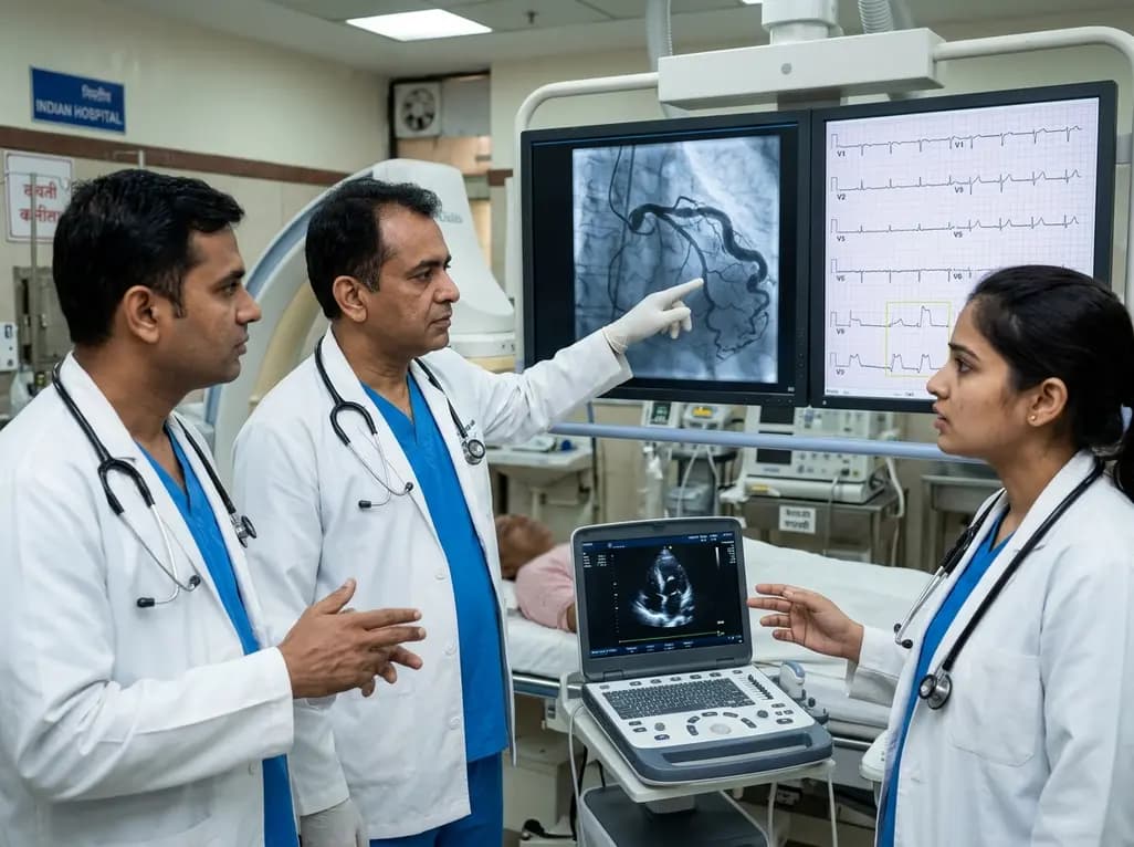
A hyperdominant left circumflex artery supplies extensive myocardial regions, creating a perilous diagnostic blind spot during acute occlusion. Standard ECGs often conceal posterior infarction, requiring clinicians to employ posterior leads and rapid echocardiography for early invasive catheterization.
Today
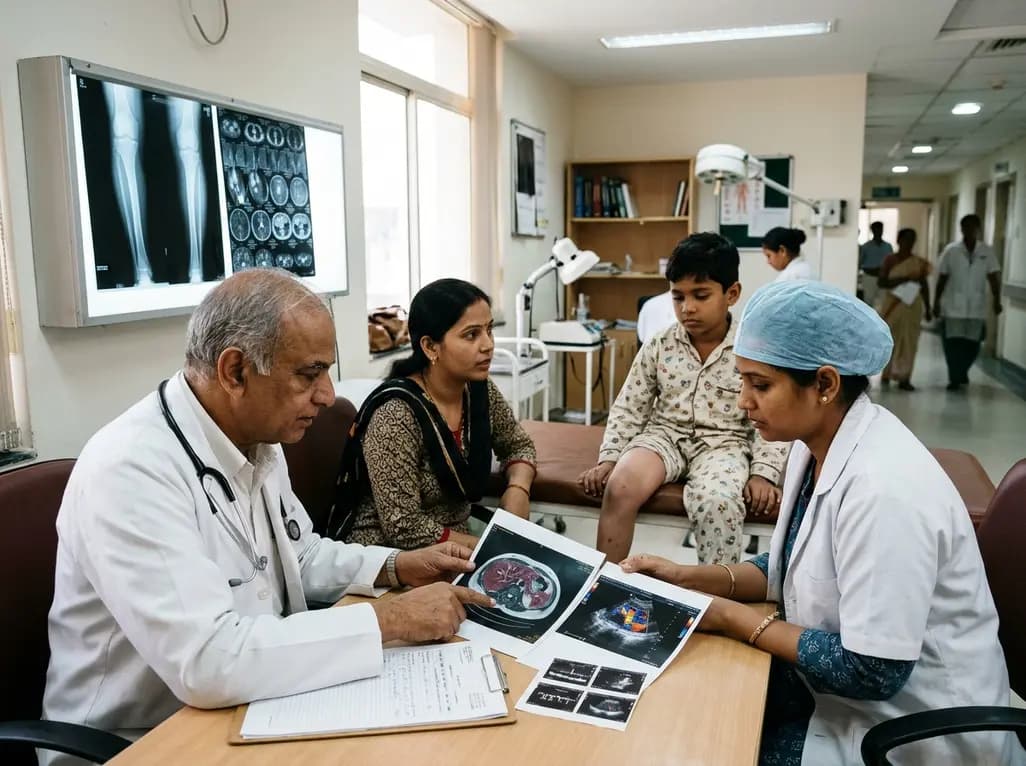
Deep intramuscular vascular malformations in pediatric Klippel-Trenaunay syndrome can mimic Maffucci syndrome. Multimodality imaging with Doppler ultrasound and MRI, along with histopathology, ensures prompt differential diagnosis, targeted surgical debulking, and optimized conservative surveillance.
Today

Discover essential clinical insights into AGLP1-type amyloidosis, a rare localized complication of glucagon-like peptide-1 analog injections. Learn how histopathology and mass spectrometry differentiate benign iatrogenic amyloidomas from systemic disease, preventing misdiagnosis and unwarranted therapies.
Yesterday
