.webp&w=384&q=75)

Loading, please wait...
Loading, please wait...

The management of primary open-angle glaucoma has evolved significantly, yet the underlying glaucomatous trabecular meshwork mechanics remain a complex frontier for medical researchers and clinicians alike. Recent studies utilize sophisticated 3D traction force microscopy to examine how cells interact with their environment in segment-resolved regions of the eye. Historically, many researchers viewed the trabecular meshwork as a uniform filter, but modern evidence confirms a highly segmental flow pattern within the conventional outflow pathway. By distinguishing between tracer-identified high-flow (HF) and low-flow (LF) regions, scientists can better understand why certain areas become more resistant to aqueous drainage over time. This research explores the delicate interplay between the cellular cytoskeleton and extracellular collagen fibers, specifically focusing on how specific pharmacological agents alter these mechanical signatures. Understanding these cellular forces is crucial for developing targeted therapies that restore normal outflow and manage intraocular pressure more effectively in clinical practice. Consequently, recognizing that the trabecular meshwork behaves differently across its circumference allows for a more nuanced approach to glaucoma pathology. This shift in perspective is essential because it moves away from a generalized model of resistance toward a localized understanding of tissue dysfunction. Therefore, analyzing these mechanics at a granular level provides a clearer picture of disease progression and therapeutic potential.
To quantify these mechanical changes, researchers used 3D traction force microscopy coupled with collagen kinematics to observe glaucomatous trabecular meshwork cells. The experimental setup involved seeding matched high-flow and low-flow cultures from a single glaucomatous donor eye onto fibrillar type I collagen gels. These gels contained fluorescent FluoSpheres, which allowed for the precise tracking of microscopic movements as the cells exerted force on their substrate. By imaging the cells hourly for 12 hours, the team could reconstruct mean traction forces and analyze how different cytoskeletal inhibitors influenced cellular behavior. Notably, the study found that in control environments, reconstructed mean traction increased consistently over the 12-hour period in both high-flow and low-flow regions. This indicates a baseline level of contractile activity that is inherent to these cells even in a diseased state. Furthermore, the use of a 3D environment is critical because traditional 2D cultures often fail to replicate the complex mechanical stresses found in the human eye. By utilizing a more physiologically relevant model, the study highlights how the extracellular matrix and the cytoskeleton work in tandem to regulate the structural integrity of the outflow pathway. This approach eventually reveals how the physical properties of the trabecular meshwork are maintained or compromised in glaucomatous conditions.
Pharmacological intervention in this study focused on three distinct cytoskeletal components: actin, microtubules, and intermediate filaments. Latrunculin B, a well-known actin disruptor, reduced cellular traction early in the observation period, though cells showed a partial recovery as time progressed. This drug is particularly interesting to clinicians because it is known to decrease outflow resistance by expanding the juxtacanalicular space. On the other hand, nocodazole was used to target microtubules. The results for nocodazole were striking, showing significant differences between high-flow and low-flow regions at the 12-hour mark. Specifically, high-flow cells treated with nocodazole maintained much higher traction and strain compared to their low-flow counterparts. Moreover, nocodazole treatment shifted the mechanical divergence toward convergence, suggesting a total reorganization of how these cells distribute force across the collagen matrix. These findings are important because they suggest that microtubule stability plays a region-specific role in maintaining the mechanical homeostasis of the trabecular meshwork. Additionally, the divergent responses between HF and LF cells highlight the underlying biological heterogeneity that exists within a single donor eye. Such variability implies that a one-size-fits-all pharmacological approach might not be the most effective way to treat segmental resistance in glaucoma patients.
Withaferin A was another critical agent used in the study, primarily targeting the intermediate filament vimentin. This compound significantly reduced traction, strain, and 'curl'—a measure of collagen fiber deformation—in both high-flow and low-flow regions by the end of the 12-hour period. Interestingly, Withaferin A also altered the dynamics of collagen orientation angles specifically within the low-flow regions. This specific mechanical response suggests that intermediate filaments are heavily involved in how low-flow cells anchor themselves and manipulate their surroundings. Because low-flow regions are often associated with higher stiffness and increased extracellular matrix deposition in glaucomatous eyes, targeting these areas specifically could be a viable therapeutic strategy. Furthermore, mixed-effects models used in the study confirmed that Withaferin A caused a measurable difference in strain change over time within the low-flow segments. This finding is significant because it points toward a distinct mechanical phenotype for low-flow cells that is sensitive to intermediate filament disruption. Consequently, therapies that can modulate these filaments might offer a new pathway for reducing the rigid mechanical state characteristic of the glaucomatous trabecular meshwork. Therefore, Withaferin A represents a promising tool for both experimental research and potential drug development aimed at the conventional outflow pathway.
For ophthalmologists in India, where the burden of primary open-angle glaucoma is substantial, understanding glaucomatous trabecular meshwork mechanics is vital for optimizing patient care. Current treatments often rely on prostaglandin analogues or beta-blockers, which primarily target aqueous production or uveoscleral outflow. However, newer classes of drugs, such as Rho-kinase inhibitors, specifically target the trabecular meshwork's contractile properties to lower intraocular pressure. The findings of this study reinforce the idea that the trabecular meshwork is a dynamic, mechanically active tissue rather than a passive sieve. By demonstrating that different cytoskeletal components control traction in a region-specific manner, the research suggests that combination therapies could be more effective. For instance, a drug that targets actin assembly might be paired with one that modulates microtubules to provide a more comprehensive reduction in tissue stiffness. Furthermore, the segmental differences identified in this study emphasize the need for precision medicine in glaucoma. If clinicians can eventually identify which regions of a patient's trabecular meshwork are most compromised, they may be able to tailor treatments to those specific areas. This level of personalization could lead to better long-term outcomes and a reduction in the need for invasive surgical procedures like trabeculectomy.
The use of a segment-resolved assay supports a comparative evaluation of cytoskeletal perturbations that was previously difficult to achieve. While this study provides a foundational understanding using a donor-matched pair, the researchers motivate the need for validation across additional donor eyes. Establishing a larger database of mechanical responses will help confirm whether these high-flow and low-flow differences are consistent across various stages of glaucoma. Additionally, future research could explore how these mechanical signatures correlate with gene expression profiles in different regions. By linking mechanical behavior to molecular signaling, scientists might uncover new biomarkers for disease progression. Moreover, the integration of 3D traction force microscopy into standard research protocols will likely accelerate the discovery of novel compounds that can safely modify the outflow pathway. As we move toward more bioengineered models of the eye, the ability to replicate and manipulate segmental flow will be essential. Ultimately, these advancements in ocular biomechanics promise to bridge the gap between benchside research and bedside clinical applications. Therefore, the ongoing study of the cellular cytoskeleton and its role in glaucoma remains one of the most promising areas for future ophthalmological breakthroughs and improved patient management strategies.
The trabecular meshwork does not drain fluid equally across its entire structure. Instead, it features high-flow and low-flow regions with distinct mechanical and molecular properties. Understanding these differences is crucial because glaucoma often causes specific areas to become more resistant than others. By targeting the unique mechanical signatures of low-flow regions, researchers hope to develop more precise therapies that restore uniform drainage and manage eye pressure more effectively.
Latrunculin B primarily disrupts actin filaments, leading to an immediate but partially reversible reduction in the traction forces exerted by trabecular meshwork cells. In contrast, nocodazole targets microtubules and produces region-specific effects. In this study, nocodazole caused a significant divergence in mechanical response between high-flow and low-flow cells at 12 hours. This suggests that while actin provides overall contractility, microtubules help maintain the specific mechanical identity of different flow segments.
3D traction force microscopy allows researchers to measure the actual physical forces that cells exert on their environment in three dimensions. This is far more accurate than traditional 2D models because the trabecular meshwork is a complex, multi-layered tissue. By using this technology, scientists can see how cells pull on collagen fibers and how drugs like Withaferin A alter those physical interactions, providing deeper insights into the biomechanical failures that lead to glaucoma.
Disclaimer: This content is for informational and educational purposes only. It is not intended as medical advice or to replace the professional judgment of a healthcare provider. Always seek the advice of your physician or other qualified health provider with any questions regarding a medical condition. Refer to the latest local and national guidelines for clinical practice.
References
Karimi A et al. Differential Cytoskeletal Control of Cell-Collagen Mechanics in High- and Low-Flow Glaucomatous Trabecular Meshwork Cells. ACS Appl Mater Interfaces. 2026 Jul 10. doi: 10.1021/acsami.6c10225. PMID: 42430182.
Raghunathan VK et al. Biomechanical properties of the human trabecular meshwork in relation to outflow facility and age. Investigative Ophthalmology & Visual Science. 2018;59(1):348-356.
Vahane N et al. Segmental outflow and trabecular meshwork stiffness in an ocular hypertensive mouse model. bioRxiv. 2024. doi: 10.1101/2024.02.05.578912.

Read summarized clinical updates, watch expert medical content, and earn CME certifications right from your smartphone.
Open your smartphone camera to scan and install.





A new study quantifies how cytoskeletal drugs like Latrunculin B and Withaferin A affect cell-collagen mechanics in high- and low-flow regions of glaucomatous trabecular meshwork cells, revealing critical segmental differences in mechanical response and potential targets for glaucoma therapy.
3 months ago

A retrospective cohort study reveals that frailty quadruples the odds of pressure injuries in hospitalized older adults. Integrating the Clinical Frailty Scale with comprehensive geriatric care is crucial for identifying vulnerability early and implementing targeted multidisciplinary prevention.
Today

Occult maternal malignancy accounts for 3.82% of persistent unreportable NIPT results with genomic noise. Multiple chromosomal aberrations, notably on chromosome 18, warrant thorough oncological evaluation and whole-body MRI to ensure timely maternal diagnosis while preserving fetal care.
Today

This article reviews the forensic and clinical complexities of acute myocarditis arising on left ventricular hypertrophy in children, highlighting early diagnostic red flags and multidisciplinary postmortem autopsy protocols.
Today
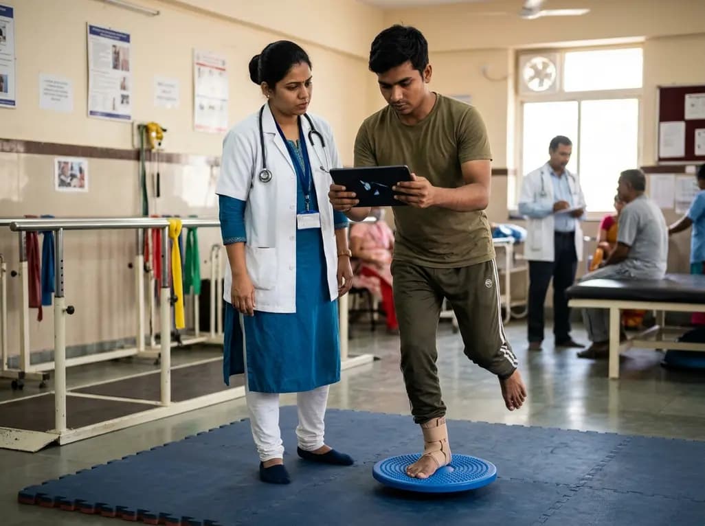
A randomized controlled trial demonstrates that an eight-week dual-task training intervention significantly improves static and dynamic postural control over standard balance exercises in individuals with chronic ankle instability, addressing critical neurocognitive deficits during athletic and daily activities.
Today

A retrospective cohort study of 7,716 ICU patients from the MIMIC-IV database demonstrates that Gradient Boosting Decision Trees significantly outperform traditional SOFA scores in predicting in-hospital mortality among patients with sepsis and diabetes, with SHAP analysis providing clinical interpretability.
Today
