.webp&w=384&q=75)

Loading, please wait...
Loading, please wait...
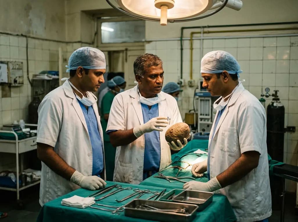
In contemporary urological practice, encountering a giant bladder stone has become an exceptional rarity. Historically common, these massive calculi now appear only when underlying pathology remains neglected for years. Recently, clinicians treated a 37-year-old male who harbored an intravesical calculus weighing 1200 grams. Computed tomography confirmed the massive stone, which surgeons successfully removed via open cystolithotomy. Consequently, this presentation highlights the importance of recognizing chronic urinary stasis early. Furthermore, it demonstrates the value of open surgery when endourology reaches practical limits.
Primary vesical calculi develop infrequently in adults with adequate nutrition. Instead, secondary factors account for the overwhelming majority of massive stone cases. Specifically, persistent bladder outlet obstruction remains the most common predisposing etiology. When benign prostatic hyperplasia, urethral strictures, or bladder neck contractures impede voiding, residual urine accumulates continuously. Consequently, chronic urinary stasis allows dissolved solutes to supersaturate, nucleate, and coalesce into crystalline matrices. In addition, chronic urinary tract infections from urease-producing microorganisms accelerate crystal aggregation. Pathogens such as Proteus mirabilis and Klebsiella species hydrolyze urea into ammonia and carbon dioxide. As a result, urinary pH rises markedly, promoting rapid precipitation of magnesium ammonium phosphate. Neurogenic bladder dysfunction further exacerbates this pathogenic cycle by blunting normal visceral sensation. Over many years, patients adapt to gradual discomfort, allowing stones to attain immense proportions. Therefore, clinicians must identify and treat the underlying mechanical or functional obstruction. Addressing this fundamental stasis remains just as vital as extracting the stone.
Patients harboring massive vesical calculi present with a diverse spectrum of lower urinary tract complaints. Initially, individuals describe progressive dysuria, frequency, urgency, and intermittent gross hematuria. Moreover, sudden terminal interruption of the urinary stream occurs when the mobile stone obstructs the bladder neck during micturition. Suprapubic pain often intensifies with physical exertion or postural changes. However, some patients experience surprisingly mild symptoms because the bladder accommodates the growing mass over decades. Accurate diagnosis requires a structured multimodal approach. A standard pelvic radiograph reveals dense radio-opaque shadows over the pelvis. Nevertheless, non-contrast computed tomography represents the definitive diagnostic imaging modality. CT scans precisely measure stone dimensions, evaluate volume, and detect secondary hydronephrosis. Furthermore, clinicians must obtain baseline serum creatinine measurements and urine cultures before intervention. Finally, diagnostic cystoscopy enables surgeons to examine the bladder mucosa directly. This step successfully excludes concurrent bladder diverticula, strictures, or malignancy.
Endourology provides reliable minimally invasive treatments for routine urinary calculi, including transurethral lithotripsy and holmium laser fragmentation. These techniques reduce hospital stays, minimize surgical trauma, and facilitate rapid patient recovery. However, their utility decreases sharply when urologists encounter an exceptionally massive stone burden. Fragmenting a giant bladder stone endoscopically requires prolonged operative time and high mechanical energy. In addition, extended manipulation increases the risk of bladder perforation, mucosal injury, and severe fluid extravasation. Therefore, open cystolithotomy remains the primary surgical standard for massive stones. A lower midline suprapubic incision allows direct, unobstructed access to the bladder cavity. Consequently, surgeons extract the complete calculus safely in a single operative session. Furthermore, open surgery allows concurrent management of associated anatomical abnormalities, such as prostatic enlargement or diverticula. In centers with limited endoscopic technology, open cystolithotomy provides an accessible, cost-effective, and definitive therapeutic solution. Thus, open surgery remains an indispensable skill in modern urological practice.
Successful open cystolithotomy requires thorough preoperative preparation and precise surgical technique. First, clinicians must administer targeted intravenous antibiotics before making the incision. This intervention significantly reduces the likelihood of urosepsis from bacteria liberated during stone mobilization. Next, surgeons expose the prevesical space through a lower midline incision without opening the peritoneal cavity. Meticulous dissection prevents accidental injury to adjacent pelvic vessels and overlying bowel loops. The surgeon then opens the anterior bladder wall extraperitoneally, away from the vascular bladder neck. Extracting a massive calculus demands gentle manual support, as rough extraction can tear friable, chronically inflamed bladder tissues. Following successful removal, copious warm saline irrigation flushes out residual crystalline debris and blood clots. Subsequently, the surgical team closes the bladder wall in two secure, watertight layers using absorbable sutures. Surgeons routinely place a closed suction pelvic drain and a large Foley catheter. These drainage devices prevent urine leakage, relieve wall tension, and accelerate uneventful mucosal healing.
Postoperative management focuses on ensuring robust bladder healing and preventing future lithiasis recurrence. During the acute recovery phase, continuous transurethral catheter drainage decompression protects the cystotomy incision for seven to ten days. Meanwhile, clinicians remove the pelvic drain once drainage volume drops below thirty milliliters daily. Because longstanding stones induce chronic tissue irritation, surgeons often perform bladder biopsies to exclude squamous metaplasia. Following hospital discharge, clinicians must implement comprehensive secondary prevention strategies. First, addressing the underlying etiology of urinary stasis remains mandatory to avert rapid stone re-formation. Surgeons must relieve bladder outlet obstruction through medical therapy or delayed surgical resection. In addition, patients must maintain high daily fluid intake to achieve dilute urine consistently. Metabolic evaluations and infrared stone composition analyses help guide personalized pharmacotherapy, such as urinary alkalinization. Finally, clinicians should schedule regular surveillance visits with renal ultrasonography and urinalysis. This vigilant follow-up preserves urinary tract integrity and ensures long-term renal health.
In modern urological practice, clinicians classify a bladder calculus as giant when its weight exceeds one hundred grams or its maximum diameter surpasses four centimeters. Although historically frequent, such immense calculi have become exceptionally uncommon today due to accessible diagnostic imaging and early clinical management. These massive calculi typically develop over years of untreated bladder outlet obstruction, urinary stasis, foreign body retention, or chronic infection with urease-producing bacteria.
Surgeons prefer open cystolithotomy when bladder stones are excessively large, extremely hard, or associated with complex structural pathology. While transurethral lithotripsy works well for smaller stones, attempting to fragment massive calculi endoscopically leads to excessive operative duration, mucosal thermal injury, and incomplete stone clearance. Furthermore, open cystolithotomy guarantees complete stone extraction in a single operative session. It also enables surgeons to simultaneously correct obstructing benign prostatic enlargement, urethral strictures, or large vesical diverticula.
Preventing recurrence requires eliminating the fundamental cause of urinary stasis, eradicating infections, and optimizing metabolic parameters. Clinicians must definitively treat underlying bladder outlet obstruction, such as benign prostatic hypertrophy or urethral strictures, to restore low-pressure, complete bladder voiding. In addition, patients must maintain high fluid intake to produce dilute urine consistently. Physicians should also perform stone composition analysis to prescribe targeted medical prophylaxis, while scheduling routine surveillance imaging to detect recurrent calculi early.
Disclaimer: This content is for informational and educational purposes only... Refer to the latest local and national guidelines for clinical practice.
References

Read summarized clinical updates, watch expert medical content, and earn CME certifications right from your smartphone.
Open your smartphone camera to scan and install.





A recent case report details the successful extraction of a 1200 g giant bladder stone in a 37-year-old male via open cystolithotomy. Learn about the etiology, diagnostic approach, operative considerations, and prevention strategies for massive vesical calculi in modern urology.
Today
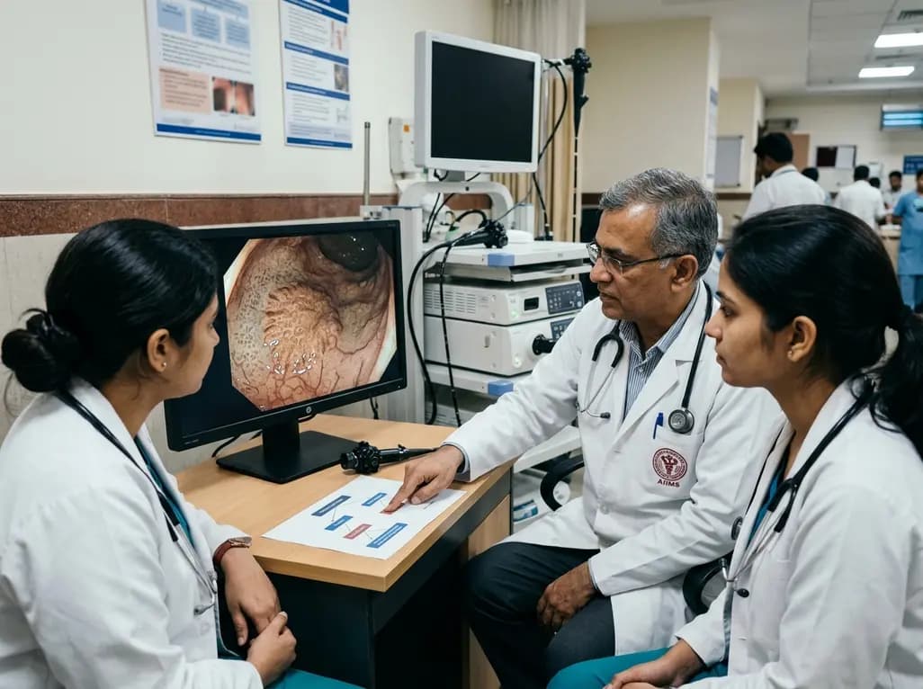
A post hoc decision tree analysis examined JNET Type 1 classification of serrated colorectal lesions. Experts prioritized ruling out anti-Type 1 features with low diagnostic entropy, whereas non-experts relied on mucosal spot patterns, highlighting key training targets for magnifying narrow-band imaging.
Today
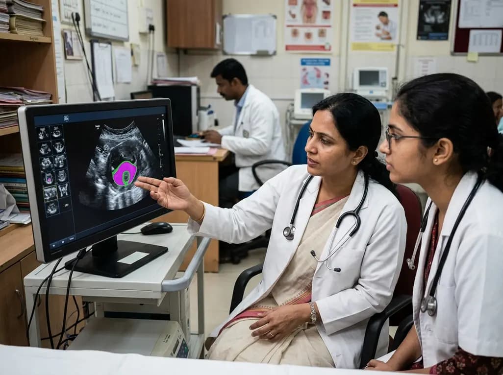
New evidence evaluates MoUNets, a static-weighted ensemble of UNet experts for automated levator hiatus segmentation in pelvic floor ultrasound. Learn how deep learning architectures perform in pelvic organ prolapse diagnostics and clinical practice.
Today

Mitochondrial deubiquitinases regulate organelle turnover by editing ubiquitin tags on damaged mitochondria. Discover how targeting these enzymes, notably USP30, offers therapeutic strategies for neurodegenerative, cardiovascular, and metabolic disorders.
Today
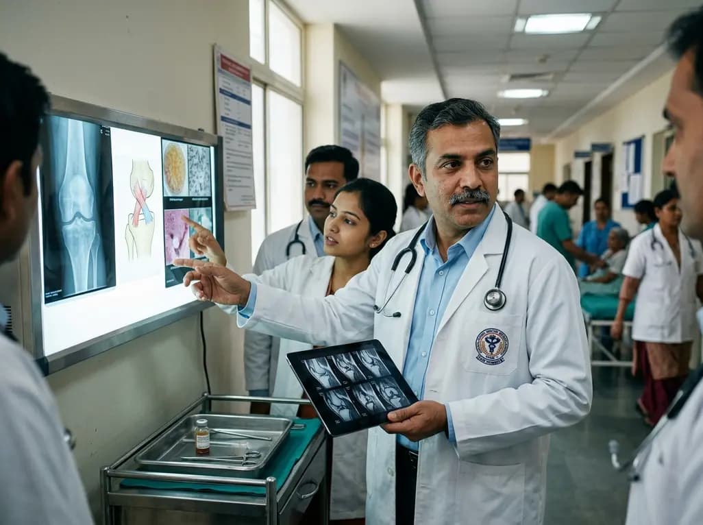
Orthobiologics in ACL surgery, including PRP, BMAC, and bridge-enhanced ACL repair (BEAR), offer biological adjuncts to enhance healing. While scaffold repair shows strong clinical parity to autografts, cell therapies remain selective adjuncts requiring standardized preparation and rigorous trial validation.
Today

A cross-sectional study reveals that elevated METS-IR is independently associated with higher myopia odds in adolescents, showing biological synergy with obesity and highlighting the need for metabolic screening in pediatric eye care.
Today
