.webp&w=384&q=75)

Loading, please wait...
Loading, please wait...

Anterior cruciate ligament ruptures present significant reconstructive hurdles for modern sports medicine clinicians. Although surgical reconstruction restores gross mechanical stability, young athletes frequently face graft re-tears and progressive osteoarthritis. Consequently, surgeons actively explore orthobiologics in ACL surgery to accelerate tissue healing and preserve native biology. These biologic adjuncts aim to enhance graft ligamentization, improve bone tunnel integration, and reduce intra-articular inflammation. Nevertheless, clinicians must critically evaluate the clinical evidence supporting these therapies before widespread adoption.
Unlike extra-articular knee ligaments, the anterior cruciate ligament exhibits virtually no spontaneous capacity for functional repair. When an athlete tears the ligament, the hostile intra-articular synovial fluid environment actively impedes normal hematoma formation. Specifically, intra-articular plasmin rapidly degrades the provisional fibrin clot before reparative cells can migrate into the wound site. Moreover, synovial fluid washes away vital growth factors that typically orchestrate soft tissue healing cascades. Because blood supply arises primarily from small endosteal vessels and the middle genicular artery, the torn midsubstance remains profoundly ischemic. Consequently, primary surgical repair historically yielded unacceptable failure rates, driving the surgical transition toward autograft and allograft ligament reconstruction. However, standard tendon grafts do not replicate the native ligament microstructure immediately upon surgical fixation. Instead, transplanted tendons must undergo a protracted biologic process known as graft ligamentization. During this prolonged remodeling phase, the graft experiences transient mechanical weakness and cellular necrosis. Therefore, biologic interventions that protect the healing matrix promote immense clinical interest among orthopedic specialists worldwide.
Platelet-rich plasma represents the most extensively researched orthobiologic agent in sports knee reconstruction today. Clinicians obtain autologous peripheral blood, centrifuge the sample, and isolate concentrated platelets containing abundant alpha-granules. These active granules subsequently release vascular endothelial growth factor, transforming growth factor-beta, and platelet-derived growth factor. Theoretically, these cytokine cascades should accelerate cellular migration, stimulate collagen deposition, and expedite tendon-to-bone integration inside femoral tunnels. Furthermore, platelet concentrates release anti-inflammatory cytokines that modulate joint effusion and mitigate postoperative synovitis. Nevertheless, published clinical trials continue to report highly conflicting results regarding functional knee recovery. Multiple randomized controlled studies demonstrate that platelet-rich plasma might improve early magnetic resonance imaging signal intensity within grafts. However, these imaging improvements rarely translate into superior patient-reported outcome measures or faster clearance for competitive sports. In addition, extreme procedural heterogeneity obscures true clinical efficacy across modern orthopedic studies. Substantial differences in leukocyte concentrations, chemical activation protocols, centrifugation speeds, and injection timing hinder comparative data synthesis. As a result, professional orthopedic societies currently classify platelet concentrates as optional biological adjuncts rather than mandatory surgical standards.
To deliver richer cellular repertoires, orthopedic investigators increasingly evaluate bone marrow aspirate concentrate. Surgeons typically harvest autologous marrow from the iliac crest or proximal tibia during operative preparation. This concentrated cellular aspirate contains mesenchymal stem cells, hematological progenitors, and crucial anti-inflammatory cytokines like interleukin-1 receptor antagonist. Therefore, researchers hypothesize that bone marrow concentrate can mitigate catabolic joint inflammation while stimulating direct tendon-bone osseointegration. Experimental animal studies certainly confirm accelerated collagen maturation and enhanced pullout strength when adding aspirates to reconstruction tunnels. Furthermore, concentrated cellular aspirates stimulate early vascular ingrowth around the graft-bone interface. In human clinical trials, however, the demonstrable advantages appear far more limited. Patients receiving bone marrow concentrate occasionally demonstrate modest early improvements in postoperative edema or graft signal homogeneity. Yet, long-term functional scores, knee stability measurements, and overall revision rates remain virtually identical to non-augmented controls. Clinicians must clearly communicate these limitations during preoperative surgical counseling. Additionally, other prospective cell therapies, such as adipose-derived stromal cells and synovium-derived stem cells, remain strictly investigational. Consequently, orthopedic practitioners cannot justify their routine clinical application without robust confirmatory randomized trials.
While cell injections produce variable outcomes, bioengineered scaffolds have fundamentally revived clinical interest in primary ligament restoration. Most notably, bridge-enhanced anterior cruciate ligament repair represents a proven breakthrough in clinical bio-restoration. This surgical technique utilizes a proprietary extracellular matrix scaffold composed of highly purified bovine collagen. During surgery, the orthopedic specialist saturates this porous scaffold with autologous whole blood and sutures it between torn ligament stumps. The scaffold stabilizes the provisional blood clot, effectively shielding fragile reparative cells from hostile synovial enzymes. Consequently, native ligament fibroblasts proliferate through the resorbable matrix and synthesize new extracellular collagen. Rigorous randomized multicenter trials show that bridge-enhanced repair achieves non-inferior clinical stability compared to standard autograft reconstruction. Furthermore, patients frequently experience superior early hamstring muscle strength restoration because the technique avoids tendon graft harvest entirely. Additionally, preserving native ligament tissue maintains valuable mechanoreceptors, which support neuromuscular control and proprioception. Thus, scaffold-based restoration establishes a viable alternative for carefully selected patients with acute proximal tears. However, surgeons must apply strict selection criteria to achieve optimal outcomes. Long-term investigations will ultimately clarify its potential to reduce post-traumatic osteoarthritis rates.
The ongoing clinical evolution of orthobiologics in ACL surgery highlights the critical balance between biologic promise and surgical reality. Currently, widespread clinical adoption faces substantial obstacles, including non-standardized preparation systems and inconsistent outcome reporting. Many existing clinical studies rely too heavily on surrogate imaging findings rather than hard endpoints like graft rupture. Therefore, future trial designs must prioritize objective knee stability, validated patient-reported metrics, and long-term joint preservation rates. Moreover, regulatory agencies increasingly demand precise characterization of cellular contents, platelet doses, and delivery vehicles. Knee specialists must recognize that biological augmentations cannot compensate for technical surgical errors such as improper tunnel placement or poor fixation. When surgeons achieve anatomic tunnel drilling and robust biomechanical fixation, selective orthobiologics may potentially optimize the intra-articular microenvironment. As regenerative technologies evolve, personalized biologic therapies tailored to patient age, activity level, and tear configuration will likely emerge. Until then, sports medicine surgeons must maintain rigorous evidence-based standards before endorsing routine biological interventions. Proper patient selection and meticulous surgical technique remain the foundation of successful knee ligament surgery. Additionally, ongoing registry data will help determine cost-effectiveness in routine clinical practice.
Currently, orthobiologic injections cannot replace standard surgical reconstruction for complete anterior cruciate ligament ruptures. Biologic agents like platelet concentrates or bone marrow aspirates lack mechanical strength to stabilize an unstable knee joint. However, scaffold-enhanced technologies like bridge-enhanced repair can restore selected acute tears without tendon grafts. Most complete midsubstance ruptures still require conventional reconstruction to re-establish knee kinematics and protect menisci from secondary degenerative tears.
Clinical evidence does not confirm that platelet-rich plasma significantly accelerates an athlete's safe return to competitive sports. While platelet concentrates enhance early cellularity and graft vascularity on postoperative magnetic resonance imaging, functional recovery depends primarily on neuromuscular rehabilitation. Furthermore, graft remodeling requires biological time that injections cannot bypass entirely. Clinicians therefore prioritize objective functional testing and dynamic knee stability assessments rather than relying on biologics to hasten clearance.
Bridge-enhanced anterior cruciate ligament repair specifically benefits young, active patients presenting with acute, proximal ligament ruptures within fifty days of injury. Suitable candidates must retain substantial, healthy native tissue attached to the tibia. Conversely, chronic tears, retracted tissue remnants, and multi-ligament injuries remain unsuitable for scaffold-based repair. For eligible individuals, this technique eliminates donor-site morbidity, accelerates hamstring strength recovery, and preserves native proprioceptive nerve fibers.
Disclaimer: This content is for informational and educational purposes only. It is not intended to be a substitute for professional medical advice, diagnosis, or treatment. Always seek the advice of your physician or other qualified health provider with any questions you may have regarding a medical condition. Refer to the latest local and national guidelines for clinical practice.
References

Read summarized clinical updates, watch expert medical content, and earn CME certifications right from your smartphone.
Open your smartphone camera to scan and install.





Orthobiologics in ACL surgery, including PRP, BMAC, and bridge-enhanced ACL repair (BEAR), offer biological adjuncts to enhance healing. While scaffold repair shows strong clinical parity to autografts, cell therapies remain selective adjuncts requiring standardized preparation and rigorous trial validation.
Today
A post hoc decision tree analysis examined JNET Type 1 classification of serrated colorectal lesions. Experts prioritized ruling out anti-Type 1 features with low diagnostic entropy, whereas non-experts relied on mucosal spot patterns, highlighting key training targets for magnifying narrow-band imaging.
Today
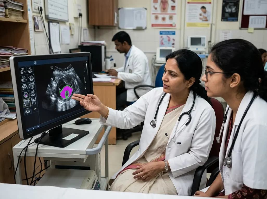
New evidence evaluates MoUNets, a static-weighted ensemble of UNet experts for automated levator hiatus segmentation in pelvic floor ultrasound. Learn how deep learning architectures perform in pelvic organ prolapse diagnostics and clinical practice.
Today
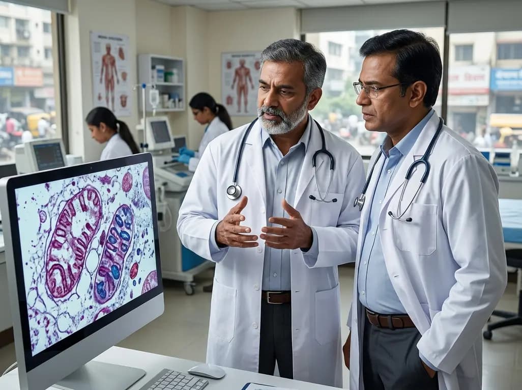
Mitochondrial deubiquitinases regulate organelle turnover by editing ubiquitin tags on damaged mitochondria. Discover how targeting these enzymes, notably USP30, offers therapeutic strategies for neurodegenerative, cardiovascular, and metabolic disorders.
Today

A cross-sectional study reveals that elevated METS-IR is independently associated with higher myopia odds in adolescents, showing biological synergy with obesity and highlighting the need for metabolic screening in pediatric eye care.
Today
