.webp&w=384&q=75)

Loading, please wait...
Loading, please wait...
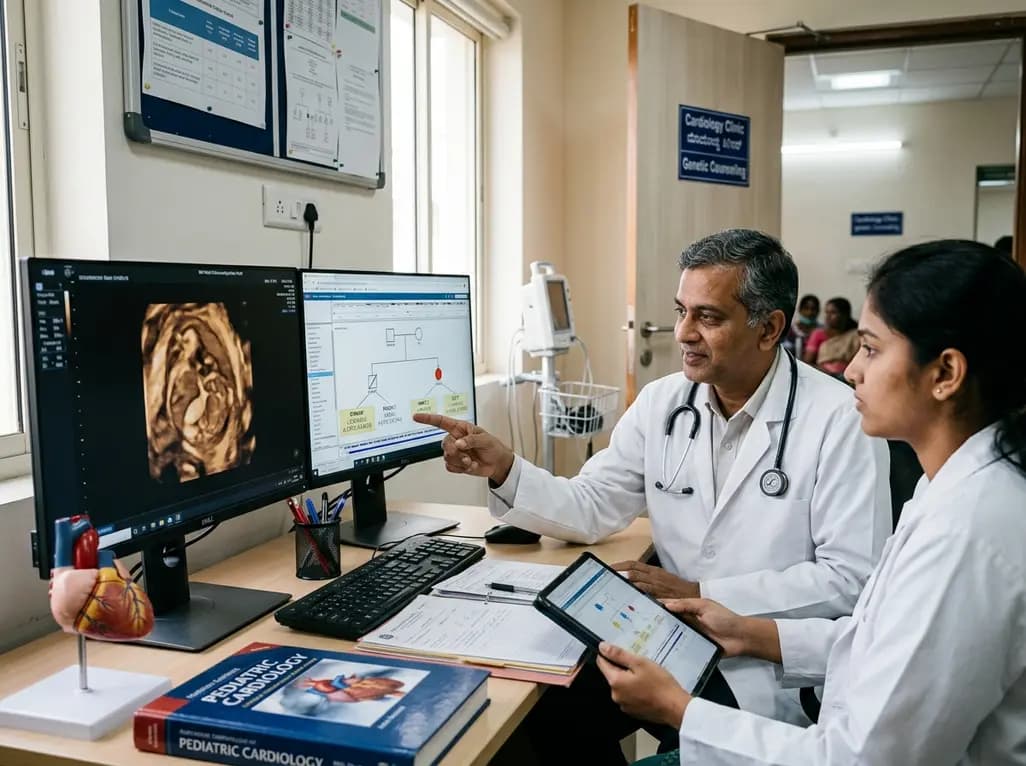
Understanding the complex genetics in heterotaxy represents a transformative leap in fetal and pediatric cardiology. Heterotaxy, also known as situs ambiguus, disrupts the normal left-right anatomical asymmetry during embryonic development. Consequently, this rare developmental disorder precipitates intricate congenital heart defects and diverse extracardiac anomalies. Because internal organs align abnormally, affected infants frequently suffer substantial morbidity and elevated neonatal mortality. Next-generation sequencing has increasingly uncovered the molecular drivers behind these laterality defects. However, standard testing still leaves a significant diagnostic gap in clinical practice. Recent multi-center studies now provide critical insights into pathogenic variants and clinical manifestations.
Next-generation sequencing technologies have revolutionized how clinicians approach congenital heart disease. Historically, conventional karyotyping and chromosomal microarray analysis resolved only a minority of heterotaxy cases. Today, trio-based whole-exome sequencing and whole-genome sequencing significantly improve diagnostic precision. Nevertheless, routine genetic screening currently identifies disease-causing variants in only 20% to 30% of affected patients. This notable diagnostic gap underscores the immense genetic heterogeneity that characterizes left-right patterning disorders.
To overcome these diagnostic hurdles, international collaborative studies have systematically analyzed cohorts of fetuses and children with laterality defects. Researchers performed comprehensive molecular investigations alongside detailed fetopathological examinations in diverse global cohorts. Furthermore, systematic literature reviews comprising over a hundred published cases have refined our understanding of disease architecture. By evaluating affected individuals alongside their parents through trio sequencing, geneticists efficiently filter benign polymorphisms from true pathogenic variants. Consequently, these robust diagnostic strategies identify novel causal mutations and enrich known disease panels. For pediatric cardiologists and medical geneticists, embracing advanced genomic sequencing is essential to illuminate the complex biological mechanisms that govern human organ laterality.
Normal vertebrate organogenesis relies on highly regulated biophysical and biochemical events at the embryonic left-right organizer, known as the primitive node. During early gastrulation, rotational movement of motile nodal cilia generates a directional fluid flow known as nodal flow. In turn, immotile sensory cilia detect this asymmetric mechanical or chemical signal. Consequently, sensory cells activate the evolutionary conserved Nodal signaling cascade strictly on the left lateral plate mesoderm.
When structural or functional defects disrupt nodal cilia, the embryo fails to establish normal visceral asymmetry. As a result, organ placement occurs randomly, producing situs ambiguus, isomerism, or classic heterotaxy. In particular, abnormal ciliary movement and defective flow perception impair downstream transcription factors that guide asymmetric cardiac looping and gut rotation. Therefore, genetic mutations in ciliary structural components or signaling proteins inevitably disturb cardiovascular morphogenesis. Geneticists broadly categorize these molecular pathways into motile ciliopathy genes, primary sensory ciliary components, and extracellular signaling molecules. Because these coordinated cascades dictate right-left orientation across multiple organs, disruptions consistently trigger extensive phenotypic variation. Understanding these embryonic mechanisms helps clinicians appreciate why identical genetic variants often cause discordant cardiac and visceral defects.
Recent genomic investigations have highlighted four pivotal genes involved in heterotaxy pathogenesis: DNAH9, PKD1L1, MMP21, and GDF1. The DNAH9 gene encodes a heavy chain subunit of outer dynein arms in motile cilia. Mutations in DNAH9 severely impair ciliary beat frequency and nodal flow generation. Consequently, biallelic variants in DNAH9 produce laterality defects and primary ciliary dyskinesia phenotypes, often accompanied by bronchiectasis and dextrocardia.
In contrast, PKD1L1 encodes a polycystin-family transmembrane protein localized to primary sensory cilia at the embryonic node. PKD1L1 forms a functional receptor-channel complex with PKD2 to sense leftward fluid flow. Pathogenic variants in PKD1L1 disrupt signal transduction, leading to complex cardiac malformations and splenic abnormalities.
Additionally, MMP21 encodes matrix metalloproteinase 21, an extracellular enzyme crucial for extracellular matrix remodeling and morphogen cleavage during axis formation. Biallelic loss-of-function variants in MMP21 strongly correlate with severe congenital heart disease, particularly conotruncal malformations and complete isomerism.
Finally, GDF1 encodes growth differentiation factor 1, a key ligand in the transforming growth factor-beta superfamily. GDF1 acts cooperatively with Nodal to establish asymmetric gene expression. Monoallelic or biallelic GDF1 mutations precipitate severe cardiovascular defects, including transposition of the great arteries and tetralogy of Fallot.
Detailed fetopathological assessments paired with comprehensive genomic data have uncovered striking genotype-phenotype correlations. Specifically, variants in MMP21 and GDF1 consistently associate with severe conotruncal malformations and complex outflow tract anomalies. Patients harboring these mutations frequently present with double outlet right ventricle, common arterial trunk, and disrupted pulmonary venous return. Furthermore, right atrial isomerism, asplenia, and anomalous systemic venous drainage frequently accompany these cardiovascular lesions.
In contrast, individuals with DNAH9 mutations often exhibit respiratory manifestations alongside heterotaxy due to primary ciliary dyskinesia. Fetopathological examinations demonstrate that extracardiac abnormalities extend well beyond splenic defects. For instance, clinicians regularly observe intestinal malrotation, biliary atresia, midline liver configuration, and abnormal pulmonary lobation. Autopsy evaluations also reveal subtle bronchial isomerism and aberrant mesenteric attachments that imaging may overlook.
Moreover, post-mortem fetopathology provides invaluable morphological validation for novel genetic variants. When prenatal ultrasound reveals complex cardiac anomalies, post-mortem examination delineates minute vascular details that correlate with genomic sequencing results. Consequently, integrating fetopathological data with high-throughput sequencing broadens the known mutational spectrum and refines variant classification under standard clinical genetics guidelines. This comprehensive pathological correlation significantly strengthens the clinical interpretation of previously uncharacterized missense or truncating mutations.
Early genetic identification in heterotaxy substantially transforms clinical management in prenatal and neonatal cardiology. When prenatal ultrasonography detects fetal laterality defects or anomalous vascular connections, rapid trio-based whole-exome sequencing delivers critical diagnostic clarity. As a result, fetal medicine specialists can offer evidence-based genetic counseling regarding long-term prognosis and recurrence risks in subsequent pregnancies.
Furthermore, precise molecular diagnosis directly guides postnatal resuscitation, surgical planning, and multidisciplinary care. For example, identifying pathogenic variants associated with asplenia alerts neonatologists to the imminent danger of life-threatening bacterial infections. Consequently, clinicians immediately initiate prophylactic antibiotic therapy and specialized vaccination schedules after birth. Additionally, identifying polysplenia prompts urgent abdominal evaluations to rule out intestinal malrotation and midgut volvulus.
Similarly, recognizing primary ciliary dyskinesia genes like DNAH9 prepares pediatric pulmonologists to manage neonatal respiratory distress and chronic airway clearance issues. In the surgical arena, understanding specific vascular anomalies prevents intraoperative complications during staged single-ventricle palliation or biventricular repair. Therefore, genomic diagnostics empower clinicians to deliver proactive, individualized medical interventions rather than reactive emergency care. These multidisciplinary collaborations ultimately improve survival and enhance the long-term quality of life for fragile newborns.
Implementing a structured diagnostic workflow is essential when evaluating suspected heterotaxy cases in clinical practice. First, clinicians should perform comprehensive fetal echocardiography and high-resolution abdominal ultrasonography to define anatomical situs and cardiovascular morphology. Second, genetic counselors should obtain detailed multi-generational family pedigrees to assess potential autosomal recessive, autosomal dominant, or X-linked inheritance patterns. Consanguinity should be carefully documented because recessive alleles frequently cause laterality defects.
Third, clinicians should utilize trio-based whole-exome sequencing or whole-genome sequencing as a first-line diagnostic modality for syndromic or complex cases. By testing both biological parents simultaneously, laboratories dramatically reduce turnaround times and minimize variants of uncertain significance. Additionally, genome-wide sequencing reliably captures copy number variations and non-coding structural variants that standard targeted panels miss.
Looking ahead, international collaborative registries and functional genomic assays will clarify the biological impact of newly discovered variants. As sequencing technologies become more accessible across global medical centers, clinicians will achieve higher diagnostic yields. Ultimately, expanding the genomic landscape of laterality defects will pave the way for targeted therapeutic strategies and optimized long-term outcomes for affected children.
Heterotaxy syndrome is a rare congenital condition where internal thoracic and abdominal organs fail to develop normal left-right asymmetry. Consequently, organs arrange abnormally across the body midline. Early diagnosis is crucial because approximately ninety percent of affected infants have complex congenital heart disease. Furthermore, early detection identifies life-threatening splenic anomalies and airway clearance problems, enabling immediate prophylactic antibiotics, targeted neonatal surgical planning, and comprehensive multidisciplinary clinical care.
Trio-based sequencing analyzes the affected individual alongside both biological parents simultaneously. This comprehensive genomic approach enables laboratories to identify de novo mutations, determine compound heterozygous variants, and evaluate phase relationships quickly. As a result, trio sequencing substantially reduces turnaround times and lowers the frequency of variants of uncertain significance. Consequently, clinicians obtain a definitive molecular diagnosis faster, facilitating precise prenatal counseling and individualized neonatal management.
Pathogenic variants in MMP21 and GDF1 strongly correlate with severe conotruncal cardiac malformations and isomerism defects. Specifically, affected infants frequently develop transposition of the great arteries, double outlet right ventricle, truncus arteriosus, and tetralogy of Fallot. In addition, these genetic alterations often coincide with total anomalous pulmonary venous return and complete atrioventricular septal defects, which require complex staged surgical interventions during early infancy.
Disclaimer: This content is for informational and educational purposes only. It is not intended to substitute professional medical advice, diagnosis, or treatment. Refer to the latest local and national guidelines for clinical practice.
References

Read summarized clinical updates, watch expert medical content, and earn CME certifications right from your smartphone.
Open your smartphone camera to scan and install.





A comprehensive review of the genetics in heterotaxy, examining key pathogenic variants in DNAH9, PKD1L1, MMP21, and GDF1, genotype-phenotype correlations, and the role of trio WES/WGS in prenatal cardiology.
last month

A Bayesian multilevel meta-analysis reveals that aerobic training combined with moderate carbohydrate restriction modestly lowers HbA1c in type 2 diabetes. However, sparse data and very low certainty leave incremental benefits over exercise or diet alone unproven, highlighting the need for individualized care.
Today

Findings from Project Viva indicate that sustained postpartum weight retention, rather than excessive gestational weight gain alone, directly worsens women's mid-life cardiovascular health, degrading blood pressure and glycemic parameters decades after delivery.
Today

A large UK Biobank study reveals that plasma proteomic signatures capture preclinical organ damage and improve multiorgan risk prediction across early CKM syndrome stages 0 to 2 beyond traditional PREVENT clinical models.
Today

A retrospective CBCT study reveals that superior TMJ space asymmetry correlates moderately with occipital condyle-atlas-axis asymmetry in unilateral TMJ osteoarthritis, highlighting an imaging correlate between jaw biomechanics and upper cervical alignment rather than a standalone diagnostic marker.
Today

A cross-sectional investigation highlights the complex pathophysiological links between serum Klotho deficiency, secondary hyperaldosteronism, and vascular damage in CKD. Discover key insights into non-invasive vascular stiffness markers and targeted therapeutic pathways for cardiorenal protection.
Today
