

Loading, please wait...
Loading, please wait...

Resection of foramen magnum meningiomas represents one of the most demanding frontiers in modern skull base surgery. These benign yet anatomically complex neoplasms arise near the cervicomedullary junction, lower cranial nerves, and vertebrobasilar circulation. Historically, surgeons relied heavily on radical transcondylar far-lateral approaches to expose ventral pathology. However, extensive bone removal frequently caused craniocervical instability and required occipitocervical fusion. Consequently, skull base teams increasingly favor less destructive posterior and posterolateral corridors. Retrosigmoid/retrocondylar and posterior midline suboccipital craniectomies provide excellent surgical access while preserving osseous structures. Recently, a rigorous PRISMA-guided meta-analysis evaluated clinical outcomes across seven retrospective surgical series. This study provides critical evidence regarding tumor resectability, recurrence rates, and neurological safety without radical condylectomy.
The craniocervical junction contains vital neural pathways that tolerate minimal surgical manipulation. Therefore, operative corridor selection requires an intricate understanding of microvascular relationships. Foramen magnum meningiomas consistently displace the brainstem, upper cervical spinal cord, and cranial nerves IX through XII. In addition, these tumors often distort the vertebral artery and the posterior inferior cerebellar artery.
Historically, neurosurgeons resected substantial portions of the occipital condyle to optimize ventral visualization. However, removing more than one-third of the condyle risks serious mechanical instability. Consequently, patients frequently faced protracted recovery times and required permanent spinal instrumentation. Furthermore, aggressive bone drilling elevates the risk of postoperative cerebrospinal fluid leaks and vertebral artery injury. Modern skull base surgery therefore emphasizes tissue preservation over radical osseous resection. By leveraging natural anatomical corridors, surgeons achieve sufficient visualization without destabilizing the craniocervical articulation.
Surgical approach selection relies heavily on tumor location and dural attachment. Retrosigmoid and retrocondylar approaches provide direct visualization for anterior and anterolateral tumors. Conversely, posterior midline suboccipital craniectomies, often combined with a C1 laminectomy, offer excellent access to posterior and posterolateral lesions.
In the pooled meta-analysis of 133 patients, surgeons used retrosigmoid/retrocondylar corridors in 55.6% of cases. Meanwhile, midline suboccipital approaches accounted for 44.4% of procedures. Importantly, surgeons restricted condylar drilling to the posterior one-third whenever bone removal was necessary. As a result, no patient developed craniocervical instability or required spinal fusion.
Nevertheless, clinicians must recognize that these two operative corridors serve different anatomical scenarios rather than interchangeable alternatives. Surgeons strategically chose retrosigmoid approaches for ventral pathology to minimize brainstem manipulation. Similarly, midline suboccipital exposure provided a safe trajectory for dorsal tumors while protecting lower cranial nerves from lateral traction.
Achieving gross total resection remains the primary surgical objective for benign craniocervical neoplasms. The pooled meta-analysis demonstrated an impressive gross total resection rate of 91% (95% CI: 84–96%) across seven studies. These results confirm that less destructive corridors provide sufficient exposure for complete tumor eradication.
Furthermore, oncologic control proved remarkably durable during clinical follow-up. The sample-size-weighted mean follow-up period spanned 58 months across the patient cohorts. During this extended timeframe, tumor recurrence occurred in only 2.3% of patients, yielding a pooled recurrence rate of 1% (95% CI: 0–4%).
Thus, conservative osseous drilling does not compromise long-term tumor clearance. In cases where the tumor capsule firmly adheres to brainstem pial vessels, surgeons intentionally leave a minute remnant. This deliberate strategy prioritizes neurological preservation over hazardous radicality. Subsequently, neurosurgeons can manage residual tumor remnants with stereotactic radiosurgery, thereby securing oncologic stability while safeguarding functional independence.
Functional preservation is an essential outcome measure following craniocervical skull base surgery. Overall, 83% of patients (95% CI: 71–93%) maintained or regained long-term functional independence. This high rate highlights the functional safety of posterior and lateral suboccipital corridors.
Nonetheless, cranial nerve morbidity represents a persistent clinical challenge. New cranial nerve deficits, transient or permanent, emerged in 21% of patients (95% CI: 13–30%). The lower cranial nerve complex—comprising the glossopharyngeal, vagus, and accessory nerves—is especially susceptible to mechanical stretching during tumor debulking. In addition, traction on the hypoglossal nerve can induce ipsilateral tongue weakness.
Fortunately, many deficits resolve partially or completely over time through compensatory neuroplasticity. However, acute deficits frequently cause severe swallowing difficulties and vocal cord paralysis. Consequently, patients face an elevated danger of aspiration pneumonia during the immediate recovery phase. Surgeons must therefore implement continuous neurophysiological monitoring intraoperatively to protect functional nerve integrity.
Postoperative cerebrospinal fluid leaks and perioperative mortality demand careful surgical consideration. In the meta-analysis, CSF leaks developed in 12% of patients (95% CI: 6–20%). Because hydrodynamics at the craniocervical junction generate high fluid pressures, watertight dural repair is essential. Surgeons must routinely combine autologous fascial grafts, vascularized muscle flaps, and dural sealants.
Moreover, the pooled perioperative mortality rate reached 3% (95% CI: 0–9%), representing seven deaths. Interestingly, two additional late deaths occurred during follow-up due to aspiration pneumonia. This finding illustrates that lower cranial nerve dysfunction can produce fatal pulmonary events months after hospital discharge.
Therefore, multidisciplinary postoperative care is vital. Early evaluation by speech-language therapists helps prevent silent aspiration. Furthermore, teams should maintain a low threshold for temporary tracheostomy or gastrostomy feeding in patients with severe bulbar palsy. Ultimately, tailoring the surgical corridor to tumor topography achieves outstanding tumor resection while minimizing patient morbidity.
Less invasive suboccipital routes provide direct visualization of the tumor while avoiding radical bone removal. By limiting condylar drilling to the posterior third, surgeons protect craniocervical stability and avoid spinal fusion. Furthermore, these techniques decrease operative duration, minimize soft-tissue disruption, and reduce blood loss while delivering excellent long-term tumor control.
Surgeons prevent instability by preserving the structural integrity of the occipital condyle and the atlanto-occipital joint. During retrocondylar approaches, drilling never exceeds the posterior one-third of the condyle. In addition, sparing the contralateral stabilizing ligaments and avoiding unnecessary facet joint disruption maintains natural biomechanics without requiring permanent metal instrumentation or occipitocervical fusion.
Cranial nerves IX, X, XI, and XII face the greatest vulnerability during dissection. These nerves often drape tightly across the ventrolateral tumor surface. Intraoperative traction or vascular compromise can trigger hoarseness, dysphagia, or tongue weakness. Consequently, neurosurgeons utilize continuous neurophysiological monitoring to identify nerve fibers and avoid catastrophic postoperative aspiration.
Disclaimer: This content is for informational and educational purposes only... Refer to the latest local and national guidelines for clinical practice.
References

Read summarized clinical updates, watch expert medical content, and earn CME certifications right from your smartphone.
Open your smartphone camera to scan and install.





A meta-analysis demonstrates that retrosigmoid/retrocondylar and midline suboccipital approaches achieve 91% gross total resection for foramen magnum meningiomas without extensive condylectomy, preserving long-term neurological function while maintaining low recurrence and acceptable perioperative complication rates.
Today

A large retrospective cohort study demonstrates that explainable machine learning models, notably XGBoost, accurately predict hospital admission at emergency triage. By analyzing routine triage parameters, hospitals can streamline bed allocation, decrease boarding times, and alleviate acute care overcrowding.
Today

A new quality-improvement initiative reveals that universal maternal mental health screening in the NICU increased screening rates from 29.5% to 95.0%, with nearly 60% of screened mothers requiring psychosocial referrals, highlighting the urgent need for embedded mental health resources.
Today
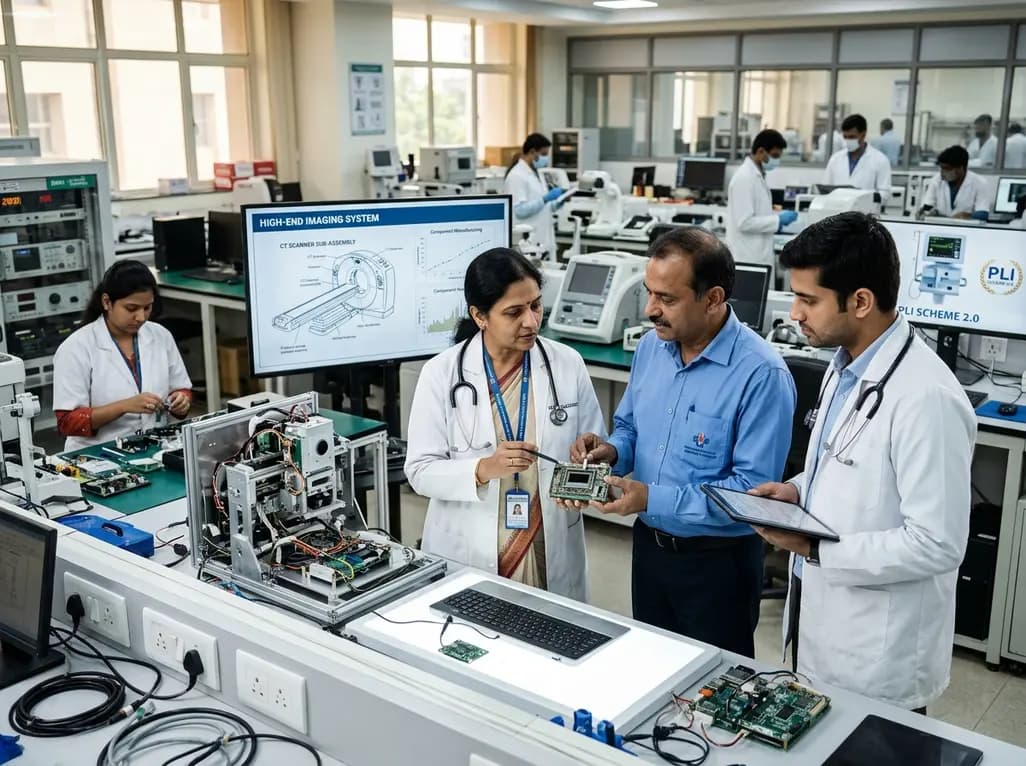
The Department of Pharmaceuticals has extended the medical devices PLI scheme to 2035-36 with a Rs 2,700 crore outlay. Featuring relaxed investment barriers and eliminated sales thresholds, the revised framework targets deep manufacturing, high-end diagnostics, critical MedTech components, and supply resilience.
Today
A metabolomic study reveals that aging delays glutathione synthesis and gamma-glutamylcysteine accumulation following muscle injury. Although precursor supplementation targets this enzymatic bottleneck, in vitro tests show no immediate effect on myogenesis, highlighting the complexity of muscle repair in older adults.
Today

A systematic review evaluates psychological interventions for smoking cessation in diabetes, showing short-term success but highlighting challenges in long-term abstinence.
Today
