.webp&w=384&q=75)

Loading, please wait...
Loading, please wait...
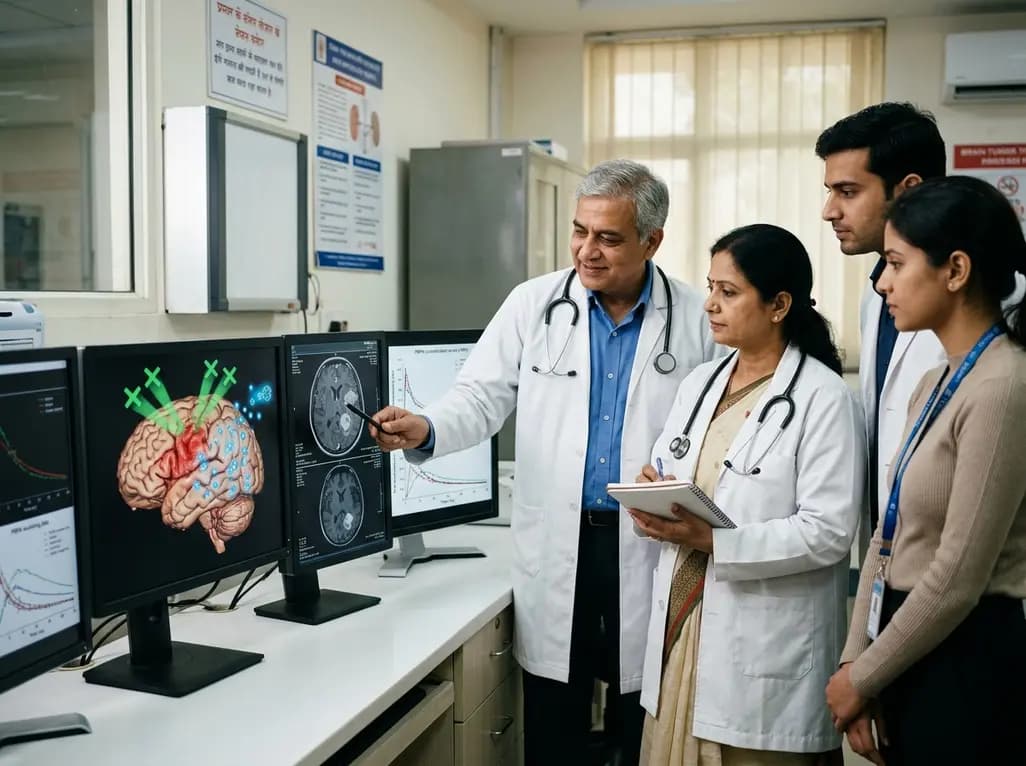
The blood-brain barrier remains a formidable anatomical bottleneck in modern neuro-oncology, severely restricting systemic chemotherapeutics, targeted biologics, and antibody-drug conjugates from reaching therapeutic intracranial concentrations. Consequently, patients with primary central nervous system malignancies and secondary brain metastases frequently experience disease recurrence due to inadequate drug delivery. In recent years, focused ultrasound combined with microbubbles has emerged as an innovative, non-invasive technique to transiently disrupt tight junctions and facilitate localized vascular extravasation. However, successful clinical adoption of focused ultrasound drug delivery has encountered substantial challenges due to an incomplete quantitative understanding of central nervous system pharmacokinetics. Therapeutic agents possess widely disparate molecular weights, clearance profiles, and transport mechanisms that govern brain tissue accumulation. Therefore, clinicians and translational researchers require robust computational frameworks to predict how acoustic parameters interact with systemic drug kinetics. By characterizing drug distribution across distinct intracranial compartments, clinicians can optimize dosing schedules, enhance therapeutic margins, and avoid subtherapeutic tissue exposure. This quantitative approach bridges the gap between laboratory ultrasound engineering and bedside oncology management, paving the way for individualized treatment regimens.
To decipher the complex biomechanical and pharmacokinetic interactions occurring during acoustic disruption, investigators developed an extended nine-compartment central nervous system physiologically based pharmacokinetic model. This advanced mathematical architecture evaluates therapeutic transport across multiple physiological sub-domains, including the systemic circulation, cerebrospinal fluid pathways, and distinct tumor microenvironments. Specifically, the framework models non-infiltrative parenchyma, the infiltrative tumor periphery with an intact vascular barrier, the sonicated infiltrative tumor zone, and the necrotic tumor core exhibiting continuous baseline disruption. Furthermore, the model explicitly incorporates parameterized variables for ultrasound-induced barrier opening, capturing onset timing, mechanical opening magnitude, and biological seal duration. Computational simulations indicate that acoustic permeability enhancements depend heavily on the systemic half-life and clearance kinetics of the co-administered agent. For instance, drugs with prolonged plasma circulation sustain convective transport across loosened endothelial tight junctions over several hours. In contrast, rapidly cleared agents require immediate spatial and temporal coordination with peak acoustic permeabilization. Therefore, physiologically based modeling provides an indispensable tool for characterizing how acoustic cavitation alters regional drug disposition across heterogeneously vascularized intracranial neoplasms.
The translational model investigated four clinically vital oncological agents: the HER2-targeted monoclonal antibody trastuzumab, two antibody-drug conjugates—trastuzumab emtansine (T-DM1) and trastuzumab deruxtecan (T-DXd)—and the standard-of-care alkylating agent temozolomide. Interestingly, simulations revealed critical pharmacological divergences between naked monoclonal antibodies and their payload-bearing counterparts. While acoustic barrier opening markedly elevated intracranial concentrations for all macromolecular therapeutics, pharmacological potency integration demonstrated distinct clinical outcomes. Specifically, trastuzumab monotherapy yielded marginal therapeutic gains despite elevated brain parenchyma accumulation because its intrinsic antitumor potency remains insufficient to overcome rapid tumor proliferation. Conversely, both T-DM1 and T-DXd demonstrated profound intracranial efficacy improvements following acoustic disruption. Their potent cytotoxic payloads—DM1 and deruxtecan—achieve decisive tumor cell eradication once acoustic energy enables sufficient macromolecular crossing. In addition, the simulations highlighted that antibody-drug conjugates benefit significantly from larger acoustic disruption magnitudes and sustained window durations. Consequently, these findings suggest that combining focused ultrasound with cytotoxic antibody payloads represents an exceptionally promising clinical strategy for treating HER2-positive breast cancer brain metastases, whereas unconjugated antibodies may offer limited added value.
In addition to large macromolecular therapies, the computational platform evaluated the small-molecule alkylating agent temozolomide, which forms the cornerstone of standard glioblastoma management. Unlike large biologics, temozolomide possesses modest baseline central nervous system penetrance; however, adequate concentrations rarely accumulate in infiltrative tumor margins where the vascular barrier remains partially intact. Model simulations established that focused acoustic intervention significantly boosts local temozolomide concentrations, but this effect demands rigorous temporal precision. Because temozolomide undergoes rapid systemic elimination with a short plasma half-life, barrier permeabilization must coincide exactly with peak systemic drug exposure. Administering ultrasound too early or too late relative to oral ingestion completely abolishes the pharmacokinetic advantage. Moreover, pharmacological potency simulations indicated that the intracranial benefit of ultrasound-augmented temozolomide is not universally uniform. Instead, heightened tumor kill is predominantly restricted to patients with favorable baseline tumor biology, particularly those exhibiting O6-methylguanine-DNA methyltransferase promoter methylation. Consequently, neuro-oncologists must account for both molecular biomarkers and precise chronopharmacology when designing clinical trials that combine acoustic barrier disruption with conventional small-molecule chemotherapy protocols.
A persistent hurdle in modern neurosurgical oncology is the management of non-enhancing infiltrative margins where microscopic malignant cells invade healthy brain parenchyma. Standard contrast-enhanced neuroimaging primarily highlights the disrupted core, leaving surrounding infiltrative zones shielded by an intact blood-brain barrier. Physiologically based simulations demonstrated that standard systemic therapies readily reach the leaky central core but fail miserably in the infiltrative periphery. By directing focused ultrasound energy specifically to these peritumoral infiltrative zones, clinicians can transiently permeabilize intact microvessels without causing collateral systemic toxicity. Furthermore, the model showed that simultaneous drug infusion and acoustic application maximizes drug deposition within infiltrative microenvironments. This localized enhancement prevents malignant clones from surviving in shielded niches and initiating fatal local recurrences. In addition, targeted permeabilization alters interstitial fluid pressure gradients, promoting outward convective flow from vascular lumens into tumor stroma. Therefore, applying focused ultrasound to the infiltrative margins transforms previously impermeable peritumoral brain tissue into an accessible therapeutic zone, addressing a primary driver of treatment failure in malignant gliomas and solitary metastases.
The quantitative insights generated by physiologically based pharmacokinetic modeling provide a definitive framework for structuring future neuro-oncology clinical trials. Clinicians can now replace empirical guesswork with computationally guided parameters, establishing rational drug-device combinations tailored to specific pharmacological classes. For large-molecule payloads like antibody-drug conjugates, clinical investigators should prioritize ultrasound protocols that maintain extended opening durations to maximize total area under the curve exposure. Conversely, for small-molecule regimens, protocols must optimize timing synchronization between drug administration and acoustic cavitation. Additionally, neuro-oncology teams can leverage these findings to refine patient selection, identifying candidates whose tumors express appropriate targets and favorable epigenetic profiles. Incorporating real-time acoustic emission monitoring with magnetic resonance guidance further ensures reproducible, safe barrier opening across diverse patient populations. Ultimately, integrating physiologically based pharmacokinetic modeling with precision ultrasound engineering represents a paradigm shift in central nervous system therapeutics. As clinical centers worldwide initiate randomized trials, this mechanistic roadmap will accelerate the clinical translation of non-invasive acoustic barrier disruption into standard oncological practice.
Focused ultrasound transiently opens tight endothelial junctions, allowing bulky antibody-drug conjugates like T-DM1 and T-DXd to cross into brain tissue. Because these macromolecular agents carry highly potent cytotoxic payloads, enhanced tissue penetration translates directly into substantial tumor cell eradication. Physiologically based pharmacokinetic models indicate this combination significantly improves intracranial efficacy for metastatic brain lesions compared to unconjugated antibodies.
Because temozolomide exhibits a rapid systemic clearance and a short plasma half-life, its therapeutic window is highly time-sensitive. Physiologically based simulations demonstrate that focused ultrasound must coincide precisely with peak systemic drug concentrations to maximize intracranial delivery. Sonicating too long before or after peak plasma levels substantially diminishes drug accumulation in infiltrative tumor zones, neutralizing the therapeutic benefit.
Malignant brain tumors feature an infiltrative margin where invading tumor cells hide behind an intact blood-brain barrier, escaping systemic chemotherapy. While the central necrotic core already exhibits vascular disruption, the peritumoral margin remains shielded. Focused ultrasound selectively permeabilizes these intact peritumoral microvessels, enabling therapeutic drug concentrations to reach invasive cells and preventing post-treatment tumor recurrence.
Disclaimer: This content is for informational and educational purposes only. It does not constitute medical advice and should not be used as a substitute for professional healthcare decision-making. Always consult a qualified medical professional for diagnosis and treatment. Refer to the latest local and national guidelines for clinical practice.
References

Read summarized clinical updates, watch expert medical content, and earn CME certifications right from your smartphone.
Open your smartphone camera to scan and install.





CNS physiologically based pharmacokinetic modeling reveals how focused ultrasound-induced blood-brain barrier opening optimizes intracranial delivery of antibody-drug conjugates and temozolomide in brain tumors.
last month

A systematic review of 41 preclinical studies reveals that baicalin dampens systemic inflammation, inhibits NF-κB and NLRP3 pathways, and improves organ preservation in sepsis models. However, substantial study heterogeneity and a complete lack of human clinical trials highlight the need for further translational research.
Today
Discover the crucial distinctions between mesonephric and mesonephric-like proliferations of the female genital tract. Learn the key histomorphologic, immunohistochemical, and molecular differences essential for accurate diagnosis and clinical management.
Today

A large UK Biobank study reveals that plasma proteomic signatures capture preclinical organ damage and improve multiorgan risk prediction across early CKM syndrome stages 0 to 2 beyond traditional PREVENT clinical models.
Today
A narrative review investigates whether the articularis genus muscle functions as an independent anatomical entity or blends with the vastus intermedius. We evaluate its morphology, role in retracting the suprapatellar bursa, and direct clinical significance in anterior knee pain and arthroplasty.
Today

A Bayesian multilevel meta-analysis reveals that aerobic training combined with moderate carbohydrate restriction modestly lowers HbA1c in type 2 diabetes. However, sparse data and very low certainty leave incremental benefits over exercise or diet alone unproven, highlighting the need for individualized care.
Today
