.webp&w=384&q=75)

Loading, please wait...
Loading, please wait...

Differentiating major depressive disorder from generalized anxiety disorder remains a formidable diagnostic challenge in outpatient mental health care. Because symptomatic overlap frequently obscures clinical presentations, psychiatrists require objective neurobiological markers to refine their diagnostic evaluations. Recent clinical research highlights how fNIRS cortical activation during cognitive activation tasks reveals distinct neurovascular profiles across affective disorders. By measuring relative hemodynamic responses across specific prefrontal regions, functional near-infrared spectroscopy provides valuable clinical insights that transcend conventional subjective symptom scales.
Clinicians encounter considerable diagnostic overlap when evaluating patients presenting with affective and neurotic distress. For instance, psychomotor agitation, poor concentration, insomnia, fatigue, and pervasive rumination appear routinely in both clinical conditions. Consequently, primary care physicians and psychiatrists frequently struggle to differentiate unipolar depression from generalized anxiety during initial interviews. Misclassification often delays evidence-based therapeutic interventions, increasing chronic disability and medication failure rates.
Moreover, comorbid anxiety commonly masks underlying major depressive episodes, compounding diagnostic complexity in busy outpatient settings. Traditional diagnostic criteria rely heavily on descriptive psychopathology and subjective clinical interviews. Although structured rating scales assist evaluation, patient recall bias and cultural interpretations frequently skew symptom reporting. Therefore, developing accessible neuroimaging biomarkers represents an urgent clinical priority. Functional neuroimaging bridges this diagnostic gap by capturing real-time physiological disruptions in affective regulation networks. Optical neuroimaging now delivers tangible neurobiological markers directly at the bedside, giving clinicians objective physiological context to support nuanced psychiatric evaluations.
Functional near-infrared spectroscopy measures localized cortical oxygenation changes by tracking fluctuations in oxygenated and deoxygenated hemoglobin concentrations. Specifically, multi-channel arrays project safe near-infrared light through cranial layers, assessing regional neurovascular coupling with remarkable temporal sensitivity. When clinicians combine optical imaging with standardized cognitive paradigms, such as the verbal fluency task, prefrontal circuits undergo direct physiological challenge.
During these semantic word-generation exercises, healthy individuals recruit the dorsolateral prefrontal cortex to orchestrate executive search strategies. In contrast, patients with affective disorders consistently exhibit blunted hemodynamic responses across frontotemporal networks. Furthermore, modern fNIRS systems tolerate minor head motion effectively, unlike cumbersome magnetic resonance imaging scanners. This unique ergonomic portability makes optical neuroimaging especially suitable for distressed psychiatric outpatients. By examining real-time task-induced oxygenation curves, clinicians evaluate top-down cognitive control mechanisms objectively. Thus, measuring fNIRS cortical activation offers a practical and robust method for evaluating executive frontocortical integrity in clinical settings.
Recent clinical data demonstrate distinct neurovascular signatures when comparing patients with depression, generalized anxiety, and healthy controls. Investigators utilized a 48-channel fNIRS apparatus to monitor regional hemodynamics across prefrontal and temporal cortices during cognitive performance. Notably, both clinical cohorts exhibited significant hypoactivation within the dorsolateral prefrontal cortex and medial prefrontal cortex relative to healthy individuals. This shared deficit confirms common frontocortical vulnerability across internalizing psychiatric syndromes.
However, comparative analyses demonstrated crucial divergence between the two patient groups. Specifically, patients with major depressive disorder showed significantly greater hypoactivation in the left dorsolateral prefrontal cortex and medial prefrontal cortex than those with generalized anxiety. In addition, localized analyses across Brodmann areas 9 and 46 revealed a progressive gradient of dysfunction, with healthy controls performing best, followed by anxiety, and then depression. Furthermore, anxiety severity scores correlated inversely with specific prefrontal hemodynamic amplitudes. These compelling physiological gradients confirm that while both conditions impair executive circuits, depression drives substantially deeper cortical suppression.
These neuroimaging discoveries provide compelling clinical utility for psychiatric assessment and therapeutic planning. First, objective hemodynamic markers assist practitioners when differentiating severe generalized anxiety from treatment-resistant unipolar depression. When diagnostic clarity remains elusive, localized prefrontal oxygenation levels can guide differential classification alongside psychometric evaluations. Consequently, clinicians can tailor pharmacotherapy and targeted psychotherapy much earlier in the disease course.
Second, identifying pronounced left prefrontal hypoactivation carries substantial therapeutic relevance for neuromodulation strategies. For example, repetitive transcranial magnetic stimulation specifically targets hypoactive dorsolateral prefrontal regions to alleviate resistant depressive symptoms. Therefore, establishing baseline cortical activation profiles may help predict which individuals will respond favorably to excitatory stimulation protocols. In addition, longitudinal optical monitoring allows clinicians to evaluate neurovascular recovery following antidepressant therapy or cognitive behavioral interventions. Ultimately, incorporating functional hemodynamic parameters transforms psychiatric diagnosis from purely qualitative description into precise neurobiological stratification.
In the Indian healthcare landscape, psychiatric epidemiology reveals an escalating burden of mixed anxiety and depressive disorders. However, high patient volumes in tertiary psychiatric centers and district hospitals limit consultation times, occasionally compromising detailed differential diagnosis. Furthermore, access to high-field functional magnetic resonance imaging remains constrained by substantial costs and long waiting periods. Under these prevailing resource realities, portable multi-channel optical systems present an economically viable diagnostic adjunct.
Because fNIRS devices require minimal physical infrastructure and operate quietly in standard consultation rooms, psychiatric departments can implement them efficiently. Nevertheless, practitioners must recognize current limitations before widespread clinical deployment. Existing studies involve modest sample sizes, demanding rigorous validation across diverse, multi-ethnic patient populations. In addition, standardizing task paradigms and machine-learning analytic pipelines remains essential to establish normative diagnostic thresholds. As clinical neuroimaging technology matures, Indian psychiatrists can harness bedside hemodynamic measurements to enhance diagnostic precision, optimize therapeutic selection, and improve long-term functional recovery.
Functional near-infrared spectroscopy distinguishes these disorders by quantifying task-induced hemodynamic responses in prefrontal regions. While both conditions exhibit reduced frontal oxygenation compared to healthy controls, major depressive disorder causes significantly deeper hypoactivation in the left dorsolateral prefrontal cortex and medial prefrontal cortex than generalized anxiety disorder.
The verbal fluency task activates executive circuits within the dorsolateral prefrontal cortex during rapid word generation. Pairing this cognitive challenge with optical neuroimaging allows clinicians to assess real-time neurovascular coupling, exposing compromised cortical reserve and impaired cognitive flexibility characteristic of specific mood and neurotic disorders.
Portable optical neuroimaging cannot replace comprehensive clinical evaluations, detailed psychiatric interviews, or validated diagnostic criteria. Instead, fNIRS functions as an objective physiological biomarker that complements clinical assessment, assisting physicians in resolving diagnostic ambiguities, monitoring therapeutic responses, and guiding targeted neuromodulation interventions.
Disclaimer: This content is for informational and educational purposes only. It is not intended as medical advice, diagnosis, or treatment recommendations. Clinical decisions should always be made by qualified healthcare professionals based on individual patient assessment and prevailing clinical guidelines. The authors and publishers disclaim any liability for decisions made based on this information. Refer to the latest local and national guidelines for clinical practice.
References
Jialin A et al. Cortical activation patterns in generalized anxiety and major depressive disorders measured by multi-channel near-infrared spectroscopy. J Affect Disord. 2025 Jun 15. doi: 10.1016/j.jad.2025.02.116. PMID: 40044089.
Takizawa R, Fukuda M, Kawasaki S, et al. Neuroimaging-aided differential diagnosis of the depressive state. Neuroimage. 2014;85 Pt 1:498-507. doi: 10.1016/j.neuroimage.2013.05.126.
Ho CS, Lim LJ, Lim AQ, et al. Diagnostic and predictive applications of functional near-infrared spectroscopy for major depressive disorder: a systematic review. Front Psychiatry. 2020;11:378. doi: 10.3389/fpsyt.2020.00378.

Read summarized clinical updates, watch expert medical content, and earn CME certifications right from your smartphone.
Open your smartphone camera to scan and install.





A multi-channel fNIRS study reveals that major depressive disorder exhibits significantly greater prefrontal hypoactivation than generalized anxiety disorder during cognitive challenges, providing an objective hemodynamic biomarker for neurobiological differentiation in clinical psychiatric practice.
Today

A multicity study highlights that over 80% of acute respiratory infections remain outside hospitals. Patients frequently choose community pharmacies and digital platforms. Integrating digital health data and retail pharmacy sales can fill critical surveillance gaps and improve early outbreak detection.
Today

Scandinavian register evidence reveals that neonatal birth length strongly predicts adult health and mortality, whereas birth weight dictates cognitive capacity. Symmetric growth restriction causes far more severe long-term deficits than asymmetric restriction, underscoring the need for comprehensive anthropometry.
Today
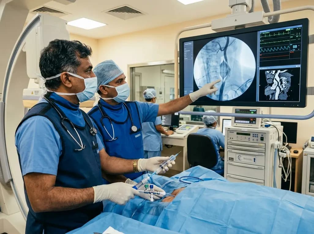
Discover how rescue cutting balloon angioplasty successfully modifies heavily calcified carotid stenosis, enabling stent delivery without baroreceptor instability and ensuring favorable long-term neurologic outcomes.
Today

A retrospective CBCT study reveals that superior TMJ space asymmetry correlates moderately with occipital condyle-atlas-axis asymmetry in unilateral TMJ osteoarthritis, highlighting an imaging correlate between jaw biomechanics and upper cervical alignment rather than a standalone diagnostic marker.
Today

Data from the 2026 Workforce Health Index reveals that 38.7% of urban employees and dependents exhibit abnormal glycemic control. Emerging at increasingly younger ages, this metabolic shift intersects with severe vitamin deficiencies, highlighting the urgent need for early workplace screenings and targeted clinical care.
Today
