.webp&w=384&q=75)

Loading, please wait...
Loading, please wait...
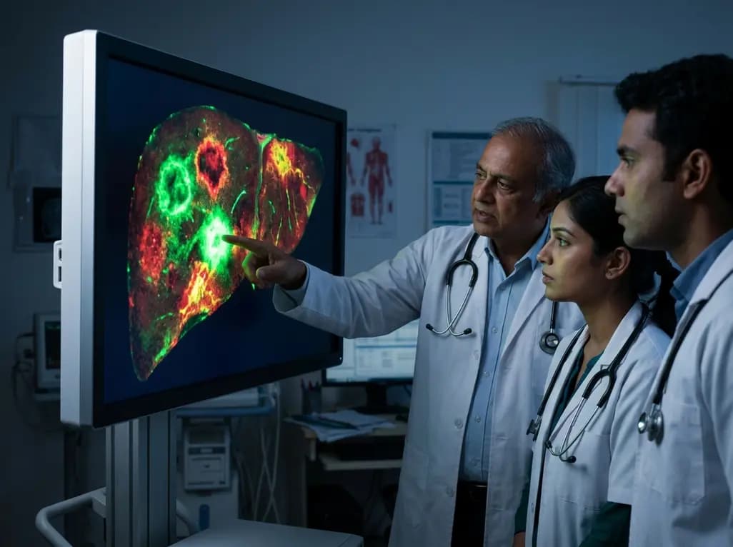
Liver diseases remain a significant global health challenge, affecting millions of patients through conditions such as hepatocellular carcinoma, hepatic fibrosis, and metabolic dysfunction-associated steatotic liver disease (MASLD). Early and accurate diagnosis is critical for improving patient outcomes; however, conventional diagnostic methods often face significant limitations. For example, tissue biopsies provide definitive results but are inherently invasive and carry risks of complications. On the other hand, non-invasive imaging modalities like ultrasound or CT often lack the spatial resolution required to detect early micro-lesions. Consequently, researchers are turning toward fluorescence imaging for liver applications as a transformative solution. This technology offers high spatiotemporal resolution and allows for real-time monitoring of pathological progression at a molecular level. By utilizing specialized fluorescent probes, clinicians can visualize metabolic changes and micro-environmental shifts before they manifest as gross anatomical alterations. This shift toward molecular-level visualization represents a paradigm change in how we approach chronic liver conditions. Furthermore, the integration of nanoscale materials and small-molecule probes provides a versatile platform for personalized diagnostics. This article explores the innovative design strategies that make these probes effective and their specific roles in managing various liver pathologies.
Developing effective probes for fluorescence imaging for liver requires a deep understanding of both chemical engineering and hepatic physiology. One of the primary hurdles in this field is the depth of tissue penetration. To address this, scientists have focused on spectral optimization, particularly moving from the visible spectrum into the near-infrared (NIR) windows. Specifically, the second near-infrared window (NIR-II), spanning 1,000 to 1,700 nm, has emerged as a game-changer. This wavelength range experiences significantly lower light scattering and tissue autofluorescence, allowing for much clearer images of deep-seated liver tissues. In addition to spectral shifts, nanomaterial engineering plays a vital role in enhancing probe performance. Researchers utilize molecular optimization to ensure that probes remain stable in the complex biochemical environment of the liver. Moreover, the development of stimuli-responsive mechanisms allows these probes to "turn on" only when they encounter specific pathological markers, such as high levels of reactive oxygen species or specific enzymes. Consequently, this smart design significantly reduces background noise and improves the signal-to-noise ratio. By tailoring the biocompatibility of these materials, scientists ensure that the probes can be safely administered and effectively cleared by the body after the diagnostic procedure is complete.
Hepatocellular carcinoma (HCC) is one of the most lethal malignancies, often because clinicians detect it only in advanced stages. Fluorescence imaging offers a promising avenue for identifying early-stage tumors and defining surgical margins with unprecedented precision. Modern fluorescent probes target biomarkers that are overexpressed in malignant cells, such as alpha-fetoprotein or specific membrane receptors. Once the probe binds to these targets, it provides a bright signal that distinguishes the tumor from healthy parenchyma. This capability is particularly useful during fluorescence-guided surgery, where surgeons can resect sub-visible micro-lesions that traditional white-light inspection might miss. Furthermore, the real-time nature of this imaging allows for immediate assessment of the surgical site. Recent studies have demonstrated that using NIR-II probes can detect HCC lesions as small as a few millimeters in size. This level of sensitivity is essential for preventing recurrence and ensuring the complete removal of malignant tissue. Additionally, researchers are exploring the use of activatable probes that respond to the acidic microenvironment of the tumor. These probes remain silent in healthy tissue but light up upon entering the cancerous zone. Such specificity not only improves diagnostic accuracy but also reduces the potential for false-positive results during complex surgical procedures.
Hepatic fibrosis is a wound-healing response to chronic liver injury that can eventually lead to cirrhosis and liver failure. Because fibrosis is a dynamic and potentially reversible process, tracking its progression is vital for clinical management. Traditional monitoring often relies on periodic biopsies, which are uncomfortable and provide only a snapshot of a specific tissue area. Fluorescence imaging provides a non-invasive alternative that can map the distribution of collagen and other extracellular matrix components across the entire liver. Probes designed for fibrosis often target activated hepatic stellate cells or specific enzymes like lysyl oxidase, which are key drivers of tissue scarring. By quantifying the fluorescence intensity, clinicians can estimate the degree of fibrotic deposition and monitor the effectiveness of therapeutic interventions. Moreover, the use of ratiometric probes allows for self-calibration, which reduces the impact of varying probe concentrations or tissue thickness. This technical advancement ensures that the measurements remain consistent over time, providing a reliable longitudinal record of the disease state. Consequently, this allows for more agile treatment adjustments. In addition to monitoring, these probes help in the early identification of patients at high risk for progressing to cirrhosis, enabling early preventive measures.
Metabolic dysfunction-associated steatotic liver disease (MASLD), formerly known as NAFLD, is increasingly prevalent due to rising global obesity rates. Diagnosing the transition from simple steatosis to metabolic dysfunction-associated steatohepatitis (MASH) is a major clinical challenge. Fluorescence imaging provides a unique window into the metabolic activity of the liver, allowing researchers to track lipid accumulation and oxidative stress in real time. Specifically, probes that respond to intracellular lipid droplets or reactive oxygen species (ROS) can visualize the metabolic "stress" levels within hepatocytes. These stimuli-responsive probes are particularly valuable because they provide functional information rather than just structural data. For instance, a probe might change its emission color as the concentration of fat within a cell increases. This color shift provides a direct readout of the disease's severity. Furthermore, the high resolution of fluorescence microscopy combined with these probes allows for the study of MASLD at the single-cell level. This level of detail helps scientists understand the heterogeneity of the disease across different liver lobes. By establishing solid connections between probe design and these pathological characteristics, researchers are creating a robust toolkit for managing the metabolic health of patients more effectively and with less reliance on invasive testing.
Despite the significant progress in laboratory settings, several hurdles remain before the widespread clinical adoption of advanced fluorescent probes. One of the primary concerns is the long-term safety and toxicity of nanomaterials. While many probes show excellent biocompatibility in animal models, human clinical trials are necessary to confirm their safety profiles and clearance rates. Furthermore, the standardization of imaging equipment across different hospitals is essential for consistent diagnostic results. Scientists must also work on refining the cost-effectiveness of these technologies to ensure they are accessible in diverse healthcare settings, including in developing regions. However, the future looks incredibly promising as researchers continue to bridge the gap between probe design and disease-specific requirements. The next generation of probes will likely feature multi-modal capabilities, combining fluorescence with magnetic resonance or photoacoustic imaging for a comprehensive diagnostic approach. Such hybrid systems would allow for both deep structural visualization and high-resolution molecular mapping. Additionally, the development of targeted delivery systems could allow these probes to serve as therapeutic agents, creating a "theranostic" platform for liver health. By continuing to optimize these liver-specific tools, the medical community moves closer to a future where liver diseases are caught early and treated with precision.
Fluorescent probes significantly enhance the detection of early-stage hepatocellular carcinoma by targeting specific molecular markers that appear long before structural changes are visible on traditional scans. These probes attach to malignant cells and emit a high-contrast signal, allowing clinicians to see micro-lesions as small as two millimeters. This precision is vital for defining clear surgical margins and ensuring that all cancerous tissue is removed, thereby reducing the risk of recurrence and improving patient survival.
Stimuli-responsive mechanisms, often called "smart" probes, offer the advantage of high specificity by remaining inactive until they encounter a specific pathological microenvironment. In liver disease, these probes might respond to unique enzyme levels, acidic pH, or oxidative stress markers. Because they only emit light at the site of the disease, they dramatically reduce background noise and false positives. This focused signaling provides clearer images and more reliable data regarding the actual metabolic activity of the diseased tissue.
Spectral optimization involves shifting the light emitted by probes into the near-infrared windows, specifically the NIR-II range. Traditional visible light is easily absorbed and scattered by thick liver tissue, which limits imaging depth. However, NIR-II light passes through biological tissues with much less interference and minimal autofluorescence. This allows for high-resolution imaging at depths of several centimeters, enabling clinicians to visualize internal liver structures and deep-seated lesions without the need for invasive surgical exposure or biopsies.
Disclaimer: This content is for informational and educational purposes only. It is not intended as a substitute for professional medical advice, diagnosis, or treatment. Always seek the advice of your physician or other qualified healthcare provider with any questions you may have regarding a medical condition. Refer to the latest local and national guidelines for clinical practice.
References
Yuan G et al. Fluorescence imaging for liver diseases: probe design strategies and diagnostic applications. Nanoscale. 2026 Jul 06. doi: 10.1039/d6nr01285j. PMID: 42405446.
Wang F, Zhong Y, Bruns O, et al. In vivo NIR-II fluorescence imaging for biology and medicine. Nature Photonics. 2024;18(3):214-225.
Ishizawa T, Saiura A, Kokudo N. Clinical application of indocyanine green-fluorescence imaging during hepatectomy. Journal of Surgical Oncology. 2020;122(6):1115-1125.

Read summarized clinical updates, watch expert medical content, and earn CME certifications right from your smartphone.
Open your smartphone camera to scan and install.





Explore the latest advancements in fluorescence imaging for liver diseases. This review highlights innovative probe design strategies for detecting hepatocellular carcinoma, hepatic fibrosis, and MASLD with high spatiotemporal resolution.
3 months ago

Explore vital recurrent stroke predictors in lacunar stroke patients managed with intensive blood pressure lowering. This analysis examines trial findings from SPS3 to uncover residual vascular risks, metabolic factors, and targeted secondary prevention strategies for high-risk neurological patients.
Today
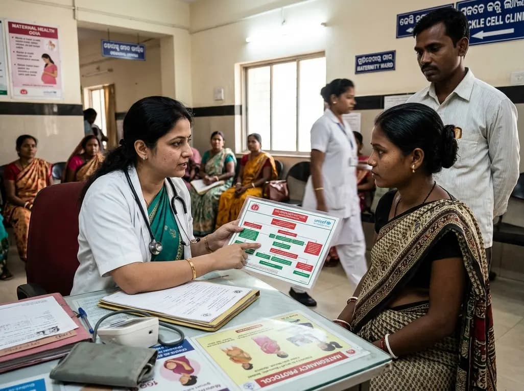
Odisha has established India's first state-level clinical protocol specifically tailored for pregnant women with sickle cell disease. Developed with NHM and UNICEF, the framework provides structured antenatal screening, crisis mitigation, standardized referral pathways, and specialized interdisciplinary care.
Today

India is advancing preventive healthcare by integrating genomics, artificial intelligence, and quantum technologies. With the SANGAM programme sequencing one million Indian genomes, clinicians can unlock personalized therapies, detect cardiometabolic risks early, and curb the burden of diabetes and heart disease.
Today
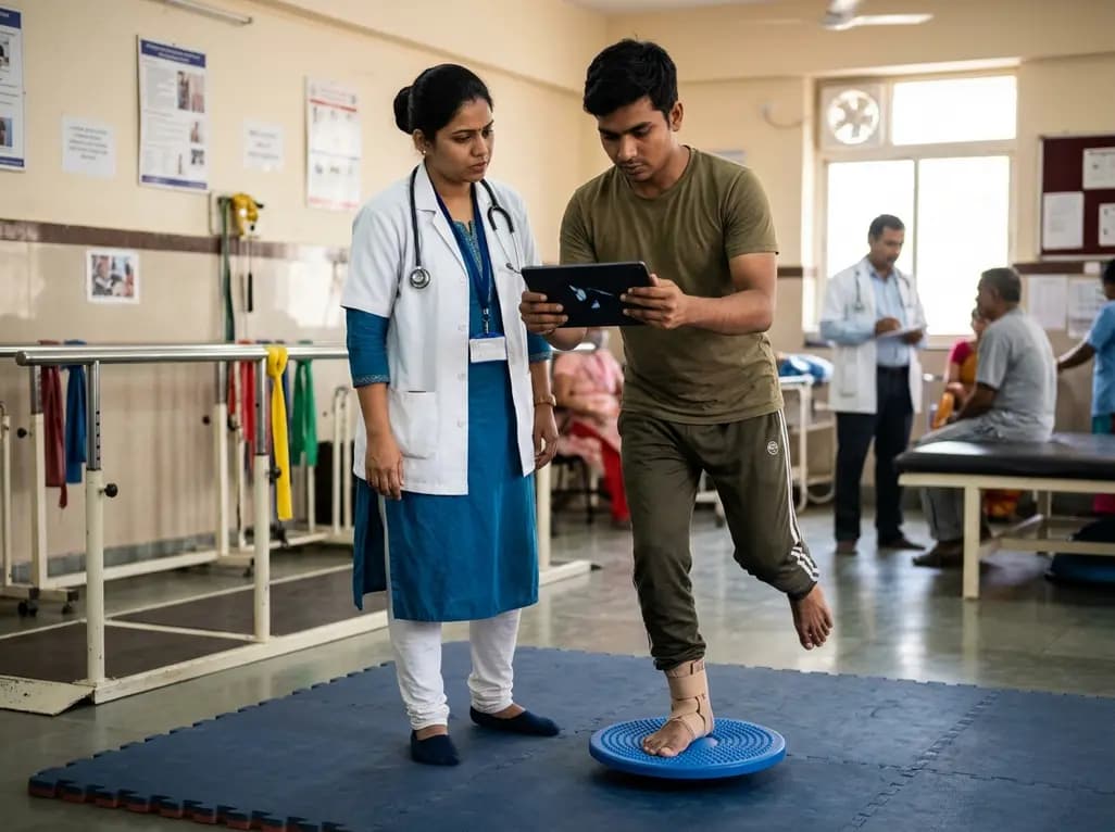
A randomized controlled trial demonstrates that an eight-week dual-task training intervention significantly improves static and dynamic postural control over standard balance exercises in individuals with chronic ankle instability, addressing critical neurocognitive deficits during athletic and daily activities.
Today
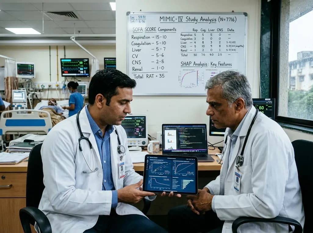
A retrospective cohort study of 7,716 ICU patients from the MIMIC-IV database demonstrates that Gradient Boosting Decision Trees significantly outperform traditional SOFA scores in predicting in-hospital mortality among patients with sepsis and diabetes, with SHAP analysis providing clinical interpretability.
Today
