

Loading, please wait...
Loading, please wait...

Maxillofacial surgeons and oral pathologists frequently encounter non-odontogenic radiolucencies in the anterior maxilla. Among these anomalies, the nasopalatine duct cyst represents the most common developmental, non-neoplastic jaw cyst. Historically, classical embryology attributed this pathology to entrapped epithelial remnants derived from the median epithelial seam. However, recent histological investigations by Morita and colleagues provide revolutionary insights into fetal development. Specifically, their observations demonstrate that the nasopalatine duct arises primarily from descending nasal epithelial protrusions rather than palatal seam entrapment.
Understanding the precise genesis of the incisive canal remains vital for head and neck specialists. In this landmark study, researchers evaluated frontal histological sections from 25 human fetuses spanning 9 to 40 gestational weeks. At 9 weeks of gestation, investigators identified the median epithelial seam alongside a distinct nasal epithelial protrusion. This cellular protrusion formed an organized epithelial cell trabecula extending toward the lower midface. Furthermore, between 10 and 14 weeks, the nasal protrusion systematically enlarged and migrated toward the incisive fossa. Consequently, this downward elongation occurred without any cellular contribution from the median epithelial seam itself. Therefore, the findings fundamentally challenge the traditional fissural entrapment hypothesis that dominated oral pathology textbooks for decades. Instead, the nasopalatine duct develops as a primary downward extension of nasal respiratory tissue. Additionally, this developmental timeline coincides with the expansion of the primary and secondary palatal shelves. As the anatomical structures expand, they establish the framework for future neurovascular conduits.
Midfacial embryogenesis demands precise mechanical and spatial coordination between adjacent skeletal structures. Specifically, the descending nasal septum plays a critical mechanical role during this developmental window. As the cartilaginous nasal septum moves inferiorly, it narrows the anterior nasal floor. Consequently, this bilateral compression forces the nasal epithelium to project downward into the developing incisive canal. The resulting epithelial cord elongates continuously until it reaches the incisive fossa on the oral aspect. Moreover, this downward projection maintains close anatomical relationships with descending branches of the sphenopalatine artery and nasopalatine nerve. In many mammals, this conduit houses an open oronasal communication associated with the accessory olfactory vomeronasal organ. However, human development deviates significantly from this primitive mammalian archetype. In human fetuses, the descending epithelial duct functions largely as a transient developmental vestige. Thus, mechanical forces driving septal descent directly influence epithelial protrusion morphology. When these cells fail to degenerate completely, they leave isolated cellular clusters within the bony canal.
After 14 weeks of gestation, distinct histological structures known as oral epithelial pearls appear consistently across fetal specimens. Histologists categorize these spherical cellular nests into median, paramedian, and lateral pearls based on spatial distribution. Interestingly, the paramedian pearls demonstrate direct interaction with the descending nasal epithelial protrusion. In specimens examined at approximately 30 weeks of gestation, researchers observed direct contiguity between the protruding nasal cavity and paramedian pearls. For example, one specimen exhibited nasal ciliated columnar epithelium connecting seamlessly with the stratified squamous lining of a paramedian pearl. In another specimen, the paramedian pearl epithelium merged completely with the overlying oral mucosal epithelium. Consequently, these transitional junctions demonstrate how an intact nasopalatine duct forms between the nasal cavity and the oral cavity. Rather than originating from trapped palatal seams, the duct forms through active epithelial fusion at these paramedian loci. Furthermore, the complex histological interface explains why mature duct remnants display both respiratory and squamous characteristics. These morphologic observations clarify longstanding debates regarding epithelial heterogeneity in anterior maxillary cysts.
In most human fetuses, the descending nasopalatine duct does not remain an open, continuous lumen. Instead, the epithelial cord undergoes progressive fragmentation and programmed cellular involution during late gestational development. Morita and colleagues identified distinct fragmentation patterns in six fetal specimens older than 16 weeks of gestation. During this regression phase, continuous epithelial tubes break apart into isolated islands, solid nests, and microcystic spaces. Furthermore, these residual epithelial rests persist embedded within the dense collagenous stroma of the incisive canal. Pathologists frequently observe striking cellular diversity within these dormant rests. Depending on their vertical level within the incisive canal, the remnants exhibit pseudostratified ciliated columnar, cuboidal, or stratified squamous epithelium. Additionally, mucous-secreting goblet cells and minor seromucous salivary gland elements frequently surround the ductal remnants. Later in adult life, spontaneous proliferation or chronic local irritants can stimulate these dormant epithelial nests. Once fluid accumulates within the lumen, cystic expansion begins. This fragmentation timeline directly confirms that embryonic remnants serve as candidate precursor lesions for subsequent pathology.
These anatomical findings hold substantial clinical relevance for dental practitioners, maxillofacial surgeons, and otolaryngologists. Because epithelial rests can persist along any segment of the incisive canal, cysts exhibit variable vertical positioning. Cysts arising superiorly near the nasal floor frequently display ciliated respiratory lining. Conversely, lesions positioned inferiorly near the incisive foramen feature stratified squamous epithelium. Clinicians must recognize that these benign lesions frequently masquerade as endodontic periapical pathology. Consequently, unaware dentists sometimes perform unnecessary root canal therapy on vital maxillary central incisors. Careful pulp sensibility testing readily distinguishes developmental cysts from odontogenic lesions. Furthermore, modern cone-beam computed tomography provides exquisite three-dimensional visualization of the incisive canal cortex. Preoperative volumetric imaging helps surgeons delineate the cystic boundaries from the nasal floor and neighboring root apices. When lesions expand or cause recurrent infections, conservative surgical enucleation represents the gold standard intervention. Surgeons carefully detach the palatal flap to expose the incisive fossa while safeguarding adjacent dental roots. Ultimately, understanding fetal duct dynamics allows clinicians to optimize surgical planning and prevent iatrogenic neurovascular complications.
Most patients remain completely asymptomatic while clinicians detect the lesion incidentally during routine dental radiographs. However, symptomatic individuals frequently report swelling in the anterior midline palate behind the central incisors. Patients occasionally report intermittent dull pain, salty drainage, or localized pressure sensations. Additionally, secondary bacterial infections can produce significant tenderness, mucosal erythema, and fistula formation, prompting immediate oral and maxillofacial surgical consultation. Furthermore, clinicians must verify pulpal vitality.
Cone-beam computed tomography and periapical radiographs typically reveal a well-demarcated, heart-shaped or round radiolucency between the maxillary central incisors. Crucially, the adjacent anterior teeth maintain pulp vitality unless concurrent endodontic pathology exists. In contrast, radicular cysts directly associate with non-vital pulp and show loss of the lamina dura. Furthermore, anatomical superimposition of the radiolucency over tooth roots often causes diagnostic confusion without thorough vitality testing.
Surgical enucleation via a palatal flap remains the definitive standard of care for symptomatic or expanding lesions. Clinicians preserve neurovascular bundles when feasible, although minimal transient sensory changes to the anterior palate can occur. Moreover, marsupialization offers an alternative approach for exceptionally large lesions encroaching upon the nasal floor. Overall, the long-term prognosis remains outstanding, with published recurrence rates remaining well under ten percent following complete surgical excision.
Disclaimer: This content is for informational and educational purposes only... Refer to the latest local and national guidelines for clinical practice.
References

Read summarized clinical updates, watch expert medical content, and earn CME certifications right from your smartphone.
Open your smartphone camera to scan and install.





A landmark embryological study clarifies how the persistent nasopalatine duct develops from nasal epithelial protrusions. The findings reshape surgical understanding of nasopalatine duct cyst formation, incisive fossa anatomy, and non-odontogenic palatal pathologies in maxillofacial practice.
Today
A systematic review and meta-analysis evaluates the clinical outcomes of metformin in patients with advanced chronic kidney disease, assessing all-cause mortality, cardiovascular endpoints, end-stage kidney disease progression, and the true incidence of lactic acidosis in clinical practice.
Today

The GESS-AEEM consensus establishes a clear clinical implementation framework for testosterone therapy in postmenopausal women with hypoactive sexual desire disorder, focusing on transdermal dosing, precise laboratory monitoring, and safety communication.
Today

A Bayesian component network meta-analysis of 130 RCTs reveals that self-monitoring and gamification provide the clearest positive effects in mHealth physical activity interventions, while social comparison may have adverse psychological trade-offs.
Today
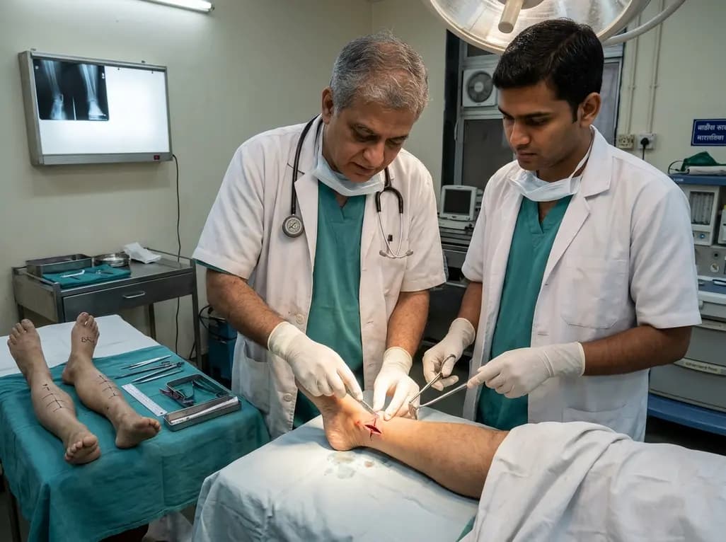
A paired cadaveric study compared triple and quintuple percutaneous Achilles tendon lengthening. While quintuple hemisection showed numerically greater elongation, differences were not statistically significant.
2 days back
A randomized pilot trial shows that consuming a prebiotic functional soda significantly reduces gastrointestinal symptom burden and improves gut tolerance in adults undergoing GLP-1-based weight management therapy.
Today
