.webp&w=384&q=75)

Loading, please wait...
Loading, please wait...

Extradural anterior clinoidectomy remains an indispensable skull base technique for managing paraclinoid and complex anterior circulation vascular lesions. Removing the clinoid process unroofs the optic canal, securing proximal vascular control. However, neurosurgeons frequently debate the isolated neuro-ophthalmic sequelae of bone removal on uncompromised optic pathways. A recent clinical study provides crucial insights into structural, functional, and electrophysiological optic changes following this procedure.
Neurosurgeons frequently encounter paraclinoid aneurysms that demand expanded exposure and proximal vascular control. Resecting the anterior clinoid process creates a generous corridor into the opticocarotid triangle and parasellar space. Traditionally, skull base surgeons debated whether an intradural or extradural trajectory offered greater cranial nerve safety. The extradural route provides earlier vascular identification before dural opening while facilitating extensive optic canal decompression. Nevertheless, manipulating bony structures adjacent to fragile neural elements carries operative hazards. Surgeons have raised concerns regarding direct instrument compression, local thermal injury, and microvascular compromise of delicate pial vessels. Historically, differentiating surgical manipulation injury from pre-existing aneurysmal compression proved challenging. By specifically analyzing cohorts without baseline canal pathology or visual compromise, investigators isolated the technique's true safety profile. Consequently, contemporary skull base teams increasingly deploy specialized cold rongeurs to execute a no-drill bone resection. This mechanical approach eliminates drill-related heat dissipation while maintaining structural exposure. Therefore, determining baseline neuro-ophthalmic outcomes in uncompressed pathways provides a critical benchmark for neurovascular decision-making across global tertiary neurosurgical centers.
Evaluating optic pathway integrity after skull base surgery requires comprehensive diagnostic tools beyond simple visual acuity charts. The referenced single-center investigation evaluated sixteen consecutive adult patients who underwent microsurgical aneurysm clipping with no-drill extradural anterior clinoidectomy. To eliminate confounding variables, investigators systematically excluded patients presenting with baseline optic canal distortion, mass effect, or pre-existing visual pathology. Follow-up examinations occurred between six and twelve months postoperatively, ensuring sufficient duration for retrograde axonal degeneration to manifest. Researchers conducted formal functional assessments using automated perimetry to establish the overall visual field index. Additionally, high-resolution optical coherence tomography quantified peripapillary retinal nerve fiber layer thickness across all anatomical quadrants. Electrophysiological testing via pattern-reversal visual evoked potentials measured P100 wave latency to identify subclinical conduction delays along myelinated axons. By rigorously comparing the operated ipsilateral eye against the unoperated contralateral eye, the study design established an ideal internal control. This multimodal framework provided an objective and reproducible blueprint for detecting subclinical optic neuropathy. Consequently, the data furnish reliable evidence regarding how unroofing alters cranial nerve physiology in pristine neural tissue.
Optical coherence tomography provides unmatched precision when assessing axonal survival following cranial base bone work. In this cohort, global retinal nerve fiber layer thickness remained statistically comparable between the operated ipsilateral eye and the contralateral control eye. Specifically, the ipsilateral eyes demonstrated an average thickness of 95.8 micrometers, whereas contralateral eyes measured 99.2 micrometers. Paired analytical comparisons confirmed that this modest difference lacked statistical significance. However, quadrant-specific evaluations revealed notable microscopic variations that deserve surgical scrutiny. A repeated-measures analysis demonstrated a distinct trend toward preferential nerve fiber thinning within the superior peripapillary quadrant. Anatomically, superior retinal axons correspond directly to the superior optic nerve surface exposed during anterior clinoid resection. Clinicians suggest that localized devascularization of small pial feeding branches during dural peeling may induce subtle focal axonal loss. Although statistical corrections eliminated significant pairwise differences between specific quadrants, this superior predilection highlights potential microvascular vulnerability. Fortunately, widespread structural atrophy did not occur across other axonal bundles. Therefore, no-drill clinoid unroofing preserves macroscopic structural integrity while producing only minimal, localized microscopic changes that spare overall optic nerve volume.
Functional preservation directly dictates patient quality of life after neurovascular intervention. Notably, high-contrast visual acuity was preserved in one hundred percent of the treated patients. Furthermore, automated perimetry revealed overall stability in the visual field index, showing no statistically significant discrepancy between eyes. Nevertheless, detailed perimetric mapping uncovered discrete subclinical visual field abnormalities in approximately nineteen percent of the patient cohort. One individual developed a distinct inferior nasal quadrantanopia, while two individuals exhibited mild blind-spot enlargement. These functional deficits correspond anatomically to the superior axonal thinning observed on retinal imaging. Electrophysiological assessments provided reassuring confirmation of preserved neural conduction velocity across anterior visual pathways. The visual evoked potential P100 latency averaged 114.8 milliseconds in ipsilateral eyes compared to 113.8 milliseconds in contralateral controls. This marginal difference lacked statistical significance, proving that salutary myelination and major saltatory conduction pathways remained intact. Furthermore, no major neurovascular or cerebrospinal fluid complications occurred during clinical monitoring. These functional and electrophysiological findings demonstrate that while gross visual function remains remarkably robust, surgeons must recognize the subtle risk of peripheral visual field changes following anterior clinoidectomy.
These clinical observations provide vital operative benchmarks for neurosurgeons managing paraclinoid vascular and neoplastic pathologies. Because extradural bone removal exposes superior optic nerve fibers, surgical teams must handle adjacent dural bands with extreme gentleness. Devascularization of the pial arterial plexus remains an underestimated mechanism of subclinical visual field deficits. Therefore, surgeons should minimize excessive bipolar electrocoagulation along the optic sheath and avoid unneeded traction during dural unroofing. Additionally, adopting cold instrumentation techniques or ultrasonic bone aspirators prevents detrimental thermal diffusion to delicate myelinated fibers. Skull base centers should routinely incorporate baseline and postoperative optical coherence tomography alongside visual field perimetry for high-risk neurovascular cases. This standardized surveillance protocol detects subclinical axonal degradation early, permitting prompt neuroprotective strategies. Moreover, neurosurgeons should explicitly counsel patients regarding potential peripheral visual field blind spots, even when central visual acuity remains fully preserved. By integrating precise surgical anatomy with rigorous neuro-ophthalmological monitoring, teams can optimize aneurysm obliteration while safeguarding essential cranial nerve functionality. Ultimately, this balanced approach ensures optimal vascular exposure without imposing silent morbidity on the anterior visual pathways.
The anterior clinoidectomy procedure can mechanically disturb delicate pial capillary networks supplying the superior optic nerve surface. Even without thermal drill injury, microvascular devascularization or gentle manipulation may induce subclinical micro-ischemia. Consequently, neurosurgeons observe localized axonal alterations or subtle visual field defects despite preserved central visual acuity and stable global nerve fiber thickness.
Mechanical drilling near the optic canal generates substantial thermal energy and micro-vibrations, which risk irreversible nerve injury. Ultrasonic bone curettes and manual rongeurs effectively eliminate heat transfer to adjacent nervous tissue. Furthermore, this cold instrumental approach enables rapid bone resection and decreases direct mechanical trauma while safely exposing paraclinoid aneurysm necks.
Clinicians should combine optical coherence tomography, visual evoked potentials, and automated visual field testing for objective post-surgical surveillance. Optical coherence tomography detects microscopic retinal nerve fiber layer thinning before clinical deficits appear. Simultaneously, visual evoked potentials evaluate subclinical conduction delays, providing comprehensive structural and electrophysiological data to evaluate nerve integrity.
Disclaimer: This content is for informational and educational purposes only... Refer to the latest local and national guidelines for clinical practice.
References
Aladdam M et al. Evaluation of functional, structural, and electrophysiological optic nerve changes following extradural anterior clinoidectomy in patients without preoperative optic canal compression. Neurosurg Rev. 2026 May 09. doi: 10.1007/s10143-026-04320-y. PMID: 42104188.
Giannantoni NM, Dobran M, Nasi D, Iacoangeli M. Extradural anterior clinoidectomy and optic canal unroofing for paraclinoid and basilar aneurysms: Usefulness of a no-drill instrumental method. Acta Neurochir (Wien). 2021;163(4):1131-1136.
Kulwin C, Matsushima K, Kohyama S, Bohnstedt BN. Intradural versus extradural anterior clinoidectomy: Comparison of visual loss in patients with paraclinoid aneurysms. World Neurosurg. 2024;185:e245-e251.

Read summarized clinical updates, watch expert medical content, and earn CME certifications right from your smartphone.
Open your smartphone camera to scan and install.





A clinical study evaluates optic nerve safety following no-drill extradural anterior clinoidectomy in uncompressed canals, showing preserved visual acuity and stable global RNFL thickness alongside subtle visual field alterations.
Today

An Australian cohort of 34,449 adults in an unsubsidized tirzepatide digital weight loss service showed that peer-referred patients achieved significantly higher 6-month adherence (48% vs 42%) and greater weight loss (16.16% vs 14.06%) than matched non-referred peers, highlighting social support in obesity care.
Today
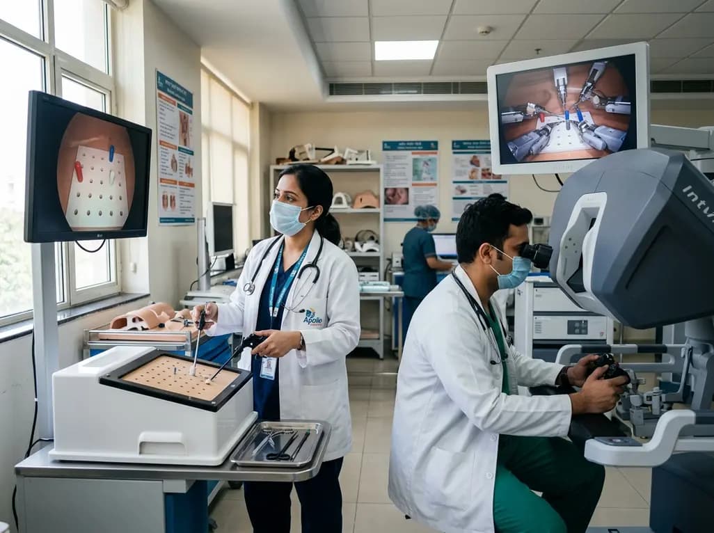
A randomized crossover study demonstrates that conventional and robotic laparoscopy require distinct neuropsychological and psychomotor skills. While trainees master conventional laparoscopy faster, robotic proficiency demands individualized, adaptive curricula tailored to specific learner profiles.
Today
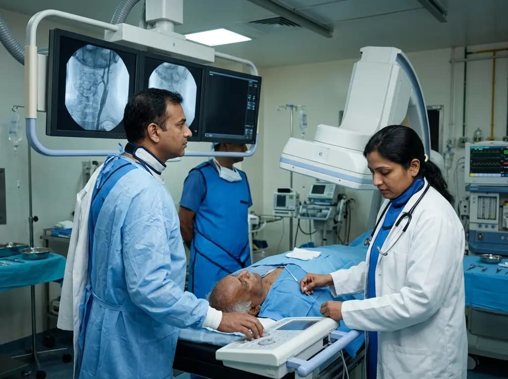
A retrospective study of 109 patients undergoing elective carotid artery stenting evaluated radiation exposure on biplane flat-panel systems. Standardized workflows and dedicated low-dose protocols provide crucial baseline data to establish neurointerventional diagnostic reference levels and optimize patient safety.
Today

A scoping review of 51 studies reveals that spinal manual therapy on the lumbo-pelvic area is commonly used for isolated lower extremity pain, predominantly knee conditions. While thrust manipulation is frequent, reporting deficits highlight the need for standardized technique descriptions to guide practice.
Today

The Supreme Court has reserved its judgment on FSSAI's proposed front-of-pack nutritional warning labels for packaged foods. With debates over compliance timelines, added versus total sugars, and ultra-processed food definitions, this landmark decision carries profound public health implications for India.
Today
