

Loading, please wait...
Loading, please wait...
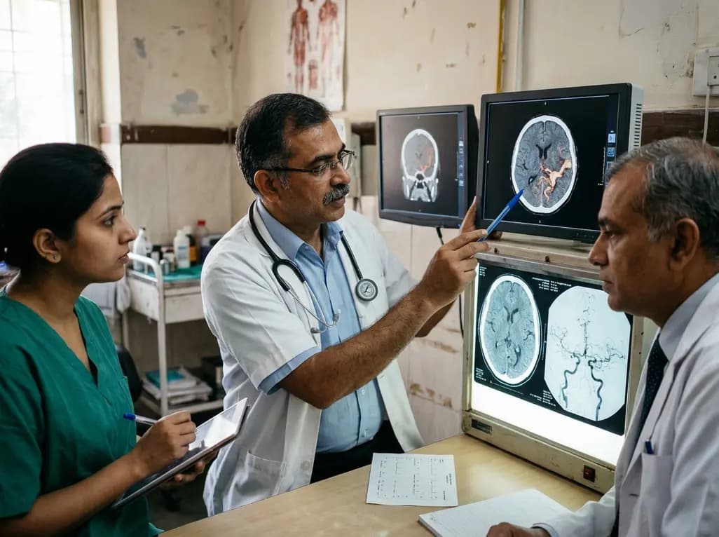
Brain arteriovenous malformations represent complex vascular lesions that carry substantial neurological risks. Unruptured lesions frequently present with recurrent epileptic seizures, which impact approximately twenty to thirty percent of newly diagnosed patients. However, neurosurgeons have historically struggled to pinpoint which specific hemodynamic configurations heighten this epileptogenic potential. To address this clinical uncertainty, investigators developed the Shankar-Krings score. This practical imaging metric uses detailed angioarchitecture to stratify seizure vulnerability. A newly published ambispective study now provides robust external validation of this classification tool.
The Shankar-Krings score evaluates specific morphological markers derived from diagnostic cerebral angiography. Originally, researchers formulated this predictive system by identifying three reproducible anatomical parameters. First, the scoring model assesses venous outflow restriction. Outflow stenosis impairs the physiological drainage of blood through congested parenchymal pathways. Second, it accounts for nidus location. Malformations located within the frontal, temporal, or parietal cortices yield significant cortical irritation compared to deep or infratentorial lesions. Third, the system notes the presence of a long pial draining vein. Consequently, such elongated veins generate expansive focal hemodynamic stress along the adjacent cerebral cortex. Clinicians assign a binary value of zero or one to each of these three angioarchitectural variables. Therefore, an individual patient receives an aggregate score ranging from zero to three. Furthermore, this simple summation allows neurointerventionalists to rapidly quantify seizure risk without requiring elaborate hemodynamic computational simulations. By translating intricate catheter angiography findings into standardized numerical metrics, the framework bridges advanced neurovascular imaging and routine clinical epileptology. Thus, clinicians gain an objective foundation for evaluating patient-specific presentation dynamics.
Understanding why brain arteriovenous malformations trigger seizures requires examining microvascular and metabolic alterations. High-flow arteriovenous shunting directly robs oxygen from surrounding neural tissue. Consequently, this vascular steal syndrome produces chronic perinidal ischemia. In addition, restricted venous outflow exacerbates local tissue congestion. Elevated venous pressures impede capillary perfusion and disrupt regional microcirculation. As a result, the adjacent cortical parenchyma experiences recurrent metabolic failure and impaired cerebrovascular reactivity. Furthermore, congested and tortuous draining veins can induce mechanical mass effect upon sensitive neocortical circuits. Gliosis develops around the vascular nidus over time. Concurrently, repeated microhemorrhages release neurotoxic hemosiderin into the surrounding neuropil. Iron deposits catalyze free radical generation and accelerate cortical excitability. Additionally, altered blood-brain barrier permeability permits inflammatory mediators to penetrate neural parenchyma. These biochemical insults disrupt normal gamma-aminobutyric acid neurotransmission while augmenting excitatory glutamatergic signaling. Ultimately, these combined vascular, mechanical, and neurochemical disruptions transform stable cerebral tissue into an irritable epileptogenic focus. Recognizing these angioarchitectural triggers enables physicians to understand the true biological origin of patient symptoms.
To confirm generalizability, researchers conducted an ambispective cohort study at an academic neurovascular centre. The investigators screened 118 consecutive individuals diagnosed with vascular malformations between 2013 and 2024. However, they strictly excluded ruptured presentations to eliminate confounding effects from acute hemorrhage. Ultimately, the team enrolled 45 patients with verified unruptured brain arteriovenous malformations into the final analytical cohort. Among these eligible participants, 19 individuals, representing 42 percent of the cohort, presented primarily with clinical seizures. Neuroradiologists systematically evaluated all baseline catheter digital subtraction angiograms. Specifically, the imaging specialists remained blinded to the clinical seizure history during image analysis. They meticulously scored each vascular lesion according to the original Shankar-Krings criteria. In addition, the research team recorded comprehensive demographic, clinical, and anatomical variables for each subject. Multivariable logistic regression subsequently examined which morphological parameters independently predicted seizure presentation. Through this rigorous methodology, the authors prevented classification bias and ensured robust data integrity across the ten-year evaluation period.
The external validation demonstrated remarkable predictive capability for the grading model. In multivariable regression models, nidus location within frontal, parietal, or temporal lobes remained independently associated with seizure presentation. Moreover, the risk of experiencing seizures escalated steeply alongside increasing composite scores. Patients with a score of zero experienced no seizures, yielding a presentation rate of zero percent. Conversely, patients with a score of one exhibited a 25 percent seizure incidence. Furthermore, patients scoring two points showed a 47 percent seizure rate. Most remarkably, every single patient who achieved a maximum score of three presented with seizures, representing a 100 percent occurrence rate. The model achieved an outstanding area under the receiver operating characteristic curve of 0.845. Consequently, this high discrimination confirms that the angioarchitectural features reliably distinguish seizure-prone malformations from asymptomatic lesions. Therefore, this independent investigation proves the reproducibility of the scoring system across distinct institutional populations. The compelling statistical concordance validates hemodynamic architecture as an authentic driver of seizure onset.
Accurate risk stratification fundamentally refines how multidisciplinary teams manage unruptured vascular lesions. Historically, the landmark ARUBA trial emphasized medical management over intervention for unruptured malformations. However, persistent seizures severely impair daily functionality, vocational independence, and overall quality of life. In clinical practice, subclinical seizure activity frequently escapes timely medical recognition. Therefore, applying an angioarchitecture-based score helps clinicians identify high-risk individuals before debilitating generalized convulsions occur. When patients present with high scores, neurologists can initiate targeted electroencephalographic monitoring and early antiepileptic drug therapy. In addition, neurosurgeons and interventional neuroradiologists can better weigh procedural risks against neurological benefits. Specifically, targeted embolization of fistulous components or surgical resection may alleviate cortical venous congestion and reduce seizure frequency. Furthermore, understanding precise angioarchitectural drivers guides tailored counseling for affected families. Physicians can clearly explain why specific vascular arrangements promote electrical instability. Ultimately, integrating structural hemodynamic scores into routine clinical workflows enhances personalized neurovascular medicine and improves patient outcomes.
The score evaluates three specific angioarchitectural features identified on diagnostic cerebral angiography. These include venous outflow restriction, lesion location within the frontal, temporal, or parietal lobes, and the presence of a long pial draining vein. Clinicians assign each feature one point, yielding a total score from zero to three.
Venous outflow restriction elevates intravascular pressure within draining veins and adjacent capillaries. Consequently, this hemodynamic congestion impedes microvascular perfusion and produces chronic local ischemia in surrounding brain parenchyma. This metabolic stress, combined with secondary tissue gliosis and regional cortical hypoxia, markedly enhances neuronal excitability and triggers recurrent seizures.
The score helps clinicians identify patients harboring high-risk angioarchitecture who face elevated seizure likelihood. Consequently, care teams can initiate proactive antiepileptic therapy and targeted neuroimaging surveillance. Furthermore, neurovascular specialists may consider targeted interventions, such as embolization or surgical resection, to relieve venous congestion and optimize long-term seizure control.
Disclaimer: This content is for informational and educational purposes only... Refer to the latest local and national guidelines for clinical practice.
References

Read summarized clinical updates, watch expert medical content, and earn CME certifications right from your smartphone.
Open your smartphone camera to scan and install.





An independent study validates the Shankar-Krings angioarchitecture score for predicting seizure presentation in unruptured brain arteriovenous malformations. Evaluating venous outflow restriction, lobar location, and long pial draining veins, the score achieved an outstanding AUC of 0.845.
Today

A case-control study demonstrates significant lipid profile alterations in myocardial infarction patients, highlighting elevated total cholesterol, triglycerides, and LDL-C alongside decreased HDL-C, emphasizing early lipid screening and aggressive secondary cardiovascular prevention.
Today

A new study evaluates a multilingual digital health platform combining rule-based cervical cancer risk assessment, retrieval-augmented generation AI, and geospatial navigation. The tool dramatically reduces clinical hallucinations while providing actionable health guidance for underserved populations.
Today

A landmark UK Biobank study by Karolinska Institutet researchers reveals that optimal rest mitigates neurodegeneration in cardiometabolic illness. Patients maintaining high sleep quality demonstrated superior cognitive scores and larger brain volumes, highlighting sleep optimization as an actionable clinical strategy.
Today
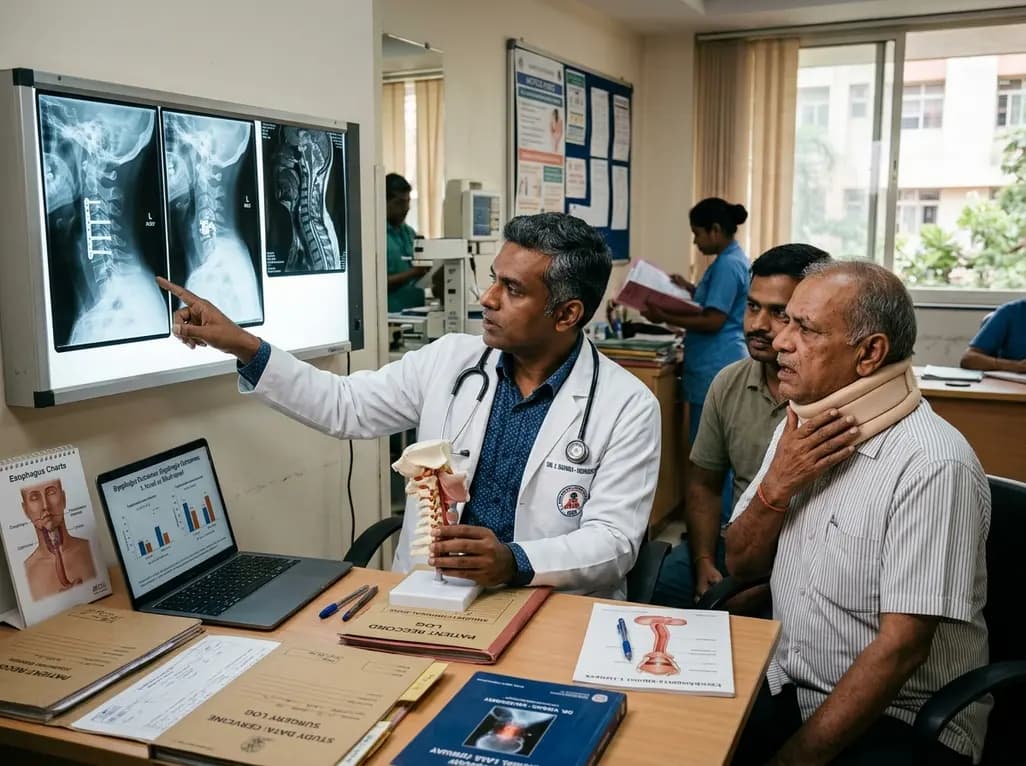
A large registry study reveals that postoperative dysphagia occurs more frequently and persists longer after multilevel anterior cervical spine surgery than single-level procedures. Key risk factors include baseline dysphagia, diabetes, upper cervical segment involvement, and medical comorbidities.
Today
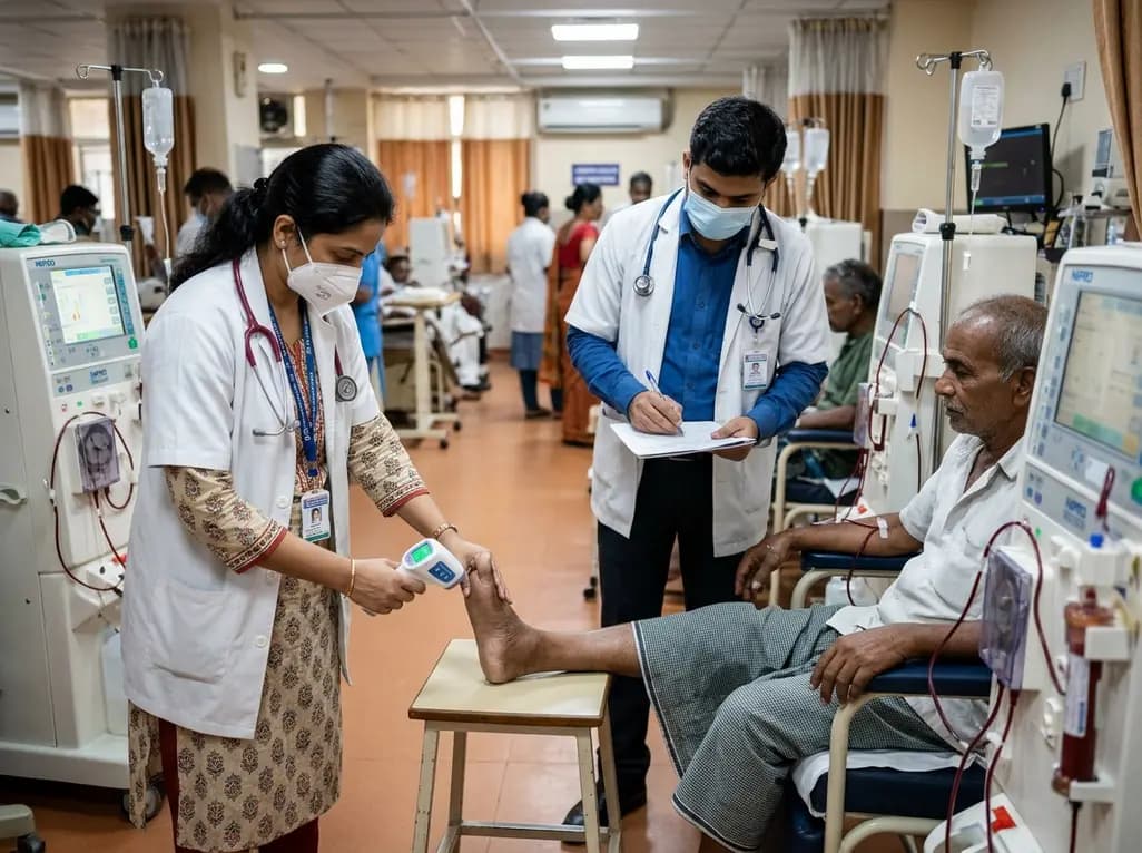
A prospective study reveals that tracking intradialytic foot temperature gradients offers a noninvasive approach to screen for severe peripheral artery disease in hemodialysis patients.
Today
