

Loading, please wait...
Loading, please wait...

Acute ischemic stroke management continues to evolve beyond conventional therapeutic windows. Historically, strict time cutoffs excluded many individuals with large vessel occlusion from receiving mechanical reperfusion. However, physiological tissue viability matters more than elapsed chronometric time. Emerging clinical data highlight that endovascular thrombectomy beyond 24 hours offers substantial neurological benefits for carefully triaged patients. By identifying persistent ischemic penumbra and collateral blood flow through advanced neuroimaging, clinicians can salvage eloquent brain tissue days after symptom onset. This paradigm shift expands treatment options for late-presenting patients who previously had limited therapeutic pathways beyond supportive medical therapy.
For decades, acute stroke protocols enforced rigid time windows to prevent reperfusion injury and futile recanalization. Landmark clinical trials initially established the efficacy of intervention up to 24 hours based on advanced CT or MR perfusion profiles. Nevertheless, patients presenting past the 24-hour mark frequently faced therapeutic exclusion, receiving only best medical management. Clinicians increasingly recognize that collateral circulation dictates ischemic core growth far more effectively than chronological clocks alone. Slow progressors often retain significant penumbral tissue days after vessel occlusion because leptomeningeal collaterals sustain capillary beds. Consequently, denying reperfusion based solely on time arbitrarily harms patients who possess salvageable cerebral parenchyma. Modern neurointerventional practice therefore demands reliable, accessible imaging biomarkers that identify viable brain parenchyma. Magnetic resonance imaging provides physiological insights without requiring high doses of iodinated contrast or complex automated post-processing software. Therefore, evaluating vascular flow dynamics on non-invasive sequences helps clinicians make confident, timely decisions for delayed presentations.
Fluid-attenuated inversion recovery vascular hyperintensities, known as FVH, represent slow or retrograde blood flow through leptomeningeal collateral arteries. Conversely, diffusion-weighted imaging, or DWI, precisely delineates the irreversibly infarcted core. When FVH extends significantly beyond the geographic boundaries of the acute DWI lesion, clinicians observe a classic FVH-DWI mismatch. This neuroimaging phenomenon indicates substantial tissue hypoperfusion supported by sluggish collateral flow that remains viable yet vulnerable. Unlike automated perfusion software, assessing FVH-DWI mismatch does not require specialized licensing or intravenous gadolinium contrast administration. As a result, community hospitals and resource-conscious healthcare systems can readily implement this imaging evaluation protocol. Furthermore, magnetic resonance sequences evaluate parenchymal salvageability rapidly while simultaneously ruling out completed catastrophic infarctions or occult hemorrhagic conversion. Clinicians can immediately visualize the collateral reserve and identify candidates who will benefit from arterial recanalization. Consequently, this imaging mismatch functions as a robust surrogate for physiological viability in delayed presentations.
Recent results from the prospective TRACK-LVO Late multicenter cohort provide compelling real-world evidence for extended intervention. The study screened 648 patients presenting beyond 24 hours from last known well and identified 247 eligible individuals harboring confirmed FVH-DWI mismatch. Among these patients, 132 individuals underwent mechanical reperfusion while 115 received best medical management. To account for confounding factors, researchers utilized inverse probability of treatment weighting-adjusted logistic regression models. Strikingly, the endovascular thrombectomy group demonstrated a more than threefold increase in the odds of achieving 90-day functional independence compared to medical management alone. Specifically, patients who achieved successful mechanical recanalization experienced superior motor and cognitive recovery. In contrast, patients receiving only conservative medical therapy experienced progressive functional decline and prolonged hospitalization. Furthermore, subgroup assessments revealed consistent functional gains across varied anatomical occlusion sites, including carotid terminus and middle cerebral artery segments. These robust findings strongly indicate that mechanical intervention effectively halts irreversible core expansion even during extremely delayed presentations.
Safety remains a paramount clinical consideration when performing recanalization on mature ischemic infarcts. Delayed endovascular thrombectomy often raises legitimate concerns regarding severe reperfusion injury, breakdown of the blood-brain barrier, and symptomatic intracranial hemorrhage. Fortunately, data from the multicenter investigation demonstrated that mechanical intervention maintained an acceptable safety profile when clinicians adhered to strict neuroimaging criteria. The occurrence of symptomatic intracranial hemorrhage did not show a statistically prohibitive surge in the interventional arm compared to standard medical management. In addition, 90-day all-cause mortality rates remained comparable between the two treatment cohorts. These neutral safety outcomes demonstrate that carefully selecting patients through tissue-based imaging prevents catastrophic reperfusion into extensively infarcted brain territories. Adequate microvascular collateral circulation preserves endothelial integrity and capillary bed structure, thereby reducing microvascular rupture during pressure restoration. Therefore, neurointerventionalists can confidently pursue endovascular thrombectomy in select late presenters without inflicting disproportionate procedural harm.
The clinical implications of these findings hold substantial significance for stroke management across diverse healthcare environments. In developing healthcare systems like India, acute stroke patients frequently present well beyond standard therapeutic windows due to geographic barriers, transit delays, and limited initial awareness. Moreover, many secondary and tertiary healthcare centers lack proprietary automated perfusion software or specialized perfusion CT processing capabilities. Because standard magnetic resonance scanners are widely available across urban and semi-urban Indian medical centers, evaluating FVH-DWI mismatch offers an exceptionally practical triage strategy. Clinicians can readily interpret conventional FLAIR and DWI sequences at bedside without requiring external cloud analysis or expensive contrast agents. Furthermore, identifying patients with robust collateral flow enables targeted interventional referrals, optimizing cath lab utilization and public health expenditure. Implementing standardized MRI-based triage pathways in regional stroke networks will allow interventionalists to rescue viable cerebral tissue in late-arriving patients who would otherwise face lifelong debilitating neurological disability.
FVH-DWI mismatch describes fluid-attenuated inversion recovery vascular hyperintensities extending beyond the boundaries of diffusion-weighted imaging lesions. This radiologic finding signifies viable penumbral tissue receiving retrograde collateral blood flow. It demonstrates that salvageable cerebral parenchyma persists despite prolonged large vessel occlusion, helping clinicians identify ideal candidates for late mechanical recanalization.
Endovascular thrombectomy beyond 24 hours significantly improves functional independence at 90 days in appropriately triaged patients. Restoring arterial blood flow salvages hypoperfused penumbra, preventing secondary neurological deterioration and reducing long-term disability. Clinical evidence confirms that patients selected via collateral imaging mismatch achieve superior functional recovery compared to medical therapy alone.
When clinicians select candidates using strict tissue viability biomarkers such as FVH-DWI mismatch, delayed mechanical thrombectomy does not significantly increase symptomatic intracranial hemorrhage or 90-day mortality. Preserved microvascular collaterals maintain capillary wall integrity. Consequently, targeted revascularization remains safe even when performed well beyond traditional 24-hour therapeutic windows.
Disclaimer: This content is for informational and educational purposes only and should not be considered as medical advice or used for treatment or diagnosis. Healthcare professionals should exercise their independent clinical judgment. Refer to the latest local and national guidelines for clinical practice.
References

Read summarized clinical updates, watch expert medical content, and earn CME certifications right from your smartphone.
Open your smartphone camera to scan and install.





A multicenter cohort study reveals that endovascular thrombectomy beyond 24 hours significantly improves functional independence in acute ischemic stroke patients selected via FLAIR vascular hyperintensity-DWI mismatch compared to medical management alone.
Today

A complex clinical case reveals how adipsic hypernatremia after traumatic brain injury, combined with hyperosmolar hyperglycemic state, triggers life-threatening dehydration and extensive venous thromboembolism, emphasizing the vital role of meticulous fluid management and vigilance.
Today

Large genomic databases like gnomAD and All of Us offer crucial insights into the population prevalence of partial RHD genotypes. Identifying these variants helps clinicians mitigate anti-D alloimmunization risks in transfusion medicine and obstetric care across diverse ancestral populations.
Yesterday
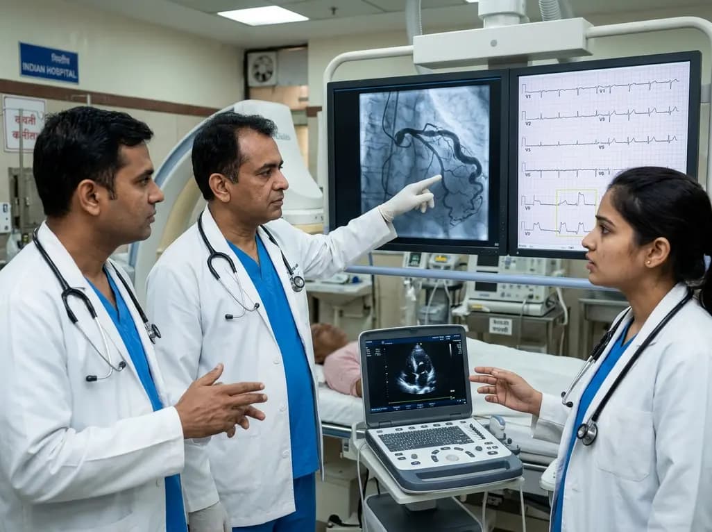
A hyperdominant left circumflex artery supplies extensive myocardial regions, creating a perilous diagnostic blind spot during acute occlusion. Standard ECGs often conceal posterior infarction, requiring clinicians to employ posterior leads and rapid echocardiography for early invasive catheterization.
Today
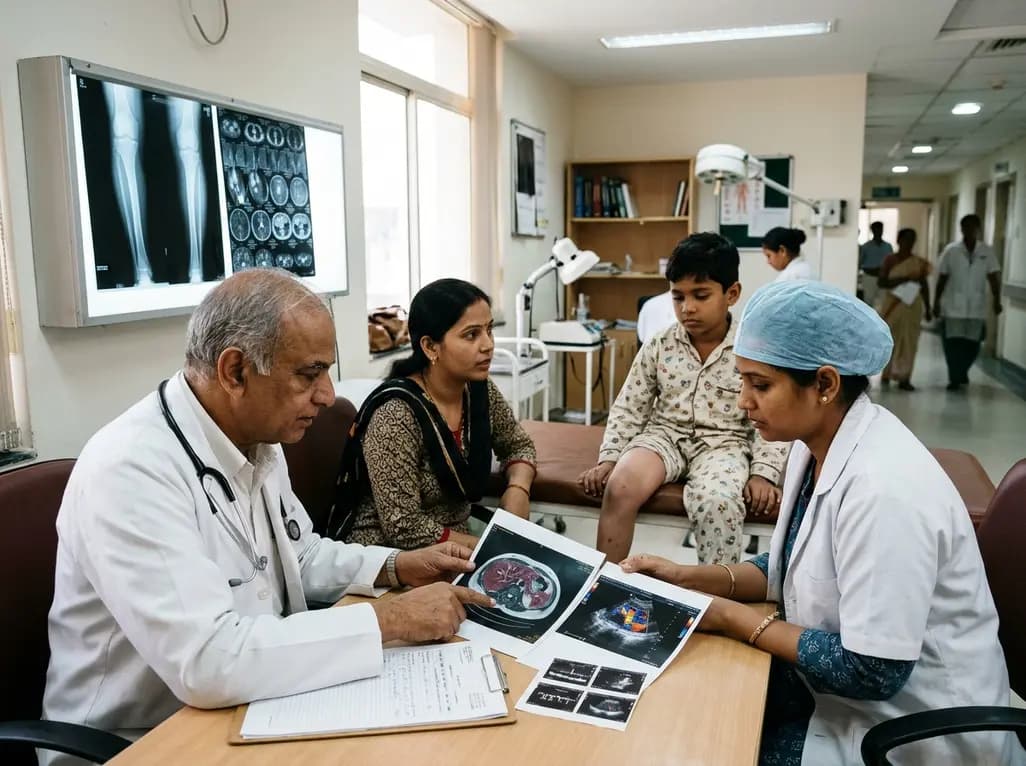
Deep intramuscular vascular malformations in pediatric Klippel-Trenaunay syndrome can mimic Maffucci syndrome. Multimodality imaging with Doppler ultrasound and MRI, along with histopathology, ensures prompt differential diagnosis, targeted surgical debulking, and optimized conservative surveillance.
Today

Discover essential clinical insights into AGLP1-type amyloidosis, a rare localized complication of glucagon-like peptide-1 analog injections. Learn how histopathology and mass spectrometry differentiate benign iatrogenic amyloidomas from systemic disease, preventing misdiagnosis and unwarranted therapies.
Yesterday
