.webp&w=384&q=75)

Loading, please wait...
Loading, please wait...

Temporal encephaloceles (TEs) are increasingly identified as primary epileptogenic lesions in patients suffering from drug-resistant temporal lobe epilepsy. While traditional craniotomies were once the standard, endonasal temporal encephalocele resection has emerged as a viable, minimally invasive alternative. This approach is particularly effective when the lesion herniates into the sphenoid sinus. However, clinicians often face challenges in defining the exact extent of the resection required to ensure seizure freedom without direct electrophysiological confirmation.
A recent illustrative case involved a 41-year-old right-handed man presenting with focal impaired consciousness seizures that were resistant to medical therapy. Initial MRI imaging identified a right-sided TE herniating into the lateral recess of the sphenoid sinus. Furthermore, scalp EEG localized the seizure onset to the right anterior temporal region, showing no signs of a broader temporal network involvement. Consequently, the surgical team opted for an endoscopic endonasal transpterygoid approach to expose the encephalocele and the adjacent temporal dura mater.
To confirm the site of epileptogenicity, surgeons placed depth electrodes directly into the encephalocele and the surrounding cortex. These recordings demonstrated frequent spikes in both areas, providing the necessary electrophysiological evidence for a tailored resection. Following this confirmation, the surgical team resected the encephalocele along with a 7-mm margin of the adjacent cortex. Moreover, they performed a multilayer skull base reconstruction to prevent cerebrospinal fluid leaks. The patient remained seizure-free for a follow-up period of 48 months with no surgical complications.
The success of this case highlights the benefits of using endonasal depth EEG to guide surgical margins. In appropriately selected patients, particularly those lacking a widespread mesial temporal network, this technique avoids the morbidity of a full craniotomy. Additionally, it supports the use of tailored lesionectomy over more extensive temporal lobectomies. This case suggests that achieving durable seizure freedom is possible through targeted, minimally invasive interventions when guided by intraoperative electrophysiology.
Intraoperative depth EEG provides direct confirmation of epileptogenicity within the herniated tissue and the adjacent cortex. This allows surgeons to precisely define resection margins, ensuring the removal of the epileptogenic zone while sparing healthy brain tissue.
Ideal candidates are patients with drug-resistant focal epilepsy and imaging evidence of a temporal encephalocele herniating into the sphenoid sinus. It is most suitable for those whose electroclinical data does not suggest a widespread or mesial temporal epileptogenic network.
The endoscopic endonasal approach is less invasive, reduces recovery time, and avoids the risks associated with large scalp incisions and bone flaps. It offers a direct route to lesions in the lateral recess of the sphenoid sinus with minimal brain retraction.
Disclaimer: This content is for informational and educational purposes only and does not constitute medical advice or a professional relationship. Always seek the advice of a qualified healthcare provider regarding any medical condition. Refer to the latest local and national guidelines for clinical practice.
References
Ohtani K et al. Endoscopic endonasal depth EEG-guided resection of a temporal encephalocele resulting in seizure freedom: illustrative case. J Neurosurg Case Lessons. 2026 May 18. doi: undefined. PMID: 42150197.
Ishii A, et al. Endonasal endoscopic surgery for temporal lobe epilepsy associated with sphenoidal encephalocele. Surg Neurol Int. 2021;12:379. doi: 10.25259/SNI_438_2021.
Khoudari H, et al. Seizure outcomes after resection of temporal encephalocele in patients with drug-resistant epilepsy: a systematic review and meta-analysis. Epilepsia. 2025.
"
Read summarized clinical updates, watch expert medical content, and earn CME certifications right from your smartphone.
Open your smartphone camera to scan and install.





A 41-year-old patient achieved 48-month seizure freedom after an endoscopic endonasal depth EEG-guided resection of a temporal encephalocele....
5 months ago
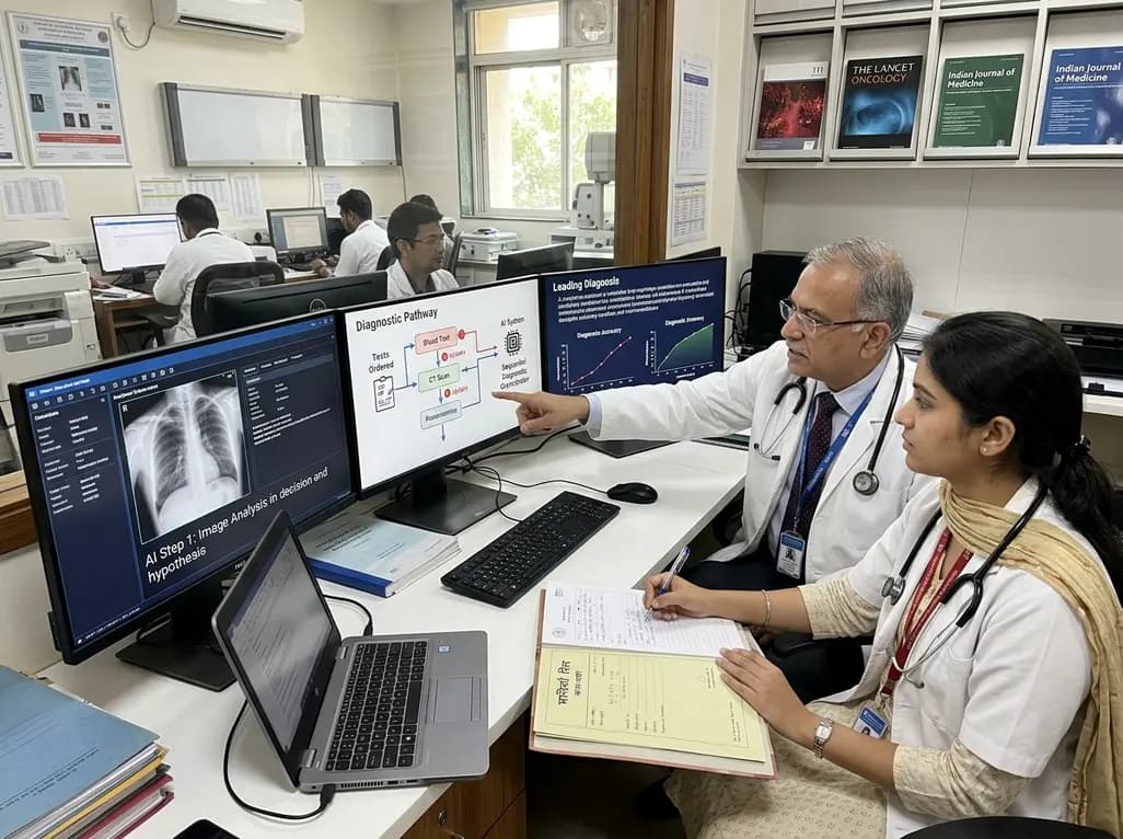
A breakthrough peer-reviewed study demonstrates that a sequential AI diagnosis system can mirror real-world clinical reasoning. By ordering stepwise tests and updating hypotheses dynamically, this multi-agent orchestrator achieved superior diagnostic accuracy while reducing investigative costs.
Today

A prenatal case highlights severe glutaric acidemia type II presenting as bilateral hyperechoic cystic kidneys at 18 weeks. Whole-exome sequencing revealed a novel homozygous ETFDH frameshift variant, demonstrating the power of rapid genetic testing in fetal renal anomalies.
Today

Review how tailored exercise programs benefit frail older patients with heart failure. Explore pathophysiological mechanisms, individualized training prescriptions, and strategies to overcome adherence barriers in clinical practice.
Yesterday

This comparative study evaluates posterior labral tears with and without glenohumeral instability, highlighting distinct demographic patterns, MRI underestimation in stable shoulders, and successful arthroscopic repair outcomes.
Yesterday

A clinical study demonstrates that early intervention with tocilizumab, either as monotherapy or bridged with corticosteroids, achieves significant proptosis reduction and resolves active inflammation in treatment-naive thyroid eye disease.
Yesterday
