.webp&w=384&q=75)

Loading, please wait...
Loading, please wait...
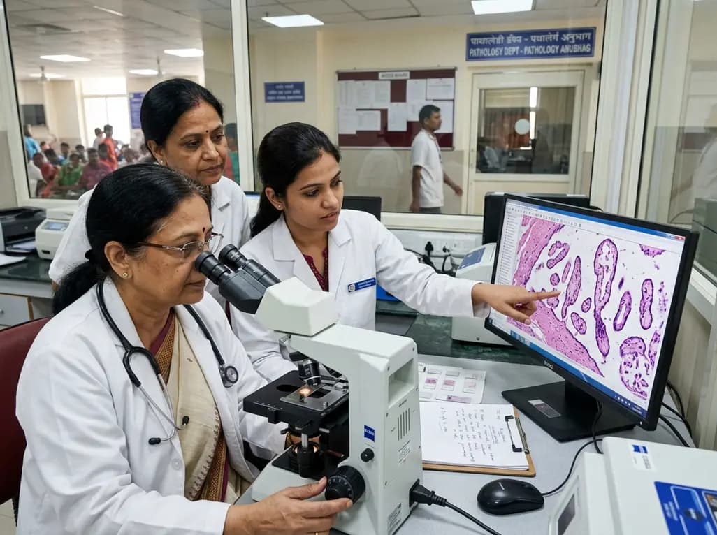
Placental examination provides vital insights into intrauterine compromise, fetal development, and unexpected perinatal loss. Historically, pathologists associated dysmorphic chorionic villi primarily with early pregnancy failures, genetic aneuploidies, and partial hydatidiform moles. However, emerging clinicopathological evidence demonstrates that these structural abnormalities can persist into later gestation. Dysmorphic villi exhibit irregular contours, stromal dysmorphism, abnormal trophoblastic invaginations, and persistent villous hypermaturity or immaturity. Consequently, identifying these anomalies in late pregnancy challenges traditional diagnostic paradigms.
Recent investigations reveal that dysmorphic chorionic villi appear across all three trimesters, showing an unexpected prevalence peak during the second trimester. Furthermore, when these abnormal villous features persist into the third trimester, their pathophysiological significance changes substantially. Pathologists frequently uncover profound feto-placental vascular disturbances rather than classic chromosomal aneuploidies. Therefore, clinicians must re-evaluate third-trimester placental morphology to anticipate potential neonatal complications. Recognizing this unique morphological phenotype enables obstetricians and perinatologists to understand underlying mechanisms of late fetal compromise. Ultimately, systematic placental histopathology bridges the gap between antenatal sonographic findings and postpartum neonatal trajectories.
Fetal vascular malperfusion represents a severe category of placental injury caused by thrombosis, vascular stasis, or umbilical cord compromise. Pathologists define this condition by hallmark features such as villous stromal-vascular karyorrhexis, avascular terminal villi, and stem vessel luminal obliteration. Additionally, extensive mural or occlusive thrombi within chorionic plate vessels frequently accompany these parenchymal changes. When blood flow through the fetal placental circulation ceases, distal villi gradually lose their functional capillary networks. Consequently, the surrounding stromal core undergoes progressive hyalinization and sclerotic transformation.
Importantly, the coexistence of fetal vascular malperfusion and dysmorphic chorionic villi creates a compounding hypoxic environment. The architectural irregularities of dysmorphic villi increase local vascular resistance, which subsequently predisposes fetal vessels to localized thrombosis and endothelial injury. Moreover, chronic cord compression or structural cord abnormalities can accelerate this downstream vascular collapse. Pathologists grading fetal vascular lesions evaluate both the distribution and severity of avascular villous clusters. Thus, accurate recognition of these overlapping histological patterns establishes a precise morphological roadmap. This detailed evaluation clarifies whether placental hypoperfusion stems from maternal vascular disease or primary fetal circulatory failure.
The clinical consequences of combined villous dysmorphism and fetal vascular malperfusion in the third trimester are profound. Fetuses subjected to chronic vascular occlusion frequently exhibit severe fetal growth restriction, oligohydramnios, and non-reassuring intrapartum cardiotocographic patterns. Furthermore, extensive fetal thrombotic vasculopathy directly correlates with an increased risk of intrauterine fetal demise and unexpected stillbirth. Because the placenta loses significant functional reserve, the fetus cannot tolerate normal uterine contractions during labor. Consequently, emergency cesarean deliveries occur at a substantially higher rate in these affected pregnancies.
In addition to acute perinatal risks, neonates face long-term systemic and neurodevelopmental sequelae. Fetal vascular thromboemboli originating in the chorionic vessels can dislodge into the systemic circulation, potentially damaging vulnerable fetal end-organs. Notably, clinicians observe strong correlations between high-grade fetal vascular lesions and neonatal encephalopathy, cerebral palsy, and renal tubular injury. Therefore, detecting these morphological hallmarks provides indispensable diagnostic clarity for pediatricians managing complicated neonatal courses. Multidisciplinary communication between pathologists and neonatologists ensures targeted clinical monitoring during the critical postpartum transition. Hence, comprehensive placental analysis acts as an essential post-delivery investigative tool.
Differentiating primary fetal vascular insults from maternal vascular malperfusion remains a critical diagnostic priority for anatomical pathologists. Maternal vascular malperfusion typically manifests with accelerated villous maturation, syncytial knots, distal villous hypoplasia, and decidual arteriopathy. In contrast, dysmorphic chorionic villi feature irregular villous shapes, aberrant stromal cellularity, and altered branching architecture that reflect intrinsic developmental derangements. However, both pathological processes can coexist in high-risk pregnancies, creating overlapping histological phenotypes that complicate diagnostic interpretation.
Fortunately, standardized morphological consensus criteria help pathologists reliably categorize these distinct lesions. While maternal vascular lesions reflect inadequate spiral artery remodeling and impaired uterine blood supply, fetal vascular malperfusion directly involves fetal vessels. Therefore, evaluating vessel patency, endothelial integrity, and distal villous stromal karyorrhexis provides definitive clues. Furthermore, immunohistochemical markers, such as CD34 endothelial staining, facilitate the detection of subtle or early vascular fragmentation. As a result, pathologists can distinguish primary feto-placental circulatory collapse from extrinsic maternal hypertensive disorders. This clear distinction directly guides maternal counseling and informs therapeutic strategies for subsequent pregnancies.
Optimizing clinical outcomes requires standardized placental submission protocols and coordinated multidisciplinary care pathways. Clinicians should routinely submit the placenta for pathological examination following pregnancies complicated by growth restriction, abnormal Doppler velocimetry, or unexplained fetal distress. Moreover, pathologists must document the presence, grade, and extent of both dysmorphic chorionic villi and vascular malperfusion lesions. Standardized synoptic reporting formats ensure that all essential histological parameters reach the clinical team promptly.
Following the diagnosis of significant fetal vascular pathology, clinicians should tailor future maternal and neonatal management plans accordingly. Because certain maternal thrombophilias, autoimmune disorders, and structural cord anomalies increase recurrence risks, targeted maternal screening is often warranted. Additionally, pediatric teams should integrate placental findings into long-term developmental surveillance protocols for high-risk infants. In subsequent pregnancies, maternal-fetal medicine specialists should implement serial growth ultrasonography and comprehensive umbilical artery Doppler assessments from early gestation. Thus, integrating placental histopathology into ongoing clinical decision-making significantly improves maternal and neonatal health outcomes across successive gestations.
Dysmorphic chorionic villi are structurally abnormal placental villi featuring irregular contours, stromal alterations, and atypical trophoblastic branching. Although clinicians historically linked them to first-trimester chromosomal aneuploidies, their persistence into the third trimester strongly correlates with fetal vascular malperfusion. Consequently, their identification in late pregnancy serves as an important histopathological marker for severe feto-placental circulatory impairment, fetal growth restriction, and adverse perinatal outcomes.
Fetal vascular malperfusion impairs oxygen and nutrient transfer while promoting localized thrombosis within fetal placental vessels. Furthermore, microthrombi can embolize into fetal circulation, potentially causing ischemic injury to vital organs such as the brain and kidneys. Clinicians observe increased frequencies of neonatal encephalopathy, intrapartum distress, and long-term neurodevelopmental deficits. Therefore, identifying these lesions alerts pediatricians to conduct rigorous neurological evaluations during neonatal follow-up.
Following a confirmed diagnosis of severe placental vascular malperfusion, clinicians recommend evaluating the mother for underlying prothrombotic or autoimmune conditions. In subsequent pregnancies, obstetricians should implement intensive fetal surveillance, including early serial growth scans, umbilical Doppler assessments, and biophysical profile monitoring. Additionally, proactive management of maternal vascular risk factors and detailed postpartum placental examination help optimize outcomes and prevent recurrent adverse perinatal events.
Disclaimer: This content is for informational and educational purposes only and is not intended as medical advice. Healthcare professionals should exercise their independent clinical judgment. Refer to the latest local and national guidelines for clinical practice.
References

Read summarized clinical updates, watch expert medical content, and earn CME certifications right from your smartphone.
Open your smartphone camera to scan and install.





A recent study reveals that dysmorphic chorionic villi in third-trimester placentas are strongly associated with fetal vascular malperfusion (72.4%), underscoring critical implications for fetal growth restriction, stillbirth risk, and neonatal management.
last month

This clinical review examines the dangerous intersection of climate change and antimicrobial resistance (AMR). It highlights thermal selection, altered transmission pathways, and the imperative for One Health and Planetary Health adaptation frameworks to preserve therapeutic efficacy.
Today
Discover the crucial distinctions between mesonephric and mesonephric-like proliferations of the female genital tract. Learn the key histomorphologic, immunohistochemical, and molecular differences essential for accurate diagnosis and clinical management.
Today

A large UK Biobank study reveals that plasma proteomic signatures capture preclinical organ damage and improve multiorgan risk prediction across early CKM syndrome stages 0 to 2 beyond traditional PREVENT clinical models.
Today

A narrative review investigates whether the articularis genus muscle functions as an independent anatomical entity or blends with the vastus intermedius. We evaluate its morphology, role in retracting the suprapatellar bursa, and direct clinical significance in anterior knee pain and arthroplasty.
Today

A Bayesian multilevel meta-analysis reveals that aerobic training combined with moderate carbohydrate restriction modestly lowers HbA1c in type 2 diabetes. However, sparse data and very low certainty leave incremental benefits over exercise or diet alone unproven, highlighting the need for individualized care.
Today
