.webp&w=384&q=75)

Loading, please wait...
Loading, please wait...
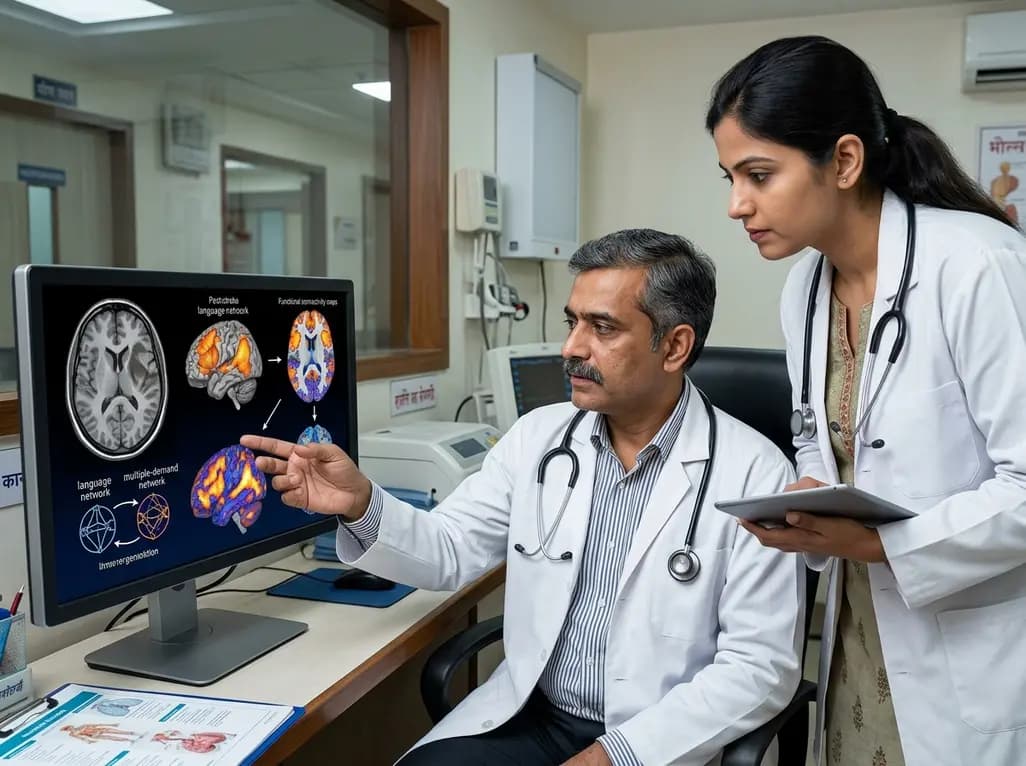
Post-stroke aphasia presents a complex clinical challenge that impairs communication and significantly diminishes quality of life. Clinicians recognize that language restoration is not a uniform trajectory but rather a heterogeneous biological process. Recent neuroimaging research highlights that post-stroke aphasia recovery relies heavily on adaptive network reorganization rather than isolated regional repair. Damage to core left-hemisphere language nodes triggers widespread functional shifts across both language-dedicated and domain-general cognitive architectures. Consequently, tracking these neuroplastic alterations over time provides crucial insights into how neural circuits reorganize to re-establish linguistic abilities. Understanding these dynamic causal interactions across acute, subacute, and chronic post-stroke phases empowers clinicians to design more targeted, lesion-specific neurorehabilitation strategies.
Historically, neuroscientists focused primarily on localized cortical activity when studying speech restitution following cerebral infarction. However, contemporary connectomics confirms that post-stroke aphasia recovery depends on effective connectivity across distributed neural circuits. In a longitudinal functional neuroimaging investigation, researchers tracked stroke survivors across acute (≤1 week), subacute (1-2 weeks), and chronic (6 months) recovery intervals. By employing Dynamic Causal Modelling during auditory sentence comprehension tasks, the investigators mapped the changing directional influences among cortical hubs. They found that effective connectivity fluctuates substantially across these distinct temporal windows. Consequently, the brain recruits secondary neural pathways to compensate for disrupted primary circuits. In the acute phase, rapid shifts in inter-hemispheric communication occur, whereas the subacute stage fosters consolidated connectivity among residual language zones. Therefore, recovery reflects a dynamic interplay between dedicated language modules and supporting cognitive networks, rather than static perilesional compensation.
A crucial discovery in modern cognitive neurology is the pivotal contribution of the domain-general multiple-demand network. This bilateral system encompasses the dorsolateral prefrontal cortex, anterior insula, pre-supplementary motor area, and dorsal anterior cingulate cortex. During speech processing, this network manages executive control, cognitive flexibility, working memory, and selective attention. The longitudinal study demonstrated that stroke survivors exhibit increased task-related connectivity directed from the multiple-demand network toward residual language regions. Interestingly, this facilitation mimics the baseline architecture observed in healthy individuals, indicating an adaptive attempt to restore normal operational dynamics. Moreover, early facilitatory drive from multiple-demand regions to canonical language areas directly correlated with superior long-term language improvements. Thus, domain-general cognitive control systems act as a critical compensatory scaffolding that supports failing language-specific processors during the acute and subacute recovery phases.
The neural architecture of recovery varies dramatically depending on the anatomical site of vascular disruption. Patients with left frontal infarctions exhibit distinct neuroplastic reorganization pathways compared to those with left temporo-parietal lesions. In frontal stroke cohorts, acute functional imaging revealed compensatory facilitatory input originating from the intact right-hemisphere inferior frontal cortex to the undamaged left frontal regions. As these patients transitioned into the chronic phase, their brains established sustained facilitatory modulation from the right dorsolateral prefrontal cortex. Conversely, patients suffering from temporo-parietal strokes demonstrated a different compensatory strategy. In the chronic stage, these individuals predominantly integrated the supplementary motor area and dorsal anterior cingulate cortex into their comprehension network. Hence, neural reorganization is strictly constrained by lesion location, showing that anterior and posterior language lesions recruit divergent cognitive control hubs over time.
These neuroimaging findings provide a biological blueprint for refining non-invasive brain stimulation protocols. Historically, repetitive transcranial magnetic stimulation (rTMS) and transcranial direct current stimulation (tDCS) have yielded variable clinical efficacy in aphasia trials. This heterogeneity largely stems from standardized, one-size-fits-all stimulation targets that fail to account for post-stroke chronometry or lesion topology. Because facilitatory connections evolve from acute right-hemispheric homologue recruitment to chronic prefrontal and cingulate integration, neurostimulation protocols must adapt accordingly. For example, stimulating the right dorsolateral prefrontal cortex may offer substantial therapeutic benefit for chronic frontal stroke patients, whereas targeting medial frontal structures could prove superior for temporo-parietal lesions. Therefore, transitioning toward connectome-guided, patient-tailored neuromodulation protocols holds immense promise for maximizing linguistic gains in neurorehabilitation clinics.
Integrating network-level insights into day-to-day clinical practice can fundamentally refine stroke rehabilitation paradigms. Neurologists, physiatrists, and speech-language pathologists must recognize that linguistic recovery draws upon domain-general executive resources. Consequently, speech therapy should not operate in isolation from general cognitive training. Combining structured linguistic exercises with executive function tasks may reinforce the facilitatory connections streaming from the multiple-demand network. Furthermore, monitoring functional connectivity dynamics allows clinicians to stratify patient prognosis more accurately during early recovery stages. Early engagement of compensatory frontoparietal pathways signals favorable long-term language gains, whereas deficient cross-network coupling may identify candidates who require intensified neuromodulatory and behavioral interventions. Ultimately, embracing a network-centric model enables clinicians to deliver synchronized, phase-appropriate therapies that significantly enhance recovery outcomes.
The multiple-demand network is a bilateral brain system supporting domain-general cognitive functions like attention and working memory. In aphasia recovery, this network provides crucial compensatory facilitation to damaged language regions, helping maintain speech comprehension and driving long-term functional improvement.
Lesion site dictates which compensatory networks the brain recruits over time. Frontal stroke patients primarily recruit the right prefrontal cortex and right-hemisphere homologues, whereas temporo-parietal stroke patients depend more on medial frontal structures like the supplementary motor area and anterior cingulate cortex.
These findings enable personalized non-invasive brain stimulation, such as transcranial magnetic stimulation. Instead of applying generic protocols, clinicians can target specific brain regions based on the patient's exact lesion location and recovery phase, maximizing therapeutic efficacy and neuroplasticity.
Disclaimer: This content is for informational and educational purposes only and does not constitute medical advice, diagnosis, or treatment recommendations. Refer to the latest local and national guidelines for clinical practice.
References
Jiang Z et al. Dynamic reorganization of task-related network interactions in post-stroke aphasia recovery. Brain. 2025 Oct 03. doi: 10.1093/brain/awaf036. PMID: 39883566.
Kiran S, Thompson CK. Neuroplasticity of Language Networks in Aphasia: Advances, Updates, and Future Challenges. Front Neurol. 2019;10:295. doi: 10.3389/fneur.2019.00295.
Saur D, Ronneberger O, Kümmerer D, et al. Dynamics of language reorganization after stroke. Brain. 2006;129(Pt 6):1371-1384. doi: 10.1093/brain/awl090.

Read summarized clinical updates, watch expert medical content, and earn CME certifications right from your smartphone.
Open your smartphone camera to scan and install.





A landmark neuroimaging study reveals how dynamic functional interactions between the language and domain-general multiple-demand networks drive post-stroke aphasia recovery over time, offering vital clues for lesion-specific, targeted neuromodulation therapies.
last month
UK Research and Innovation is deploying £80 million across the NHS to trial nine cutting-edge technologies. The initiative features seven-minute brain MRIs, blood biomarker assays, and predictive AI tools, aiming to evaluate 92% of suspected cases within 18 weeks and overcome lengthy diagnostic backlogs.
Today

A mixed-methods study identifies key drivers of PMTCT service utilization among adolescent girls and young women aged 15-24. Highlighting age disparities, PrEP uptake, and systemic barriers, the evidence guides clinicians toward adolescent-responsive, integrated maternal HIV prevention protocols.
Today

PepsiCo and Monster Beverage have challenged the FSSAI ban on the energy drink label in Indian courts. While companies highlight supply disruptions, regulatory actions spotlight severe cardiovascular and metabolic risks associated with excessive caffeine, sugar, and taurine consumption, especially in adolescents.
Today

A narrative review investigates whether the articularis genus muscle functions as an independent anatomical entity or blends with the vastus intermedius. We evaluate its morphology, role in retracting the suprapatellar bursa, and direct clinical significance in anterior knee pain and arthroplasty.
Today

A Bayesian multilevel meta-analysis reveals that aerobic training combined with moderate carbohydrate restriction modestly lowers HbA1c in type 2 diabetes. However, sparse data and very low certainty leave incremental benefits over exercise or diet alone unproven, highlighting the need for individualized care.
Yesterday
