.webp&w=384&q=75)

Loading, please wait...
Loading, please wait...
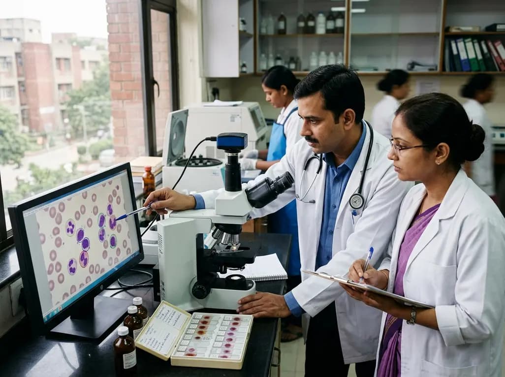
Routine clinical hematology laboratories are undergoing a major technological transformation through automated imaging platforms. Modern digital morphology analyzers are increasingly utilized for white blood cell differential counting, replacing or augmenting manual microscopy. These instruments scan stained peripheral blood smears, identify cellular targets, and generate high-resolution images. Artificial intelligence algorithms then pre-classify leukocytes into specific diagnostic categories. However, this workflow introduces distinct analytical variables that standard quality control programs do not address. Consequently, laboratory leaders must establish robust external quality assessment schemes tailored specifically to digital platforms to maintain diagnostic accuracy and regulatory compliance.
Most modern diagnostic hematology laboratories implement digital morphology analyzers as efficient frontline screening tools. The automated system scans the blood smear, captures leukocyte images, and presents a proposed differential count. Medical laboratory scientists then verify these findings on high-resolution displays before releasing clinical reports. In routine practice, laboratories establish predefined review criteria to trigger manual microscopic re-examination. For instance, when the analyzer flags immature granulocytes, blast cells, or abnormal lymphoid populations, technologists perform manual microscopic validation. Thus, digital imaging does not entirely replace human expertise. Instead, it creates an integrated workflow where software algorithms and expert examiners collaborate to optimize laboratory turnaround times while maintaining rigorous diagnostic standards.
Recent multicenter evaluation studies highlight significant variability across institutions when classifying morphologically similar leukocytes. While digital systems reliably distinguish typical segmented neutrophils, lymphocytes, and eosinophils, diagnostic discrepancies frequently emerge among complex cell lines. Specifically, differentiating band neutrophils from segmented neutrophils or distinguishing reactive lymphocytes from atypical lymphocytes presents persistent classification hurdles. Furthermore, automated image pre-classification frequently struggles with rare or abnormal cell populations, including dysplastic monocytes and circulating blasts. Examiner verification significantly mitigates these errors, but subjective human interpretation still introduces examiner-level variability. Therefore, clinical hematology networks must standardize cell definitions and conduct continuous morphologic competency training across all testing sites to minimize reporting divergence.
A critical finding in digital morphology performance evaluation is the substantial proportion of unclassified images and cellular artifacts. Staining inconsistencies, slide preparation flaws, precipitate debris, and cell overlap directly degrade image acquisition quality. When an automated analyzer cannot categorize an image with high statistical confidence, it routes the object to an unclassified folder. Technologists must then manually reclassify or discard these artifacts. If slide quality deteriorates, the volume of unclassified objects expands rapidly, which burdens laboratory staff and distorts differential percentages. Consequently, external quality assessment programs must account for pre-analytical slide preparation quality. Standardized staining protocols remain vital prerequisites for dependable digital leukocyte analysis across regional testing networks.
Traditional external quality assessment schemes predominantly evaluate quantitative performance using standard deviation index metrics. However, standard numerical scoring fails to capture critical qualitative errors in hematologic morphology. For example, a small statistical shift in neutrophil percentage might receive an acceptable numerical score, yet the failure to detect rare circulating myeloblasts carries catastrophic clinical consequences. Therefore, proficiency testing for digital platforms must evaluate abnormal cell detection sensitivity alongside numerical differentials. Proficiency providers should incorporate challenging clinical scenarios, such as acute leukemias, reactive lymphocytosis, and myelodysplastic changes. By extending assessment criteria beyond standard numerical indices, proficiency testing accurately reflects the true diagnostic capability of participating hematology laboratories.
Establishing effective quality assurance for digital hematology requires comprehensive, standardized operational protocols across all participating centers. First, external proficiency programs must distribute centrally prepared, verified blood smears alongside digital reference libraries. Second, laboratories must implement periodic internal verification protocols to track individual examiner reclassification accuracy. Third, quality managers should monitor the ratio between pre-classification algorithm outputs and final post-classification technician results. Analyzing this variance reveals algorithmic drift, camera calibration errors, and individual technician training deficiencies. Ultimately, continuous digital proficiency assessment ensures that advanced artificial intelligence tools consistently translate into safe, accurate patient care across private and hospital laboratories alike.
Digital analyzers automate slide scanning and capture high-resolution images of individual leukocytes. Sophisticated artificial intelligence algorithms pre-classify each cell based on morphological features. Technologists review and verify these digital images on a monitor rather than manually scanning glass slides under a microscope. This workflow enhances operational efficiency, reduces eye strain, and facilitates rapid telepathology consultations across networked healthcare facilities.
Standard deviation index metrics focus exclusively on numerical deviations from a peer group mean value. However, clinical hematology diagnostics heavily depend on identifying rare, qualitative morphological abnormalities, such as blasts or Auer rods. A laboratory could achieve an acceptable numerical differential score while missing critical neoplastic cells entirely. Assessment schemes must therefore incorporate qualitative identification metrics alongside standard numerical counts.
Laboratories can significantly reduce unclassified images by standardizing pre-analytical blood smear preparation and automated staining protocols. Ensuring optimal cell monolayer distribution, eliminating stain precipitate, and maintaining clean optical components prevent image artifacts. Additionally, regular instrument calibration and ongoing staff training in digital reclassification help technologists efficiently process ambiguous cellular images according to established laboratory guidelines.
Disclaimer: This content is for informational and educational purposes only. It is not intended to be a substitute for professional medical advice, diagnosis, or treatment. Always seek the advice of your physician or other qualified health provider with any questions you may have regarding a medical condition. Refer to the latest local and national guidelines for clinical practice.
References

Read summarized clinical updates, watch expert medical content, and earn CME certifications right from your smartphone.
Open your smartphone camera to scan and install.





Digital morphology analyzers enhance routine white blood cell differential testing, but effective external quality assessment requires tailored frameworks, standard slide preparation, and examiner verification protocols to ensure high diagnostic precision across hematology laboratories.
last month

A systematic review of 41 preclinical studies reveals that baicalin dampens systemic inflammation, inhibits NF-κB and NLRP3 pathways, and improves organ preservation in sepsis models. However, substantial study heterogeneity and a complete lack of human clinical trials highlight the need for further translational research.
Today
Discover the crucial distinctions between mesonephric and mesonephric-like proliferations of the female genital tract. Learn the key histomorphologic, immunohistochemical, and molecular differences essential for accurate diagnosis and clinical management.
Today

A large UK Biobank study reveals that plasma proteomic signatures capture preclinical organ damage and improve multiorgan risk prediction across early CKM syndrome stages 0 to 2 beyond traditional PREVENT clinical models.
Today
A narrative review investigates whether the articularis genus muscle functions as an independent anatomical entity or blends with the vastus intermedius. We evaluate its morphology, role in retracting the suprapatellar bursa, and direct clinical significance in anterior knee pain and arthroplasty.
Today

A Bayesian multilevel meta-analysis reveals that aerobic training combined with moderate carbohydrate restriction modestly lowers HbA1c in type 2 diabetes. However, sparse data and very low certainty leave incremental benefits over exercise or diet alone unproven, highlighting the need for individualized care.
Today
