.webp&w=384&q=75)

Loading, please wait...
Loading, please wait...

Glioblastoma remains one of the most lethal central nervous system malignancies, characterized by aggressive recurrence and profound biological heterogeneity following standard chemoradiation. While anti-vascular endothelial growth factor (anti-VEGF) agents like bevacizumab provide symptom relief and radiographic improvement, clinical trials show variable survival benefits across unselected patient cohorts. Consequently, neuro-oncologists require robust noninvasive biomarkers to stratify patients before initiating targeted therapies. Emerging evidence highlights that diffusion MRI glioblastoma signatures can identify a distinct subgroup of recurrent tumors destined for favorable clinical outcomes under anti-VEGF regimens. By decoding the underlying biophysical properties of these imaging phenotypes, clinicians can better understand tumor biology and optimize personalized therapeutic interventions.
Recent investigations have demonstrated that diffusion-weighted imaging provides valuable prognostic information beyond simple structural assessment. Specifically, histogram analysis of the apparent diffusion coefficient (ADC) within contrast-enhancing tumor regions categorizes recurrent lesions into distinct prognostic subsets. Researchers establish a validated threshold using the lowest peak of the double Gaussian distribution of ADC values, designating lesions at or above 1240 µm²/s as high-ADC phenotypes. Historically, patients harboring high-ADC tumors exhibit significantly superior progression-free and overall survival when treated with anti-VEGF therapies compared to their low-ADC counterparts.
However, the biological mechanism underpinning this differential therapeutic sensitivity previously remained poorly defined. A landmark investigation evaluated anti-VEGF-naïve patients with IDH-wildtype glioblastoma who completed standard chemoradiotherapy. By examining comprehensive multiparametric MRI datasets, investigators systematically unpacked the pathophysiological landscape distinguishing high-ADC from low-ADC tumors. The analysis confirmed that these phenotypes do not differ simply by chance or anatomical location. Instead, they represent intrinsically distinct microenvironmental states. Therefore, quantitative diffusion assessment offers an objective window into therapeutic vulnerability, enabling clinicians to identify candidates who will truly benefit from vascular endothelial growth factor inhibition.
Dynamic susceptibility contrast perfusion MRI provides essential insights into tumor vascularity and microcirculatory integrity. When evaluating recurrent post-chemoradiation glioblastoma, perfusion metrics reveal striking physiological differences between diffusion-stratified subgroups. Specifically, the study demonstrated that high-ADC glioblastoma lesions display significantly lower relative cerebral blood volume (rCBV) compared to low-ADC lesions, with mean values of 1.02 versus 1.28, respectively.
This hemodynamic disparity reveals crucial biological details about tumor vascular architecture. High-ADC lesions feature lower microvascular density or diminished aberrant hypervascularity, reflecting a less chaotic angiogenic network. In contrast, low-ADC tumors exhibit elevated rCBV, which signals dense cellularity combined with robust microvascular proliferation. Consequently, one might intuitively expect highly vascular tumors to respond best to anti-angiogenic therapy. Nevertheless, clinical evidence shows the exact opposite phenomenon. Tumors with moderate or normalized baseline perfusion experience superior vascular remodeling and improved oxygenation following bevacizumab administration. Conversely, highly hypervascular, densely cellular lesions with low ADC values often harbor severe intrinsic hypoxia and complex resistance mechanisms. Thus, perfusion imaging complements diffusion metrics by identifying distinct vascular phenotypes that guide treatment selection.
Beyond perfusion and diffusion alone, advanced molecular MRI techniques characterize the complex metabolic microenvironment of post-treatment gliomas. Chemical exchange saturation transfer (CEST) MRI targeting amine protons at 3 ppm reflects intracellular and extracellular pH as well as mobile protein accumulation. In this clinical study, high-ADC lesions exhibited significantly higher magnetization transfer ratio asymmetry values (2.36% versus 2.10%) compared to low-ADC tumors.
Additionally, quantitative relaxometry revealed key tissue alterations between these groups. High-ADC tumors demonstrated significantly prolonged quantitative T1 relaxation times, measuring 114.8 milliseconds compared to 100.9 milliseconds in low-ADC lesions. Prolonged T1 relaxation times generally indicate increased interstitial water content, expanded extracellular space, or lower cellular packing. Furthermore, the elevated amine CEST signal points toward an altered extracellular biochemical milieu, potentially reflecting different metabolic byproducts or variations in tissue acidosis. Importantly, investigators found no significant differences in T2* relaxation times, contrast-enhancing tumor volume, or subtraction map dynamics between the groups. Therefore, these imaging alterations do not stem from gross tumor size discrepancies. Instead, they highlight subtle biochemical and architectural differences within viable enhancing tissue.
Accurate patient stratification remains paramount in neuro-oncology because recurrent glioblastoma lacks universal second-line standards of care. Currently, clinicians frequently administer bevacizumab to reduce cerebral edema and lower steroid dependence. However, unselected clinical trials failed to establish a universal overall survival advantage for anti-VEGF monotherapy. These advanced imaging findings provide compelling evidence that molecular and biological phenotypes dictate therapeutic efficacy.
Notably, the study discovered that conventional clinical and molecular markers failed to differentiate high-ADC from low-ADC phenotypes. Patient sex, MGMT promoter methylation status, and EGFR amplification status showed no statistically significant correlation with ADC classifications. Furthermore, neither subgroup showed an anatomical predilection for specific cerebral lobes or deep brain structures. Consequently, standard molecular pathology panels and baseline clinical demographics cannot substitute for physiological neuroimaging when assessing anti-VEGF responsiveness. Because traditional genomic profiling misses these microenvironmental states, multiparametric MRI serves as an indispensable adjunct for precision oncology. By implementing quantitative ADC and rCBV thresholds, multidisciplinary tumor boards can objectively identify individuals most likely to achieve true disease stabilization.
Integrating advanced neuroimaging protocols into clinical workflows demands standardized acquisition protocols and robust post-processing pipelines. To replicate these predictive diffusion metrics, radiology departments must employ standardized diffusion-weighted sequences alongside calibrated dynamic susceptibility contrast perfusion. Automating double Gaussian histogram analysis of ADC values helps neuro-radiologists eliminate interobserver variability during tumor segmentation.
Moreover, incorporating physiological MRI into routine follow-up protocols addresses the pervasive challenge of distinguishing true recurrence from radiation-induced pseudoprogression. While conventional anatomical scans merely display expanding gadolinium enhancement, multiparametric MRI differentiates hypervascular viable tumor from hypovascular radiation injury. Combining ADC quantification with rCBV and quantitative relaxometry enhances diagnostic certainty during critical decision points. For neuro-oncologists, having objective imaging criteria allows for confident early therapeutic pivots. Clinicians can confidently recommend bevacizumab to patients displaying high-ADC and moderate rCBV profiles while reserving alternative clinical trials, novel immunotherapies, or re-irradiation for low-ADC phenotypes. Ultimately, adopting these advanced imaging techniques bridges the gap between molecular diagnostics and individualized neuro-oncology treatment strategies.
A high ADC phenotype, defined by an apparent diffusion coefficient threshold at or above 1240 µm²/s, indicates lower tumor cellularity, expanded extracellular space, and lower relative cerebral blood volume. Clinically, this imaging signature identifies post-chemoradiation glioblastoma patients who experience superior survival outcomes following anti-VEGF therapy.
High-ADC glioblastomas feature lower baseline relative cerebral blood volume and increased interstitial fluid, reflecting less aggressive microvascular proliferation. Consequently, anti-VEGF agents normalize this aberrant vasculature more effectively, improving regional oxygenation and therapeutic delivery without triggering the rapid hypoxic escape mechanisms commonly observed in hypercellular, low-ADC lesions.
No, standard molecular biomarkers such as MGMT promoter methylation status and EGFR amplification status do not correlate significantly with diffusion MRI phenotypes. In addition, patient sex, tumor volume, and anatomical tumor location show no group-specific associations, making quantitative physiological neuroimaging an independent predictive biomarker for therapy.
Disclaimer: This content is for informational and educational purposes only and should not be considered medical advice. Always consult a qualified healthcare professional regarding any medical condition or treatment. Refer to the latest local and national guidelines for clinical practice.
References

Read summarized clinical updates, watch expert medical content, and earn CME certifications right from your smartphone.
Open your smartphone camera to scan and install.





Post-chemoradiation glioblastomas with high apparent diffusion coefficient (ADC) phenotypes predict favorable anti-VEGF response. These lesions exhibit distinct biophysical profiles, including lower cerebral blood volume, elevated amine CEST signals, and prolonged T1 relaxation times.
Today

A multicity study highlights that over 80% of acute respiratory infections remain outside hospitals. Patients frequently choose community pharmacies and digital platforms. Integrating digital health data and retail pharmacy sales can fill critical surveillance gaps and improve early outbreak detection.
Today

Scandinavian register evidence reveals that neonatal birth length strongly predicts adult health and mortality, whereas birth weight dictates cognitive capacity. Symmetric growth restriction causes far more severe long-term deficits than asymmetric restriction, underscoring the need for comprehensive anthropometry.
Today
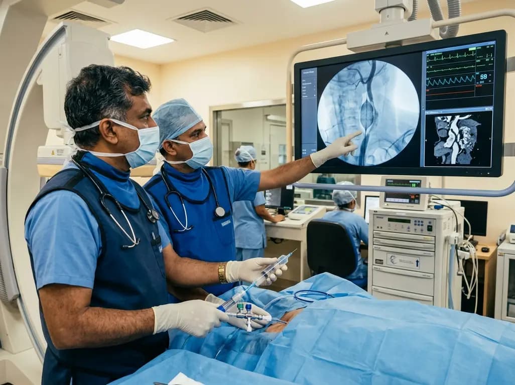
Discover how rescue cutting balloon angioplasty successfully modifies heavily calcified carotid stenosis, enabling stent delivery without baroreceptor instability and ensuring favorable long-term neurologic outcomes.
Today
A retrospective CBCT study reveals that superior TMJ space asymmetry correlates moderately with occipital condyle-atlas-axis asymmetry in unilateral TMJ osteoarthritis, highlighting an imaging correlate between jaw biomechanics and upper cervical alignment rather than a standalone diagnostic marker.
Today

Data from the 2026 Workforce Health Index reveals that 38.7% of urban employees and dependents exhibit abnormal glycemic control. Emerging at increasingly younger ages, this metabolic shift intersects with severe vitamin deficiencies, highlighting the urgent need for early workplace screenings and targeted clinical care.
Today
