

Loading, please wait...
Loading, please wait...

Glioblastoma represents the most aggressive primary malignant brain tumor encountered in neurosurgical oncology. Despite macroscopic gross total resection of contrast-enhancing tissue, microscopic malignant invasion persists deep within adjacent parenchymal margins. Consequently, local recurrence remains nearly inevitable. Predicting the precise anatomical trajectory of recurrence represents a longstanding clinical challenge. Recent multicenter research demonstrates that preoperative diffusion anisotropy can reliably forecast temporal glioblastoma progression. By tracking microstructural white matter alterations, advanced diffusion tensor metrics provide critical spatial intelligence for neurosurgeons before entering the operating room.
Temporal lobe glioblastomas present unique technical considerations for neurosurgical teams. Because the temporal lobe contains critical functional networks, aggressive surgical margins carry substantial neurological risks. Language centers, visual pathway radiations, and memory circuits converge within tight anatomical confines. Therefore, surgeons must balance maximal cytoreduction against postoperative functional deficits. Standard surgical protocols currently emphasize complete resection of contrast-enhancing tumor margins on T1-weighted post-contrast magnetic resonance imaging. However, glioblastoma cells readily infiltrate along organized white matter tracts far beyond visible enhancements.
Historically, clinicians relied on fluid-attenuated inversion recovery sequences to evaluate peritumoral non-enhancing infiltration. Yet, peritumoral FLAIR hyperintensity cannot reliably distinguish vasogenic edema from active neoplastic invasion. Furthermore, standard FLAIR imaging fails to indicate the directional vectors of migratory glioma cells. As a result, recurrence frequently occurs along predictable structural pathways that remain invisible on routine structural scans. Identifying preoperative biomarkers that pinpoint these vulnerable anatomical corridors offers a transformative opportunity for targeted surgical planning and tailored postoperative radiotherapy.
Diffusion tensor imaging measures the translational displacement of water molecules in three-dimensional space. In cerebral tissue, this technique yields two fundamental, complementary components known as isotropic diffusion (DTI-p) and diffusion anisotropy (DTI-q). The isotropic component represents non-directional, random water movement, which typically increases in areas of bulk vasogenic edema and extracellular expansion. Consequently, DTI-p often parallels the non-specific findings observed on conventional FLAIR sequences.
In contrast, diffusion anisotropy captures the highly directional movement of water molecules along structured axonal bundles. Glioblastoma cells exploit these organized white matter pathways for rapid parenchymal migration. As invading neoplastic cells disrupt axonal packing and myelin integrity, directional diffusion values change measurably before gross structural destruction occurs. Therefore, DTI-q serves as a sensitive biophysical probe for early, tract-oriented tumor infiltration. By separating directional tract disruption from diffuse non-directional edema, DTI-q delivers superior anatomical specificity regarding occult tumor trajectories.
A recent multicenter investigation evaluated the predictive performance of preoperative imaging biomarkers in thirty-two patients with newly diagnosed, IDH-wildtype temporal glioblastoma. All enrolled individuals underwent complete resection of contrast-enhancing tumor tissue and received regular longitudinal follow-up imaging. Investigators mapped preoperative abnormalities on FLAIR, DTI-p, and DTI-q against subsequent radiographic recurrence locations relative to the surgical cavity.
The statistical analyses demonstrated that DTI-q significantly outperformed both conventional FLAIR and isotropic DTI-p across multiple anatomical directions. Specifically, DTI-q achieved a median predictive accuracy of 75%, compared with 50% for DTI-p and only 44% for FLAIR imaging. Furthermore, rigorous Holm-corrected paired McNemar tests confirmed that DTI-q provided superior accuracy for lateral, superior, and inferior progression patterns. Interestingly, combining multiple imaging parameters into composite models yielded no diagnostic advantage over DTI-q alone. These prospective findings demonstrate that diffusion anisotropy independently isolates the predominant directions of microscopic tumor dissemination.
These predictive imaging capabilities carry profound practical implications for neurosurgical oncology practice. In temporal lobe tumors, neurosurgeons frequently debate whether to perform an extended resection or an anterior temporal lobectomy. When a glioblastoma occupies the non-dominant temporal pole, extending resection margins laterally or anteriorly remains neurologically tolerable. However, executing such extended resections without directional evidence risks functional impairment or leaves occult microscopic disease behind in unaddressed vectors.
When preoperative DTI-q demonstrates prominent lateral or anterior invasion patterns, surgeons can confidently consider supramarginal resection or tailored lobectomy. Conversely, if DTI-q identifies predominant superior or medial infiltration toward the brainstem and Meyer loop, surgeons must prioritize functional preservation and avoid hazardous parenchymal over-resection. Thus, directional tract analysis prevents arbitrary margin extension. By incorporating these objective predictive vectors into preoperative workflow, surgical oncology teams can customize bone flaps, trajectory planning, and resection boundaries with millimeter precision.
The temporal lobe houses several major white matter fascicles that serve as conduits for invasive glioma cells. These pathways include the uncinate fasciculus, inferior longitudinal fasciculus, and arcuate fasciculus. Glioblastoma cells migrate preferentially along these dense axonal bundles because extracellular matrices provide permissive pathways for motility. Conventional imaging obscures this biological reality because vasogenic fluid signal masks microscopic invasion.
DTI-q maps resolve this limitation by tracking structural directional changes within these specific fascicles. For instance, superior tumor progression often threatens the optic radiations and motor pathways within the internal capsule. Similarly, medial progression jeopardizes the mesial temporal structures and basal ganglia. By detecting anisotropic disruption along these specific white matter tracts preoperatively, multidisciplinary teams can anticipate high-risk recurrence zones. Consequently, radiation oncologists can also utilize these spatial maps to optimize target volume delineation, boosting dose delivery to high-risk invasion vectors while sparing adjacent functional parenchyma.
Integrating advanced diffusion metrics into routine neurosurgical practice requires standardization across MRI acquisition protocols and post-processing pipelines. Modern neuro-navigation platforms already accommodate tractography data, making the incorporation of DTI-q maps technically feasible in tertiary centers. Looking ahead, combining directional diffusion biomarkers with intraoperative ultrasound and fluorescence guidance could establish a comprehensive, spatially informed surgical paradigm. As prospective validation expands across larger multicenter cohorts, DTI-q promises to redefine personalized resection strategies, helping neurosurgeons anticipate tumor recurrence before the initial incision occurs.
DTI-q measures directional water movement along organized white matter tracts. Because temporal glioblastoma cells migrate along these axonal pathways, DTI-q detects microstructural tract alterations before visible tumor enhancement develops. This directional sensitivity enables clinicians to forecast whether recurrence will progress laterally, superiorly, or inferiorly relative to the cavity.
FLAIR hyperintensity primarily reflects non-directional vasogenic edema rather than pure cellular invasion. Consequently, it cannot differentiate passive extracellular fluid shifts from active, tract-directed tumor infiltration. This non-specific signal obscures directional migration vectors, resulting in poor predictive accuracy for directional recurrence patterns compared with diffusion anisotropy imaging.
Anticipating recurrence vectors allows neurosurgeons to tailor surgical margins strategically. If DTI-q shows lateral infiltration in non-dominant temporal tumors, surgeons can safely extend resection via anterior lobectomy. Conversely, medial or superior invasion warnings alert teams to protect eloquent language tracts and visual pathways, preventing unnecessary postoperative functional morbidity.
Disclaimer: This content is for informational and educational purposes only. It should not be used as a substitute for professional medical advice, diagnosis, or treatment. Refer to the latest local and national guidelines for clinical practice.
References

Read summarized clinical updates, watch expert medical content, and earn CME certifications right from your smartphone.
Open your smartphone camera to scan and install.





Multicenter findings demonstrate that preoperative diffusion anisotropy (DTI-q) accurately predicts directional progression in temporal glioblastoma, significantly outperforming conventional FLAIR and isotropic diffusion to guide individualized surgical resection.
Today

Gastrointestinal smooth muscle cells rely on nanoscale membrane domains to integrate mechanosensitive currents. Recent insights show that nanodomain architecture, rather than channel properties alone, orchestrates motility, opening new therapeutic avenues for clinical dysmotility and gastrointestinal aging.
Today

A landmark study reveals that lipid-rich malignant ascites alters the T-cell metabolic transcriptome, causing cholesterol efflux and impaired fatty acid oxidation. This suppresses TCR signalling and blocks BiTE immunotherapy, while lipid depletion restores antitumour cytotoxicity in ovarian cancer.
Today
Novel clinical evidence demonstrates the feasibility of combining VA-ECMO with the NyokAssist self-expanding transvalvular pump for active left ventricular unloading in infarct-related cardiogenic shock, achieving successful stabilization and decannulation.
Today
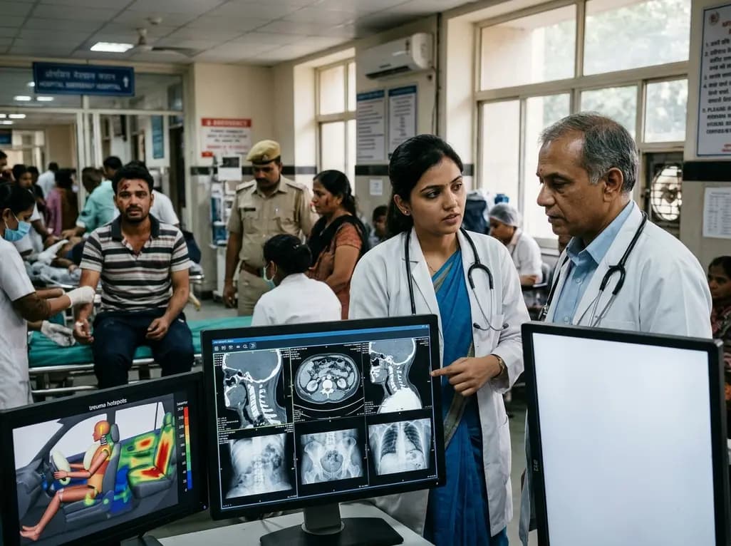
Recent crash analyses demonstrate significant variations in traffic injury severity across vehicle seating positions. Electric vehicle passengers face disproportionately higher trauma risks than drivers, particularly during airbag deployments, demanding adapted emergency assessment and clinical vigilance.
Today

Understand the causal role of Lipoprotein(a) in cardiovascular disease, why international guidelines recommend once-in-a-lifetime molar testing, and the clinical implications of recent phase 3 RNA outcome trials.
Today
