.webp&w=384&q=75)

Loading, please wait...
Loading, please wait...
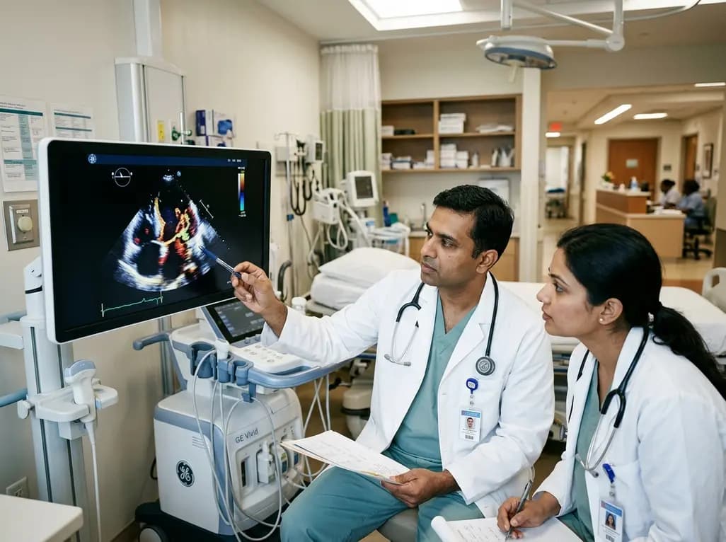
Infective endocarditis remains a formidable cardiovascular challenge associated with substantial in-hospital morbidity and mortality across global healthcare settings. Clinicians continually evaluate how invasive bacteremic triggers contribute to cardiac endothelial seeding. In particular, oral flora infective endocarditis represents a significant subset of cases where commensal viridans streptococci and related oral microbes invade bloodstream pathways during dental procedures or poor oral hygiene. A recent retrospective observational study conducted at Royal Derby Hospital examined consecutive transoesophageal echocardiography examinations to assess these clinical relationships. Researchers analyzed 133 unique confirmed clinical episodes between April 2022 and October 2024. The investigators specifically evaluated how documented dental exposures correlate with microbiological patterns and structural valve damage. Consequently, their contemporary observations provide practical insights into real-world patient vulnerability and antibiotic prophylaxis adherence in routine clinical practice.
The study cohort demonstrated demographic and clinical features consistent with modern cardiovascular disease trends. Specifically, the mean patient age was 63.3 years, and male patients comprised 66.2% of the confirmed cohort. Native-valve infective endocarditis accounted for 75.9% of all documented cases, highlighting that non-prosthetic valves remain highly vulnerable. Furthermore, transoesophageal echocardiography revealed that the aortic valve was the most frequently affected anatomical structure, involved in 62.4% of patients. In terms of microbiology, Staphylococcus aureus emerged as the single predominant causative organism, responsible for 30.8% of cases. Meanwhile, oral flora microorganisms accounted for 33.1% of the total infective burden. Overall, 19.5% of patients required surgical or device-related interventions, while in-hospital mortality reached 11.3%. Importantly, no single clinical or imaging variable significantly predicted mortality, demonstrating the multifaceted complexity of disease outcomes.
A primary focus of the investigation was evaluating the link between documented dental procedures and causative pathogen profiles. Notably, twenty-six patients had a confirmed history of dental interventions prior to disease onset. Among this group, documented dental procedures were significantly associated with oral flora infective endocarditis compared to patients without documented dental exposure. In fact, oral flora caused 50.0% of infections in the dental exposure cohort versus 28.8% in unexposed patients. Statistical analysis confirmed a significant odds ratio of 2.47 (95% confidence interval, 1.03 to 5.94; p = 0.040). These findings emphasize that transient bacteremia during dental work can directly seed susceptible cardiac tissue. Consequently, healthcare providers must recognize that even minor gingival trauma or procedural manipulation can trigger invasive bacteremic events in susceptible individuals.
Transoesophageal echocardiography serves as a cornerstone imaging modality in the timely identification of cardiac vegetations, abscesses, and leaflet perforations. While transthoracic echocardiography provides an essential baseline screen, it often lacks sufficient sensitivity to detect small vegetations or subtle perivalvular extensions. Therefore, transoesophageal echocardiography provides superior acoustic resolution, especially for evaluating native aortic valve leaflets and prosthetic hardware. In this study, clinicians used transoesophageal echocardiography systematically to establish anatomical diagnoses and assess structural complications requiring surgical intervention. Moreover, precise imaging allows clinicians to distinguish between staphylococcal destruction and the typically more indolent, yet structurally damaging, streptococcal presentations. Thus, advanced echocardiography remains indispensable for guiding both medical antimicrobial regimens and the timing of urgent surgical repair.
The study identified a striking disparity regarding the administration of preventive antimicrobial therapy. Specifically, among the 26 patients with documented invasive dental procedures, documented antibiotic prophylaxis was present in only two cases (7.7%). This low rate reflects existing debates surrounding international prophylaxis guidelines. For instance, the UK National Institute for Health and Care Excellence historically discouraged routine prophylaxis, whereas European and American guidelines recommend targeted coverage for high-risk cardiac lesions. However, this retrospective data may also highlight an underreporting issue in clinical charts rather than true omission. Therefore, physicians and dental professionals must improve shared documentation protocols. Ensuring thorough pre-procedure cardiac risk assessments and maintaining precise procedural records can bridge communication gaps between dental clinics and cardiology wards.
Preventing endovascular infection requires active collaboration among cardiologists, primary care physicians, infectious disease specialists, and dental practitioners. Because daily oral activities like brushing can also generate low-grade bacteremia, maintaining optimal daily oral hygiene is paramount for all cardiac patients. Furthermore, clinicians must educate patients with native valve pathology and prosthetic implants regarding early symptoms, such as unexplained prolonged fever and malaise. In addition, when invasive dental procedures involving gingival manipulation are scheduled, clinicians should carefully stratify patient risk according to current international guidance. By fostering seamless communication and promoting strict antiseptic dental protocols, healthcare teams can substantially reduce procedural infection risks. Ultimately, comprehensive risk stratification and vigilant clinical monitoring remain the most reliable defenses against severe endocardial infections.
Dental procedures that manipulate the gingival margins or mucosal tissue frequently disrupt local blood vessels. Consequently, normal commensal oral bacteria, such as viridans group streptococci, enter the bloodstream and cause transient bacteremia. In patients with underlying endothelial damage or valvular abnormalities, these circulating organisms adhere to platelets and fibrin deposits. Ultimately, this bacterial adherence leads to vegetation formation, localized tissue destruction, and full-blown endocardial infection requiring prolonged intravenous antimicrobial treatment.
International cardiology guidelines, including recommendations from the American Heart Association and European Society of Cardiology, advise prophylactic antibiotics for high-risk patients. These high-risk groups include individuals with prosthetic heart valves, prior endocarditis, or specific cyanotic congenital heart diseases undergoing invasive dental procedures. Conversely, routine prophylaxis is not recommended for intermediate-risk native valve disease in some guidelines. Therefore, clinicians must assess individual patient risk factors and local clinical guidelines carefully.
Transoesophageal echocardiography provides significantly higher spatial resolution because the ultrasound probe sits in the esophagus directly behind the heart. This positioning minimizes acoustic interference from chest wall tissue, lungs, and ribs. As a result, transoesophageal imaging detects smaller vegetations, identifies subtle prosthetic valve dehiscence, and visualizes perivalvular abscesses far more reliably than transthoracic echocardiography. Consequently, it is the reference standard imaging modality for diagnosing complex endocardial infections.
Disclaimer: This content is for informational and educational purposes only... Refer to the latest local and national guidelines for clinical practice.
References

Read summarized clinical updates, watch expert medical content, and earn CME certifications right from your smartphone.
Open your smartphone camera to scan and install.





A contemporary UK study evaluates the link between dental exposure and oral flora infective endocarditis using transoesophageal echocardiography. We review key findings on valvular patterns, pathogen profiles, and antibiotic prophylaxis considerations.
last month
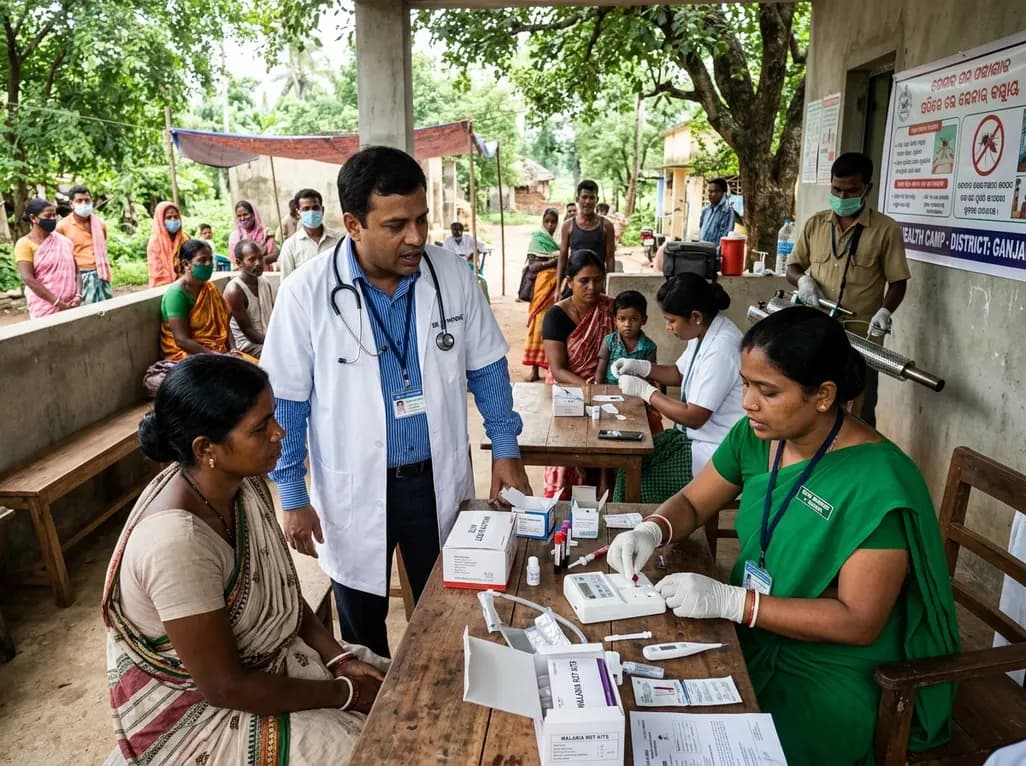
Following 37,899 reported cases and 9 deaths, Odisha has scaled up active case detection, rapid diagnostic testing, and vector control across high-burden districts. Here is a comprehensive clinical breakdown of the state's aggressive containment protocols, diagnostic pathways, and targeted field management.
Today

A retrospective study of 86,012 pregnancies demonstrates that size-selective cfDNA enrichment boosts mean fetal fraction to 18.88% and achieves a 59.20% PPV for fetal copy number variants. However, 85.83% of CNVs showed size discrepancies, underscoring the mandatory role of confirmatory microarray testing.
Today

Radiofrequency catheter ablation of the cavotricuspid isthmus successfully terminates typical atrial flutter. This article reviews the mechanisms driving coronary sinus electrogram changes, wavefront activation pathways, and clinical criteria to verify bidirectional block during electrophysiology studies.
Today
A 12-week within-subject trial compared incline and preacher dumbbell curls, finding incline curls elicited greater biceps brachii growth while preacher curls favored the brachialis and brachioradialis. Total elbow flexor hypertrophy and regional growth profiles remained comparable between the two exercises.
Today
A cross-sectional study demonstrates that increased BMI in adolescents significantly enhances mandibular length and alters salivary adipokines like adiponectin and resistin, providing novel insights into craniofacial growth and orthodontic planning.
Today
