.webp&w=384&q=75)

Loading, please wait...
Loading, please wait...
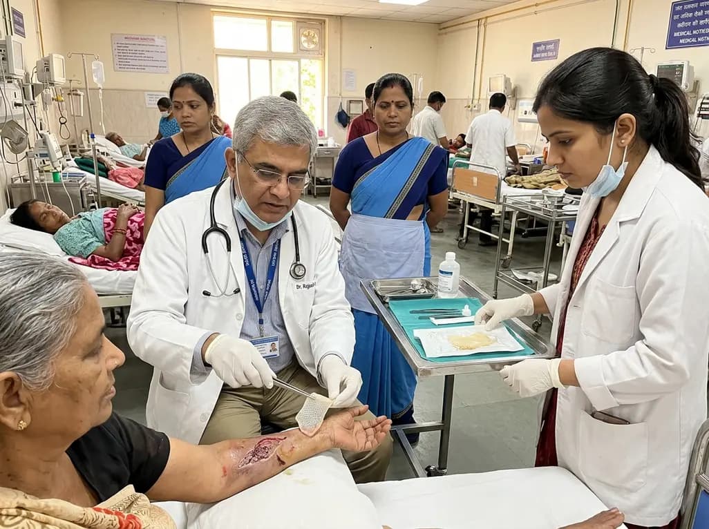
Effective dermal reconstruction requires biomaterials that replicate the extracellular matrix while guiding cellular behavior. The decellularized fish swim bladder represents a breakthrough collagen matrix that accelerates tissue repair. Preclinical investigations demonstrate that this natural marine scaffold supports cell adherence, accelerates migration, and integrates smoothly into acute and chronic wounds. Consequently, clinicians are evaluating fish-derived matrices as viable alternatives to mammalian grafts.
Marine-derived extracellular matrix biomaterials offer unique physiological advantages over terrestrial animal derivatives. Swim bladders are naturally abundant in dense, structurally intact Type I and Type III collagen fibers. In addition, they present minimal risk of zoonotic disease transmission, such as bovine spongiform encephalopathy. Processing these organs through gentle detergent protocols yields a clean acellular architecture. This acellular state eliminates native immunogenic cellular fragments while preserving bioactive glycosaminoglycans and adhesive glycoproteins. Furthermore, the decellularized matrix maintains a natural three-dimensional orientation that mimics the human papillary and reticular dermis. Therefore, host cells quickly identify the microenvironment as a native template for biological repair. This favorable structural stability provides a resilient framework capable of withstanding mechanical shearing forces in mobile wound locations.
Structural and biomechanical characteristics vary significantly among different fish species. A comparative evaluation of four distinct fish species demonstrated that the swim bladder of Rutilus frisii kutum delivers exceptional matrix properties. Specifically, this species maintains an optimal balance between tensile strength, swelling behavior, and interconnected porosity. Adequate pore size is critical because it regulates fluid exudate absorption and facilitates vascular ingrowth. If pores are excessively large, mechanical integrity drops rapidly under physiological strain. Conversely, overly dense matrices prevent endogenous cell ingress and trap inflammatory debris. The Rutilus frisii kutum scaffold exhibits balanced hydration capacity without excessive swelling. As a result, the scaffold preserves its physical dimensions when sutured or secured into full-thickness cutaneous defects.
Quantitative in vivo histological assessments show rapid cellular integration following scaffold implantation. Host fibroblasts and native vascular endothelial cells attach to the scaffold surface within twenty-four hours. Subsequently, cell migration proceeds inward across the matrix layers in a predictable timeline. By post-implantation day five, cell density and penetration depth increase significantly. This rapid cellular infiltration facilitates coordinated extracellular matrix deposition and scaffold remodeling. Moreover, the decellularized biomaterial adheres firmly to the wound bed from initial placement. This persistent contact prevents dead space formation and reduces shear disruption. Consequently, granulation tissue matures quickly, allowing accelerated neo-epithelialization across full-thickness skin margins.
Wound repair frequently stalls when excessive inflammation degrades nascent matrix proteins. Fortunately, decellularized marine matrices actively modulate local cytokine signaling. Host macrophages interacting with the fish collagen matrix transition from a pro-inflammatory M1 phenotype toward a regenerative M2 phenotype. Thus, the scaffold reduces harmful tissue proteases and sustains angiogenic growth factor concentrations. Furthermore, the absence of mammalian cellular antigens minimizes foreign body reactions. Histological analyses confirm minimal multinucleated giant cell accumulation around the graft edges. Because the matrix tempers prolonged inflammatory cascades, it effectively prevents pathological wound contracture and excessive hypertrophic scarring.
Surgical specialists and dermatologists encounter challenging tissue deficits resulting from trauma, oncological resections, and metabolic ulcers. Utilizing decellularized marine collagen matrices provides a scalable and cost-effective clinical strategy. In chronic diabetic foot ulcers or venous stasis wounds, these scaffolds protect the exposed base while stimulating microvascular formation. Similarly, reconstructive surgeons can utilize these dermal grafts beneath thin split-thickness skin grafts to enhance skin elasticity. Because the biomaterial degrades through natural enzymatic pathways, it leaves behind organized autologous dermis. Therefore, adopting optimized marine biomaterials can substantially improve reconstructive outcomes while lowering reliance on scarce donor tissue.
Marine swim bladder collagen eliminates the risk of transmitting mammalian prion diseases and viral pathogens. In addition, it carries fewer cultural and religious restrictions compared to bovine or porcine grafts. The processing method also preserves a highly organized, dense collagenous architecture that supports mechanical durability and rapid host cell infiltration during early wound repair stages.
Host cells begin adhering to the scaffold surface within the first twenty-four hours after surgical placement. Infiltration expands rapidly into the porous matrix over subsequent days. Experimental analyses demonstrate marked increases in cell density and penetration depth by day five, establishing stable tissue integration and active vascular remodeling throughout the wound bed.
The thorough decellularization process removes cellular antigens, nucleic acids, and lipid debris that typically provoke immune rejection. Consequently, host tissues tolerate the scaffold without triggering significant foreign-body reactions or excessive leukocytic infiltration. This allows host macrophages to promote tissue repair rather than chronic destructive inflammation.
Disclaimer: This content is for informational and educational purposes only and does not constitute medical advice, diagnosis, or treatment recommendations. Clinical decisions should always be made by qualified healthcare professionals based on individual patient assessments. The therapies, techniques, and materials discussed may reflect ongoing research and investigational protocols. Refer to the latest local and national guidelines for clinical practice.
References
Jalali S et al. Decellularized fish swim bladder as a dermal graft: Histological, biomechanical, and in vivo characterization. J Biomater Appl. 2026 Aug 27. doi: 10.1177/08853282261483997. PMID: 42659641.
Di Mitri M, Di Carmine A, Thomas E, et al. Ideas and innovations: Acellular fish skin grafts in reconstructive and wound healing applications. Plast Reconstr Surg Glob Open. 2023;11(9):e5244.
Bain M, Zhang J, Li J, et al. Decellularization of fish tissues for tissue engineering and regenerative medicine applications. Regen Biomater. 2024;11:rbae120.
Litowczenko J, Woźniak-Budych MJ, Staszak M, et al. A novel wound dressing material for full-thickness skin defects composed of a crosslinked acellular swim bladder. Front Bioeng Biotechnol. 2022;10:1049210.

Read summarized clinical updates, watch expert medical content, and earn CME certifications right from your smartphone.
Open your smartphone camera to scan and install.





New biomaterial research highlights the decellularized fish swim bladder as a viable collagen dermal graft. Providing superior biomechanics, moderate porosity, and rapid cell migration, this scaffold accelerates wound closure and modulates inflammation across complex surgical and dermatological wounds.
last month

A systematic review of 41 preclinical studies reveals that baicalin dampens systemic inflammation, inhibits NF-κB and NLRP3 pathways, and improves organ preservation in sepsis models. However, substantial study heterogeneity and a complete lack of human clinical trials highlight the need for further translational research.
Today
Discover the crucial distinctions between mesonephric and mesonephric-like proliferations of the female genital tract. Learn the key histomorphologic, immunohistochemical, and molecular differences essential for accurate diagnosis and clinical management.
Today

A large UK Biobank study reveals that plasma proteomic signatures capture preclinical organ damage and improve multiorgan risk prediction across early CKM syndrome stages 0 to 2 beyond traditional PREVENT clinical models.
Today
A narrative review investigates whether the articularis genus muscle functions as an independent anatomical entity or blends with the vastus intermedius. We evaluate its morphology, role in retracting the suprapatellar bursa, and direct clinical significance in anterior knee pain and arthroplasty.
Today

A Bayesian multilevel meta-analysis reveals that aerobic training combined with moderate carbohydrate restriction modestly lowers HbA1c in type 2 diabetes. However, sparse data and very low certainty leave incremental benefits over exercise or diet alone unproven, highlighting the need for individualized care.
Today
