.webp&w=384&q=75)

Loading, please wait...
Loading, please wait...
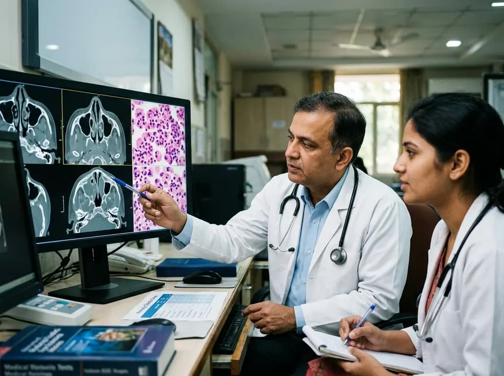
Accurately evaluating osteitis severity in CRSwNP offers critical diagnostic insight into patient endotypes prior to surgical intervention. Chronic rhinosinusitis with nasal polyps presents as a heterogeneous inflammatory condition with divergent biological drivers. Clinicians frequently encounter challenges when distinguishing eosinophilic disease from non-eosinophilic forms before formal surgical histopathology becomes available. However, objective computed tomography markers now allow otolaryngologists to evaluate underlying sinonasal inflammatory pathways non-invasively. Recent prospective evidence demonstrates that radiologic neocortical thickening and hyperostosis closely mirror the extent of tissue eosinophilia. Patients harboring dense eosinophilic infiltration typically display significantly greater bony remodeling across their paranasal sinus architecture. Consequently, quantifying bony changes provides a reliable surrogate marker for identifying type 2 inflammation. This radiological stratifier helps rhinology teams identify recalcitrant endotypes early in the diagnostic pathway. Furthermore, recognizing this high-risk phenotype assists surgeons in anticipating severe mucosal fragility and higher bleeding risks during endoscopic sinus surgery. Clinicians can also modify their postoperative follow-up regimens accordingly. Therefore, radiographic staging transforms routine preoperative imaging from a purely structural roadmap into a functional biomarker of tissue biology.
Radiologists and rhinologists traditionally quantify sinonasal disease burden using the Lund-Mackay score. This standard system assesses soft-tissue mucosal opacification and ostiomeatal complex obstruction. However, mucosal opacification alone fails to capture the chronic, destructive inflammatory signaling occurring deeper within underlying bone. In contrast, the Kennedy Osteitis Score grades bony remodeling, sclerosis, and cortical thickness on bone-window computed tomography scans. A prospective study involving 154 patients examined how both metrics perform in discerning tissue eosinophilia, defined as ten or more eosinophils per high-power field. The investigators discovered that the Kennedy Osteitis Score correlated significantly with tissue eosinophil density. Moreover, multivariable regression confirmed that osteitis severity served as an independent predictor of eosinophilic disease. While the Lund-Mackay score documents immediate disease volume, it exhibits weaker discriminatory capability regarding specific inflammatory endotypes. In comparative receiver operating characteristic curve analyses, the Kennedy Osteitis Score demonstrated superior specificity for eosinophilic disease. Thus, scoring bony remodeling provides distinct information that mucosal staging cannot offer. Incorporating osteitis assessment into routine computed tomography reporting significantly strengthens preoperative risk stratification.
Sinonasal osteitis does not merely represent a passive consequence of chronic mechanical pressure or bacterial infection. Instead, active molecular crosstalk between inflamed mucosa and underlying bone drives profound structural remodeling. Activated eosinophils release major basic protein, eosinophil cationic protein, and transformational growth factors directly into the periosteum. Furthermore, type 2 cytokines such as interleukin-4, interleukin-5, and interleukin-13 activate osteoblasts and osteoclasts simultaneously. This cytokine signaling alters normal bone turnover, resulting in neo-osteogenesis, fibrosis, and hyperostosis within the ethmoid trabeculae. Consequently, persistent mucosal eosinophilia perpetuates chronic osteitis, which in turn acts as an inflammatory nidus. This vicious cycle explains why patients with severe osteitis experience high rates of polyp recurrence after standard polypectomy. Moreover, bacterial biofilm formation frequently thrives along osteitic, irregular bone margins, compounding therapeutic recalcitrance. Recognizing this bidirectional relationship clarifies why topical medical treatments often fail when clinicians leave underlying osteitis unaddressed. Therefore, comprehensive disease management must tackle both superficial mucosal inflammation and profound osteitic remodeling to ensure durable patient recovery.
Preoperative identification of the eosinophilic endotype fundamentally changes how rhinology teams approach clinical care pathways. Eosinophilic polyps respond briskly to systemic and topical corticosteroids, yet they recur rapidly when anti-inflammatory maintenance lapses. Consequently, patients exhibiting high osteitis scores benefit substantially from individualized therapeutic regimens. Surgeons can plan comprehensive, thorough surgery, such as extensive complete ethmoidectomy and wide antrostomies, rather than minimal drainage. Complete mucosal clearance over thickened osteitic partitions reduces local inflammatory reservoirs. In addition, recognizing eosinophilic disease early enables timely discussions regarding targeted biologic therapies. Monoclonal antibodies targeting interleukin-4 receptor alpha, interleukin-5, or immunoglobulin E offer dramatic symptom relief for recalcitrant disease. However, these therapies carry significant cost implications. Identifying radiological osteitis helps otolaryngologists justify advanced biologic therapies for patients at the greatest risk of postoperative failure. Furthermore, early endotyping directs clinical teams toward intensive topical corticosteroid rinses and frequent surveillance. Thus, integrating radiologic osteitis grading into surgical planning ensures that high-risk individuals receive tailored, disease-modifying care from day one.
In Indian clinical practice, patients with chronic rhinosinusitis with nasal polyps present with distinct diagnostic and therapeutic challenges. Environmental air pollution, high allergen burdens, and recurrent microbial infections frequently cause mixed mucosal inflammatory patterns. Furthermore, many patients present late with extensive bilateral disease and significant sinonasal polyposis. Histopathology laboratories across semi-urban or peripheral centers may lack standardized protocols for counting tissue eosinophils per high-power field. Therefore, computed tomography provides an accessible, non-invasive diagnostic modality that Indian clinicians already routinely perform before functional endoscopic sinus surgery. Incorporating the Kennedy Osteitis Score into imaging protocols enables Indian rhinologists to predict eosinophilic disease without awaiting postoperative pathology reports. This non-invasive assessment assists doctors in counseling patients regarding the elevated risk of disease recurrence. Additionally, it helps clinicians formulate realistic postoperative expectations and budget for prolonged maintenance pharmacotherapy. Given that biologics remain a significant out-of-pocket expenditure in India, identifying high osteitis allows doctors to select the best surgical candidates for aggressive clearance. Ultimately, objective imaging biomarkers empower clinicians nationwide to deliver personalized, cost-effective rhinology care.
The Lund-Mackay score evaluates mucosal opacification and sinus ostial occlusion across paranasal cavities. In contrast, the Kennedy Osteitis Score grades bony remodeling, neo-osteogenesis, and hyperostosis on bone-window computed tomography scans. While Lund-Mackay reflects total inflammatory volume, it cannot reliably predict the underlying inflammatory endotype. Conversely, the Kennedy Osteitis Score directly reflects chronic tissue eosinophilia, providing superior specificity for identifying severe, recalcitrant type 2 sinonasal disease.
Identifying the eosinophilic endotype preoperatively is vital because eosinophilic polyps exhibit aggressive recurrence rates and resistance to standard conservative therapies. Recognizing this type 2 inflammatory phenotype allows otolaryngologists to adjust surgical extent, ensuring thorough removal of diseased bony partitions. Furthermore, it enables proactive postoperative management, including high-volume corticosteroid irrigations, closer endoscopic follow-up, and timely consideration of targeted biologic therapies like dupilumab to prevent rapid disease recurrence.
Pathologists diagnose eosinophilic chronic rhinosinusitis by quantifying eosinophils within resected sinonasal mucosal tissue under high-power microscopic fields. Most diagnostic consensus criteria define the eosinophilic endotype by the presence of ten or more eosinophils per high-power field across multiple representative tissue sections. Severe forms may exceed seventy eosinophils per field. This intense tissue infiltration strongly correlates with mucosal basement membrane thickening, extensive subepithelial edema, and profound radiologic osteitis.
Disclaimer: This content is for informational and educational purposes only... Refer to the latest local and national guidelines for clinical practice.
References

Read summarized clinical updates, watch expert medical content, and earn CME certifications right from your smartphone.
Open your smartphone camera to scan and install.





A prospective cohort study demonstrates that CT-based osteitis severity correlates with tissue eosinophil density in chronic rhinosinusitis with nasal polyps. Evaluating bony hyperostosis via the Kennedy Osteitis Score enables reliable, non-invasive preoperative identification of the eosinophilic endotype.
Today
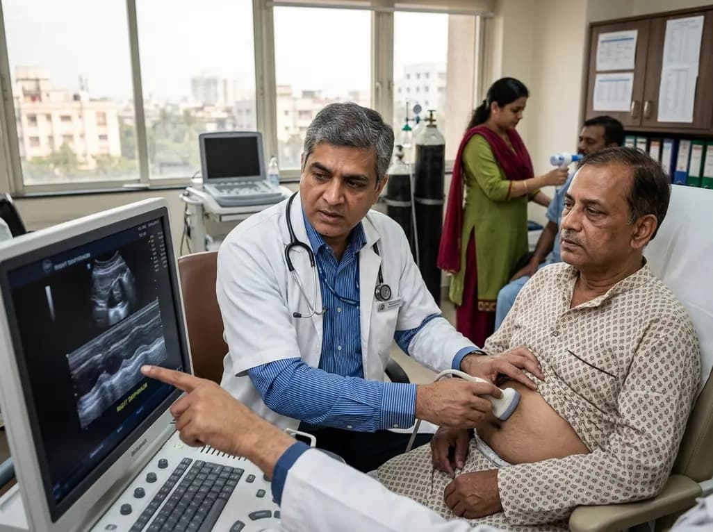
Diaphragmatic dysfunction affects over a third of chronic stroke patients, worsening sleep, mood, and functional mobility. Ultrasound assessment and inspiratory training improve outcomes.
Today

The ClinGen Prenatal Gene Curation Expert Panel evaluated 63 disease relationships across 61 genes, establishing clinical validity for severe fetal phenotypes like hydrops and stillbirth to enhance prenatal genomic interpretation and clinical care.
Yesterday
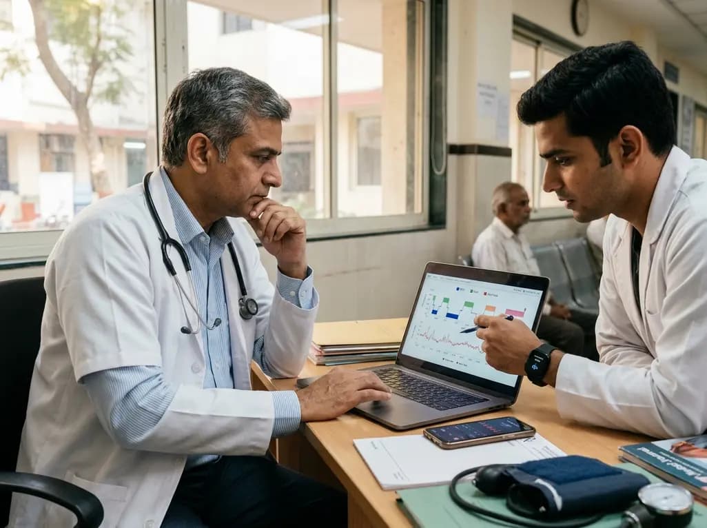
Wearable sleep health technology provides continuous biometric monitoring to augment cardiovascular risk assessment. By tracking autonomic proxies and sleep architecture, these digital tools empower clinicians to guide cardiometabolic interventions, despite persistent challenges in wake detection specificity.
Today
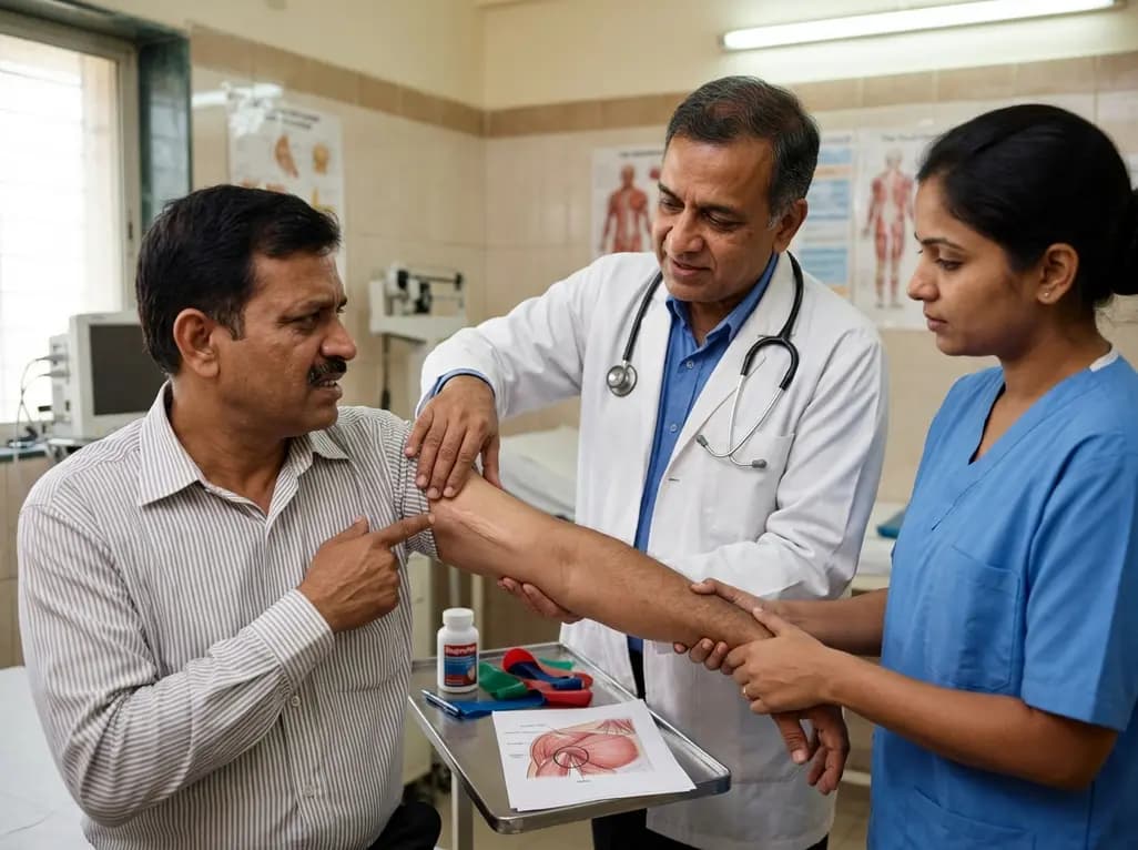
Idiopathic axillary web syndrome (IAWS) is a rare cause of shoulder pain and mobility restriction characterized by palpable axillary cording without prior surgery or trauma. Early diagnosis, nonsteroidal anti-inflammatory therapy, and physical rehabilitation lead to complete resolution of symptoms.
Today

SWEDEPAD-1 trial insights show paclitaxel-coated devices do not improve long-term limb salvage in patients with chronic limb-threatening ischemia and tissue loss. While one-year reinterventions decreased, the devices were linked to a higher risk of major amputation at three months.
Today
