.webp&w=384&q=75)

Loading, please wait...
Loading, please wait...
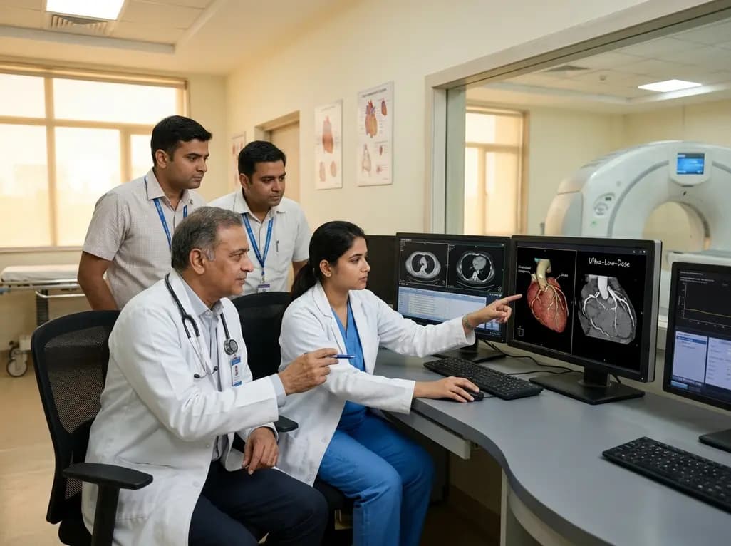
Cardiovascular disease remains the leading cause of mortality globally, making accurate cardiovascular risk stratification essential. Coronary artery calcium scoring provides a validated, non-invasive method to evaluate calcified atherosclerotic burden and guide primary preventive therapy. However, conventional computed tomography protocols expose patients to ionizing radiation, which clinicians seek to minimize. The emergence of photon-counting detector CT offers a transformative technological shift in cardiac imaging. Recent prospective investigations evaluate whether ultra-low-dose true non-contrast acquisitions or contrast-derived reconstructions offer optimal diagnostic performance without compromising clinical decision-making.
Conventional energy-integrating detectors convert incoming X-ray photons into visible light before generating electrical signals. Consequently, this multi-step conversion introduces electronic noise and limits spatial resolution. In contrast, photon-counting detector CT employs direct-converting semiconductors that measure individual photons and their energy levels. This fundamental mechanism eliminates electronic noise completely, enabling superior spatial resolution and energy selectivity at lower radiation doses.
Clinicians increasingly leverage these capabilities to resolve intricate coronary anatomy previously obscured by blooming artifacts. Because calcified plaques often create severe partial volume effects on traditional scanners, photon-counting technology substantially improves plaque characterization. Furthermore, multi-energy data acquisition allows advanced spectral reconstructions, such as virtual non-contrast and virtual non-iodine imaging, directly from contrast-enhanced scans. Consequently, cardiovascular specialists can extract vital diagnostic insights regarding vascular lumen patency and atherosclerotic calcification. Understanding how these novel platforms perform across distinct imaging modes is crucial for modern radiologic practice.
In contemporary clinical workflows, assessing coronary calcification typically requires a dedicated non-contrast scan prior to coronary computed tomography angiography. However, performing two separate acquisitions inevitably increases cumulative radiation exposure. Researchers have therefore investigated whether virtual non-contrast or virtual non-iodine series generated from post-contrast scans could obviate the need for independent non-contrast imaging.
Recent comparative investigations on dual-source photon-counting systems benchmark ultra-low-dose true non-contrast scans, virtual non-contrast images, and virtual non-iodine reconstructions against standard true non-contrast references. The standard protocol utilizes sequential acquisition at 120 kVp, whereas the ultra-low-dose approach employs high-pitch helical acquisition with tin filtration at 100 kVp. While angiography-derived spectral reconstructions demonstrate strong correlation with traditional standards, they show minor systematic deviations in total Agatston scores. Specifically, virtual non-iodine algorithms perform admirably, yet they do not fully match the extraordinary fidelity achieved by dedicated ultra-low-dose true non-contrast acquisitions. Consequently, clinicians must evaluate whether reconstructed images satisfy precision requirements for individualized patient risk classification.
Radiation stewardship represents a cornerstone of diagnostic radiology, particularly in asymptomatic populations undergoing cardiovascular screening. Traditional non-contrast calcium scoring protocols typically yield dose-length products around 70 to 80 mGy·cm. In contrast, coronary CT angiography requires considerably higher exposures, often exceeding 400 mGy·cm depending on heart rate and anatomical complexity. Therefore, identifying scanning techniques that substantially curtail dose while maintaining diagnostic accuracy remains paramount.
Recent clinical data demonstrate that high-pitch helical scanning with tin filtration on photon-counting platforms reduces the median dose-length product to approximately 7.98 mGy·cm. This represents an impressive dose reduction of nearly 90% compared to standard sequential non-contrast protocols. Tin filtration effectively removes low-energy photons that contribute to radiation burden without enhancing image contrast. Moreover, the superior detector quantum efficiency of photon-counting hardware ensures that image quality is preserved despite dramatic dose decreases. As a result, healthcare practitioners can perform routine calcification surveillance with radiation doses comparable to background environmental exposure.
Beyond radiation metrics, diagnostic concordance determines whether a novel protocol can safely guide medical management. Quantitative correlation analysis shows that ultra-low-dose true non-contrast imaging achieves an exceptional Pearson correlation coefficient of 0.996 when compared directly against standard non-contrast imaging. Furthermore, this correlation significantly outperforms virtual non-iodine reconstructions derived from coronary angiography datasets.
Bland-Altman analysis similarly demonstrates minimal bias and narrow limits of agreement between ultra-low-dose and standard acquisitions. When evaluating categorical risk strata, which divide patients into zero, minimal, mild, moderate, and severe calcium burden, ultra-low-dose protocols maintain outstanding weighted Cohen's kappa values exceeding 0.95. Consequently, patient reclassification rates remain minimal, ensuring that therapeutic interventions like statin therapy or aspirin initiation are neither inappropriately withheld nor unnecessarily prescribed. In contrast, virtual non-contrast and virtual non-iodine images occasionally underestimate Agatston scores due to spectral overlap near dense calcifications. Therefore, dedicated ultra-low-dose non-contrast acquisitions offer superior diagnostic reliability for quantitative risk stratification.
The clinical utility of coronary artery calcium scoring hinges on its predictive value for future cardiovascular events. In asymptomatic patients categorized as intermediate risk by traditional risk calculators, identifying coronary calcification shifts management towards intensive lipid-lowering therapy and lifestyle modification. Conversely, a calcium score of zero provides a powerful negative risk marker, often supporting the deferral of lifelong pharmacological therapy.
Because therapeutic recommendations depend heavily on specific Agatston score thresholds, diagnostic consistency is critical. The exceptional agreement provided by ultra-low-dose photon-counting acquisitions ensures that clinicians can reliably apply established guidelines without misclassifying patient risk. Additionally, the remarkably low radiation burden eliminates historical hesitation surrounding calcium scoring in younger cohorts or serial surveillance. For cardiology practices and primary care physicians, this technology facilitates proactive, evidence-based preventive interventions long before ischemic symptoms emerge. Integrating these ultra-low-dose scans into cardiovascular checkups enhances early detection while keeping patient safety at the forefront of clinical care.
Although ultra-low-dose true non-contrast imaging provides superior quantitative precision, angiography-derived virtual non-iodine imaging presents distinct logistical advantages. In scenarios where a patient undergoes comprehensive coronary CT angiography, generating virtual non-iodine images eliminates an additional scan acquisition. This single-scan workflow streamlines imaging protocols, reduces scanner occupancy time, and saves incremental radiation exposure.
However, radiologists and cardiologists must acknowledge technical boundaries regarding spectral material decomposition. Virtual non-iodine algorithms rely on distinct spectral attenuation profiles to subtract intravascular contrast while preserving calcified plaque. In regions with dense contrast concentration, spectral overlap can lead to slight over-subtraction or under-subtraction of calcium deposits. Consequently, when the primary clinical goal is dedicated cardiovascular risk screening, clinicians should favor dedicated ultra-low-dose true non-contrast protocols. Conversely, when comprehensive anatomical evaluation of luminal stenosis is already mandated, virtual non-iodine reconstructions serve as a practical, radiation-free adjunct. Matching the imaging modality to the specific clinical indication ensures optimal diagnostic yield and patient safety.
Photon-counting detector CT measures individual X-ray photons directly through semiconductor detectors, completely eliminating electronic noise. Additionally, implementing tin-filtered spectra removes low-energy photons that contribute to radiation exposure without enhancing image quality. Consequently, the scanner maintains an excellent contrast-to-noise ratio at substantially reduced radiation levels, allowing accurate Agatston scoring with radiation doses nearly ninety percent lower than conventional standard non-contrast CT protocols.
Virtual non-iodine reconstructions derived from coronary angiography provide strong overall correlation, but they do not entirely replace dedicated non-contrast scans. Spectral material decomposition can occasionally over-subtract or under-subtract small or dense calcifications near concentrated iodine contrast. Therefore, dedicated ultra-low-dose non-contrast acquisitions remain the preferred approach when precise quantitative calcium scoring and risk stratification are required for primary cardiovascular prevention decisions.
Ultra-low-dose calcium scoring provides exceptional diagnostic accuracy while exposing patients to minimal radiation, with dose-length products around 8 mGy·cm. This minimal exposure makes the test safer for asymptomatic screening, younger intermediate-risk individuals, and potential longitudinal monitoring. Furthermore, it maintains high categorical agreement with standard protocols, preventing inappropriate patient reclassification and ensuring accurate, guideline-directed statin and aspirin therapy decisions.
Disclaimer: This content is for informational and educational purposes only and does not constitute medical advice, diagnosis, or treatment. Healthcare professionals should exercise their independent clinical judgment when interpreting diagnostic imaging and determining patient management strategies. Refer to the latest local and national guidelines for clinical practice.
References

Read summarized clinical updates, watch expert medical content, and earn CME certifications right from your smartphone.
Open your smartphone camera to scan and install.





A new study reveals that ultra-low-dose photon-counting detector CT achieves 90% radiation reduction in coronary artery calcium scoring while maintaining excellent correlation with standard scans, outperforming angiography-derived virtual non-contrast images.
last month
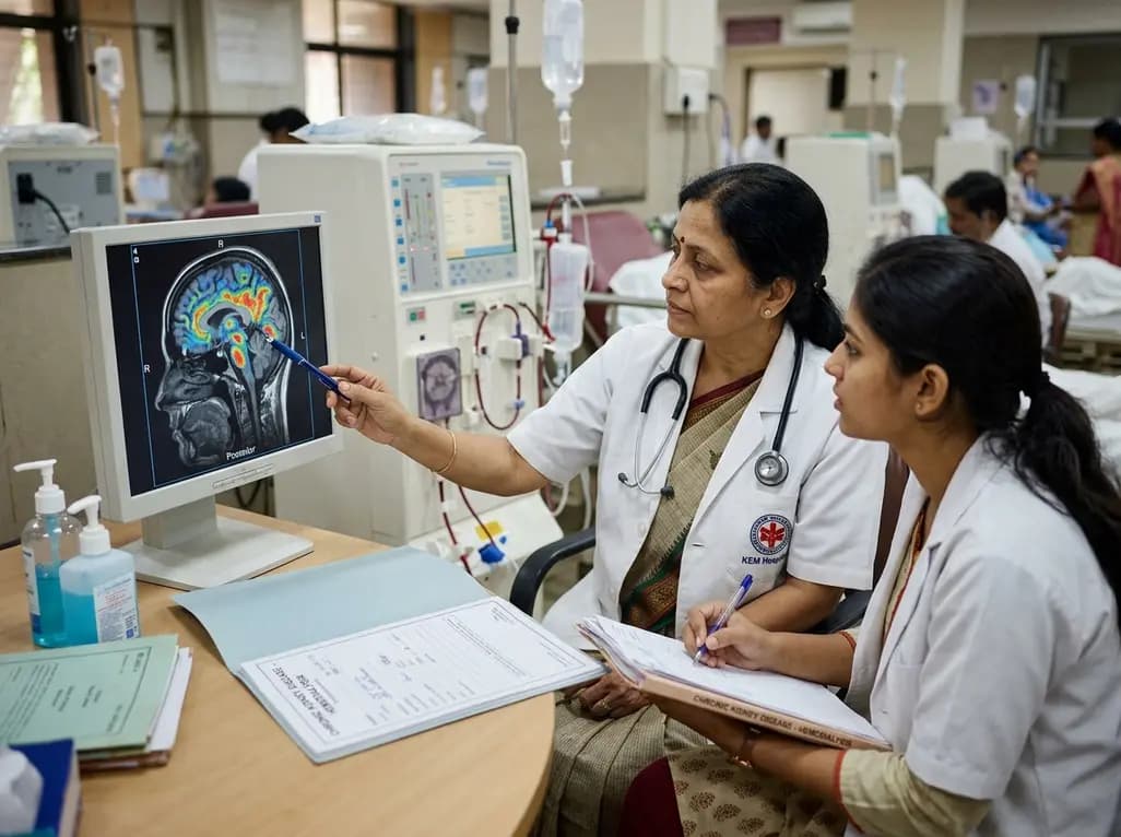
A retrospective clinical analysis reveals that hemodialysis-associated cognitive dysfunction arises from synergistic pathological mechanisms, including severe chronic anemia, worsening renal clearance, abnormal regional brain iron deposition, and an imbalance between neuroinflammatory and neurotrophic factors.
Today

A multiparametric MRI approach combining quantitative T2 shading, apparent diffusion coefficient (ADC) measurements, and morphological signs provides superior diagnostic accuracy over subjective evaluation to differentiate ovarian endometriomas from benign hemorrhagic cysts.
Today

Discover a novel laparoscopic approach for iatrogenic diaphragmatic hernia repair following pedicled omentoplasty. This modified Sugarbaker technique integrates dorsal pedicle lateralization and round ligament reinforcement to secure the diaphragmatic defect while preserving vital omental vascularity.
Today

Evaluating Hill-Sachs lesion size and the distance to dislocation is critical in recurrent shoulder instability. As bone loss concepts shift from binary on-track classifications to continuous risk assessment, clinicians must identify when isolated Bankart repair is insufficient and supplementary procedures are needed.
Today

New research reveals that M1 macrophage-derived exosomes aggravate diabetic nephropathy by transferring WTAP to stabilize S1PR2 mRNA. Silencing WTAP in these vesicles attenuates endothelial injury and renal fibrosis, pointing toward innovative nanomedicine therapies.
Today
