

Loading, please wait...
Loading, please wait...

Managing adult patients with grade II diffuse glioma requires rigorous, evidence-based multidisciplinary collaboration. Historically, neurosurgeons and neuro-oncologists debated the appropriate timing of interventions, often balancing watchful waiting against early intervention. However, scientific advances in molecular neuropathology and surgical technique have substantially transformed clinical standards. The Congress of Neurological Surgeons recently published an updated systematic review and clinical practice guideline consolidating contemporary literature. Consequently, medical specialists now possess reliable benchmarks to guide initial diagnosis, surgical resection, and subsequent adjuvant therapies for optimal patient outcomes.
Accurate baseline neuroimaging establishes the foundation for sound neuro-oncological management. Standard structural evaluation mandates high-resolution magnetic resonance imaging protocols. These protocols encompass pre-contrast and post-gadolinium T1-weighted sequences alongside comprehensive T2-weighted and fluid-attenuated inversion recovery sequences. Furthermore, clinicians perform serial surveillance imaging to track volumetric expansion and detect early malignant transformation. The emergence of new contrast enhancement typically signals transformation into higher-grade disease. However, conventional sequences alone may fail to delineate true infiltrative boundaries. Therefore, the updated guidelines suggest integrating advanced physiological imaging techniques into standard evaluation pathways. Perfusion-weighted imaging, including dynamic susceptibility contrast and dynamic contrast enhancement, clarifies tumoral microvasculature. Similarly, diffusion-weighted imaging assesses cellular density in non-enhancing parenchymal margins. Arterial spin labeling also delivers non-invasive vascular assessments without requiring intravenous contrast media. Moreover, specialized metabolic neuroimaging offers distinct diagnostic advantages. Clinicians can utilize amino-acid positron emission tomography with tracers like fluoroethyl-L-tyrosine to distinguish low-grade lesions from aggressive neoplasms. Furthermore, multi-parametric MRI helps radiation oncologists define accurate target volumes during stereotactic planning. Consequently, advanced multimodal imaging optimizes diagnostic precision, directs targeted biopsies, and informs pre-operative surgical trajectory planning.
Surgical resection serves as the primary intervention for most adult diffuse low-grade gliomas. Historically, some surgical practitioners selected prolonged observation for asymptomatic, incidentally discovered tumors. However, current evidence decisively confirms that early surgical intervention provides superior long-term survival over watchful waiting. The updated guidelines emphasize that surgical resection, whether gross total or subtotal, significantly reduces progression rates compared to biopsy alone. Extent of resection correlates directly with lengthened progression-free survival and delayed malignant transformation. Furthermore, extensive debulking dramatically improves symptom management, particularly medically refractory epilepsy. Neurosurgeons must balance aggressive tumor removal against the risk of permanent neurological deficit. Inducing a permanent neurological deficit severely impairs patient functional independence and negates survival advantages. Therefore, modern surgical teams routinely incorporate intraoperative neurophysiological mapping. Awake craniotomy with direct cortical and subcortical stimulation remains the definitive gold standard for tumors adjacent to eloquent motor and language networks. In addition, surgeons employ intraoperative neuronavigation, functional tractography, and intraoperative ultrasound to identify infiltrative tumor margins safely. Consequently, surgeons continuously refine operative strategies to maximize patient survival outcomes and preserve long-term cognitive function. Thus, neurosurgeons achieve maximal safe cytoreduction while vigilantly preserving essential neurological functions.
Accurate histopathological and genetic evaluation guides all contemporary neuro-oncological treatment decisions. Classical microscopic assessment of resected tissue remains essential for identifying glial morphology. However, molecular diagnostics have thoroughly redefined modern tumor taxonomy. The updated guidelines emphasize that pathology departments cannot classify diffuse gliomas solely through histological examination. Instead, clinicians must obtain comprehensive molecular testing on representative surgical specimens. Specifically, testing for isocitrate dehydrogenase gene mutations serves as a mandatory diagnostic and prognostic prerequisite. Patients harboring IDH mutations experience markedly superior overall survival compared to those with IDH-wildtype tumors. In addition, neuropathologists must evaluate whole-arm chromosomal 1p and 19q codeletions. Co-occurring IDH mutation and 1p/19q codeletion defines oligodendroglioma, carrying the most favorable clinical prognosis. Conversely, IDH-mutant tumors with intact 1p/19q represent astrocytomas, which commonly display ATRX loss and TP53 alterations. Furthermore, molecular profiling uncovers high-risk genetic signatures in morphologically low-grade lesions. For example, CDKN2A/B homozygous deletion in an IDH-mutant astrocytoma immediately indicates grade IV aggressive clinical behavior. Similarly, EGFR amplification or TERT promoter mutations classify IDH-wildtype neoplasms as molecular glioblastomas. Consequently, integrated molecular diagnostics dictate precise prognostic stratification and direct targeted postoperative therapies.
Postoperative therapeutic decisions depend upon residual tumor volume, patient age, and molecular risk factors. Clinicians stratify patients into low-risk and high-risk categories to establish treatment intensity. High-risk patients include individuals aged forty years or older, those with subtotal resection, or tumors measuring over four centimeters. Historically, clinicians debated whether to deliver radiation immediately or delay until progression. Contemporary evidence indicates that early fractionated radiotherapy significantly extends progression-free survival in high-risk patients. However, observation remains acceptable for selected young patients with completely resected, favorable IDH-mutant oligodendrogliomas. In addition, combining chemotherapy with radiotherapy delivers substantial overall survival advantages compared to radiation alone. For patients harboring 1p/19q-codeleted oligodendrogliomas, procarbazine, lomustine, and vincristine chemotherapy paired with radiotherapy provides outstanding long-term disease control. Alternatively, clinicians frequently prescribe temozolomide because it offers manageable hematologic toxicity and excellent patient tolerance. Furthermore, clinicians monitor complete blood counts diligently throughout chemotherapy cycles to detect early myelosuppression and prevent neutropenic complications. Moreover, oncology teams should initiate adjuvant systemic therapy within twelve weeks post-surgery to optimize therapeutic efficacy. Therefore, multidisciplinary tumor boards coordinate adjuvant regimens carefully according to surgical margins and molecular findings.
Long-term surveillance remains vital because low-grade diffuse gliomas frequently recur and transform malignantly. Neuro-oncologists schedule magnetic resonance imaging every three to six months during early postoperative years. Furthermore, advanced perfusion imaging and amino-acid positron emission tomography distinguish true tumor recurrence from radiation necrosis. When recurrence occurs, the updated guidelines emphasize that repeat surgical resection offers significant clinical benefit for eligible patients. Repeat cytoreduction decreases intracranial hypertension, alleviates neurological deficits, and secures fresh tissue for repeat molecular sequencing. For unresectable recurrences, systemic second-line chemotherapy or stereotactic re-irradiation provides meaningful localized disease control. In Indian clinical practice, implementing these guidelines requires tailored resource allocation. While major tertiary centers offer advanced intraoperative mapping and next-generation sequencing, peripheral centers face diagnostic constraints. Therefore, establishing structured regional referral networks ensures reliable central testing for IDH and 1p/19q status. Indian neurosurgeons also adopt intraoperative ultrasound as an accessible alternative to intraoperative magnetic resonance imaging. Additionally, temozolomide monotherapy offers an economical, well-tolerated systemic treatment option across diverse socioeconomic strata. Consequently, adopting standardized protocols elevates neuro-oncological care nationwide.
Maximal safe resection significantly prolongs progression-free and overall survival compared to simple stereotactic biopsy. Furthermore, cytoreductive surgery reduces mass effect, alleviates intractable seizure activity, and provides extensive tissue for essential molecular classification. Neurosurgeons prioritize cytoreduction while preserving eloquent functional areas through intraoperative mapping techniques to prevent irreversible postoperative neurological morbidity.
Pathologists must test for isocitrate dehydrogenase gene mutations and whole-arm chromosomal 1p/19q codeletions. These essential molecular biomarkers distinguish oligodendrogliomas from astrocytomas. In addition, evaluating CDKN2A/B homozygous deletions and ATRX expression refines diagnostic accuracy. Identifying these genetic alterations establishes definitive prognosis and directly guides individualized postoperative radiotherapy and chemotherapy strategies.
Clinicians initiate adjuvant treatment for high-risk patients, including individuals aged forty or older, those with subtotal resection, or tumors over four centimeters. When indicated, oncologists recommend starting adjuvant chemotherapy or chemoradiotherapy within twelve weeks post-surgery. Prompt initiation prevents early disease recurrence while maintaining patient performance status and overall functional quality of life.
Disclaimer: This content is for informational and educational purposes only... Refer to the latest local and national guidelines for clinical practice.
References
Ormond DR et al. Congress of Neurological Surgeons Systematic Review and Evidence-Based Guidelines for the Treatment of Adults With WHO Grade II Diffuse Glioma: Update. Neurosurgery. 2026 May 01. doi: 10.1227/neu.0000000000003912. PMID: 41983690.
Badve C et al. Congress of neurological surgeons systematic review and evidence-based guidelines for the role of imaging in newly diagnosed WHO grade II diffuse glioma in adults: update. J Neurooncol. 2025;174:7–52. doi: 10.1007/s11060-025-05043-8.
Olson JJ et al. Congress of Neurological Surgeons systematic review and evidence-based guidelines on the management of recurrent diffuse low-grade glioma: update. J Neurooncol. 2024. doi: 10.1007/s11060-024-04825-y.

Read summarized clinical updates, watch expert medical content, and earn CME certifications right from your smartphone.
Open your smartphone camera to scan and install.





The Congress of Neurological Surgeons has updated evidence-based guidelines for adult WHO grade II diffuse glioma, detailing advanced neuroimaging, maximal safe surgical resection, molecular profiling (IDH and 1p/19q), and tailored adjuvant radiotherapy and chemotherapy to optimize survival.
Today
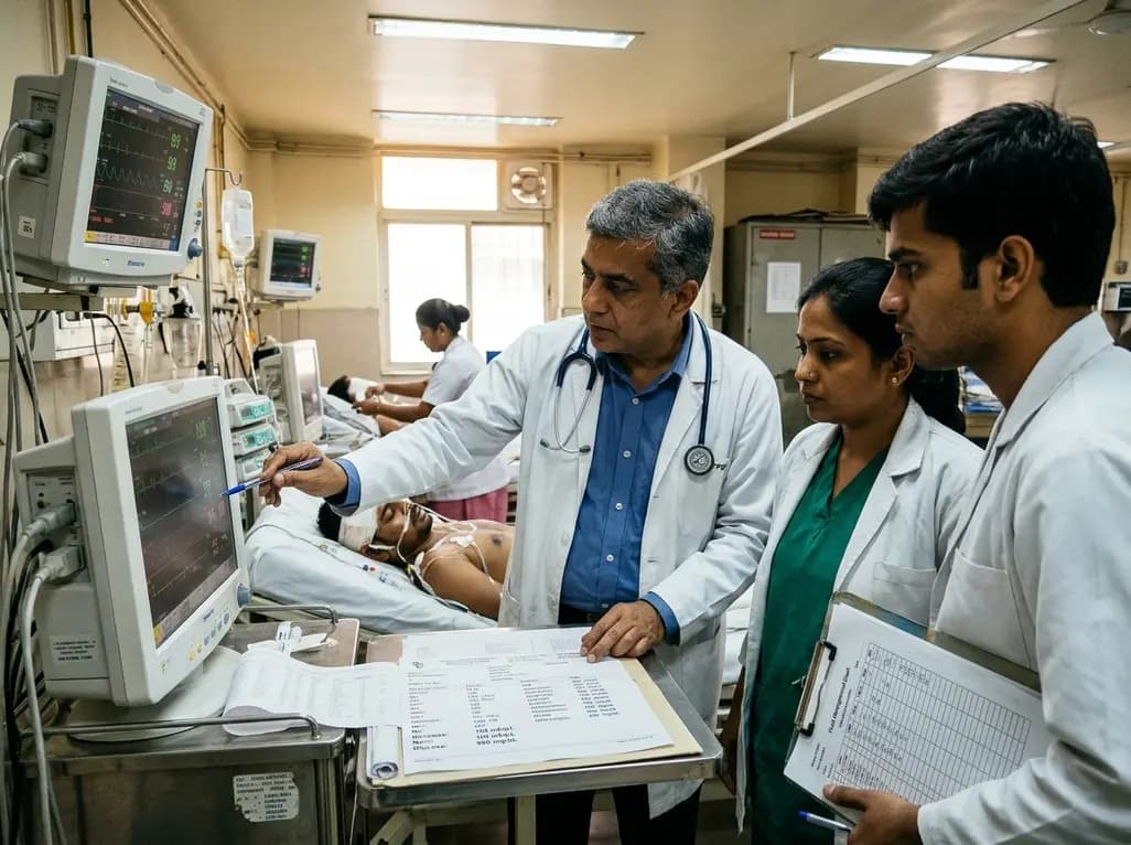
A complex clinical case reveals how adipsic hypernatremia after traumatic brain injury, combined with hyperosmolar hyperglycemic state, triggers life-threatening dehydration and extensive venous thromboembolism, emphasizing the vital role of meticulous fluid management and vigilance.
Today

Large genomic databases like gnomAD and All of Us offer crucial insights into the population prevalence of partial RHD genotypes. Identifying these variants helps clinicians mitigate anti-D alloimmunization risks in transfusion medicine and obstetric care across diverse ancestral populations.
Yesterday
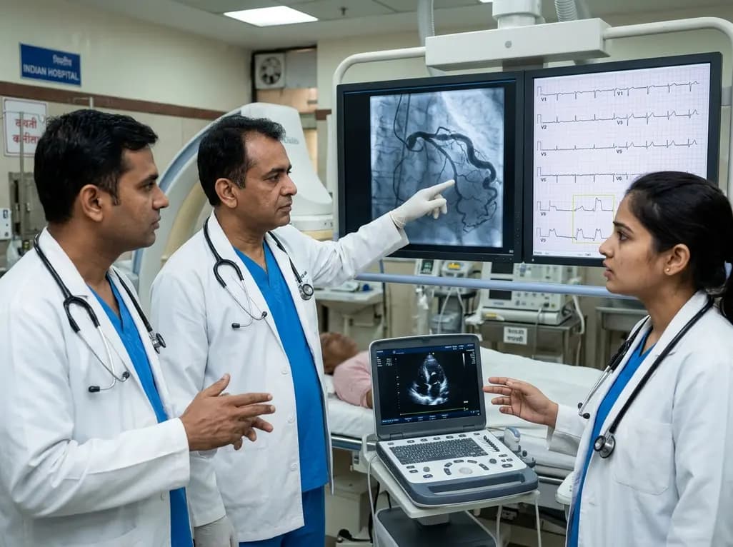
A hyperdominant left circumflex artery supplies extensive myocardial regions, creating a perilous diagnostic blind spot during acute occlusion. Standard ECGs often conceal posterior infarction, requiring clinicians to employ posterior leads and rapid echocardiography for early invasive catheterization.
Today
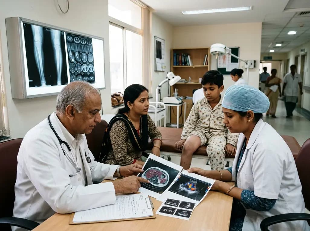
Deep intramuscular vascular malformations in pediatric Klippel-Trenaunay syndrome can mimic Maffucci syndrome. Multimodality imaging with Doppler ultrasound and MRI, along with histopathology, ensures prompt differential diagnosis, targeted surgical debulking, and optimized conservative surveillance.
Today
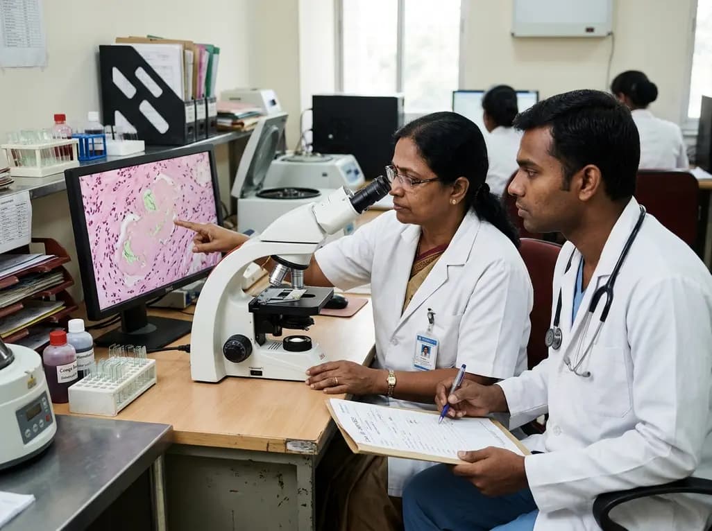
Discover essential clinical insights into AGLP1-type amyloidosis, a rare localized complication of glucagon-like peptide-1 analog injections. Learn how histopathology and mass spectrometry differentiate benign iatrogenic amyloidomas from systemic disease, preventing misdiagnosis and unwarranted therapies.
Yesterday
