.webp&w=384&q=75)

Loading, please wait...
Loading, please wait...

Accurate prenatal diagnosis of congenital spinal anomalies remains essential for obstetricians, fetal medicine specialists, and pediatric neurosurgeons. Closed spinal dysraphism represents a complex spectrum of neural tube defects characterized by continuous skin coverage over the spinal lesion. Unlike open spina bifida, these malformations lack classic cranial markers such as the lemon or banana signs. Consequently, antenatal identification requires meticulous sonographic evaluation of fetal spine anatomy. A landmark 10-year two-center cohort study provides vital evidence regarding prenatal ultrasound reliability, neurosurgical management, and long-term functional outcomes.
Prenatal ultrasound remains the frontline diagnostic modality for detecting fetal spinal malformations during routine mid-trimester structural evaluations. In this retrospective cohort study, detailed ultrasound examinations demonstrated remarkable diagnostic precision, achieving accurate anatomical characterization in nearly ninety percent of cases. Specifically, saccular malformations constituted the vast majority of presentations, accounting for almost eighty-six percent of all cohort lesions. Clinicians observed that most defects occurred in the lumbosacral or sacral spine regions. Furthermore, sonographers identified limited dorsal myeloschisis as the predominant subtype, representing nearly half of all evaluated cases. Posterior meningoceles and conus spinal cord lipomas formed the remaining saccular presentations. Because intact skin covers the posterior osseous defect, maternal serum alpha-fetoprotein levels typically remain normal. Therefore, sonographers must actively scrutinize direct anatomical indicators, such as low-lying conus medullaris, focal subcutaneous cystic masses, and disrupted posterior vertebral arches. Additionally, fetal magnetic resonance imaging serves as an invaluable complementary modality to confirm spinal cord tethering. Consequently, prospective parents receive precise anatomical information that guides timely prenatal counseling.
Standardizing anatomical terminology is essential for improving prognostic accuracy and guiding parental counseling. In this investigation, researchers categorized all prenatal spinal defects according to the updated Orphanet classification system for dysraphisms. Historically, variable classification systems caused considerable confusion during multidisciplinary case discussions. The Orphanet taxonomy effectively resolves these discrepancies by classifying lesions based on precise embryological mechanisms and anatomical characteristics. Moreover, this structured system categorizes dysraphic lesions into distinct saccular and non-saccular configurations. Saccular forms, including limited dorsal myeloschisis, conus lipomas, and meningoceles, project dorsally through the spinal defect and demonstrate recognizable sonographic features. In contrast, non-saccular lesions like filum lipomas or tight filum terminale lack external masses and frequently elude mid-trimester detection. Importantly, applying the Orphanet criteria allowed clinicians to attain an impressive postnatal diagnostic concordance of nearly ninety percent. Operative inspection or postnatal imaging required minor diagnostic reclassifications in only ten percent of managed infants. Therefore, adopting this standardized taxonomy ensures consistent communication among maternal-fetal medicine specialists, pediatric radiologists, and operating neurosurgeons.
Although closed spinal dysraphism typically spares cranial structures, associated cerebral anomalies can occur and significantly impact perinatal outcomes. In this multi-center series, clinicians identified concurrent structural brain anomalies in approximately seventeen percent of affected fetuses. The most common intracranial abnormalities included ventriculomegaly, corpus callosum dysgenesis, and distinct posterior fossa malformations. Consequently, detecting concurrent cerebral pathology heavily influenced clinical management and pregnancy trajectories. The overall cohort recorded a live birth rate exceeding eighty-four percent, reflecting generally optimistic prognoses for isolated lesions. However, approximately sixteen percent of pregnancies underwent termination, primarily driven by the presence of severe intracranial malformations. In fact, major cerebral abnormalities accounted for eight out of eleven pregnancy interruptions in this cohort. Therefore, prenatal care teams must conduct exhaustive neurosonographic assessments whenever they identify a spinal lesion. Clinicians should systematically assess the ventricular system, midline cerebral structures, and posterior fossa to rule out complex neurodevelopmental disorders. Furthermore, fetal medicine specialists should offer chromosomal microarray analysis to exclude broader syndromic conditions.
Following delivery, timely pediatric neurosurgical assessment is crucial to evaluate cord tethering and plan operative reconstruction. Among liveborn infants with documented postnatal surgical records, operating neurosurgeons performed detethering procedures in over ninety percent of cases. Surgical repair primarily aims to untether the spinal cord, excise dysplastic stalks, and reconstruct the dural sheath. Moreover, neurosurgeons carefully tailored the timing of surgical intervention according to lesion integrity and clinical presentation. Infants presenting with thin-walled cystic masses or compromised cutaneous coverings required early surgical repair to prevent central nervous system infections. Conversely, asymptomatic infants with robust skin coverage underwent scheduled detethering during early infancy. Intraoperative neurophysiological monitoring guided these meticulous microsurgical dissections, effectively safeguarding functional sacral nerve roots. Furthermore, surgical teams observed low complication rates, confirming the overall safety of elective untethering in specialized pediatric units. The high concordance between prenatal sonographic staging and intraoperative findings significantly optimized preoperative planning. Thus, proactive neurosurgical intervention successfully prevented progressive neurological deterioration in the vast majority of treated infants.
Assessing functional outcomes during early childhood provides indispensable evidence for prenatal counseling and long-term care planning. At thirty-six months of age, children demonstrated exceptionally favorable lower-extremity motor capabilities. Specifically, over ninety-four percent of toddlers achieved independent ambulation without requiring orthoses or walking aids. This excellent motor trajectory highlights a key functional advantage of closed lesions over open neural tube defects. However, long-term urological outcomes revealed a substantially different clinical reality. Nearly thirty-nine percent of children exhibited persistent neurogenic bladder dysfunction at thirty-six months. In addition, earlier evaluations at eighteen months showed that almost half of all patients demonstrated urological morbidity. Urodynamic studies commonly revealed detrusor-sphincter dyssynergia, poor bladder compliance, or elevated post-void residual volumes requiring clean intermittent catheterization. Furthermore, gastrointestinal dysfunction affected over twenty-two percent of toddlers, causing chronic constipation or soiling. Consequently, clinicians must recognize that preserved motor strength does not guarantee normal bowel and bladder autonomic innervation. Pediatric urologists must initiate proactive urodynamic monitoring during early infancy to prevent silent upper urinary tract damage.
Closed spinal dysraphism features intact skin covering over the defect and typically presents with a normal posterior fossa without the classic lemon or banana signs. Conversely, open spina bifida lacks cutaneous coverage, leaks cerebrospinal fluid into amniotic fluid, and almost universally causes Chiari II malformations. Prenatal ultrasound in closed defects focuses directly on evaluating localized subcutaneous masses, conus medullaris termination levels, and cord tethering rather than identifying indirect cranial signs.
Although motor function remains preserved in most patients, nearly forty percent develop significant neurogenic bladder dysfunction by age three. Subtle spinal cord tethering disrupts delicate autonomic neural pathways supplying the bladder and urethral sphincter. Early urodynamic testing detects elevated intravesical pressures and detrusor dyssynergia before irreversible damage occurs. Consequently, proactive interventions like clean intermittent catheterization and anticholinergic therapy effectively protect upper renal tract health and promote future urinary continence.
Postnatal management requires surgical exploration and cord detethering in over ninety percent of diagnosed infants. Pediatric neurosurgeons resect dysplastic tissue stalks, excise spinal lipomas, and reconstruct the dural sac under microscopic visualization. Stable infants with intact skin usually undergo elective surgery during the first few months of life. Furthermore, intraoperative neurophysiological monitoring guides nerve identification, successfully minimizing mechanical damage to functional sacral roots during microsurgical untethering.
Disclaimer: This content is for informational and educational purposes only and does not constitute medical advice. Refer to the latest local and national guidelines for clinical practice.
References

Read summarized clinical updates, watch expert medical content, and earn CME certifications right from your smartphone.
Open your smartphone camera to scan and install.





A 10-year cohort study highlights prenatal ultrasound accuracy and postnatal functional outcomes in closed spinal dysraphism, demonstrating favorable motor milestones alongside notable urologic morbidity.
Today

A groundbreaking study highlights how elevated ISG15 fuels RIG-I/MDA5 signaling and macrophage-microglia crosstalk during blood-brain barrier disruption in neuromyelitis optica spectrum disorder, revealing novel therapeutic targets for neuroinflammation.
Today

An updated systematic review and meta-analysis of 7,875 patients reveals that routine microaxial left ventricular support during nonemergent high-risk PCI does not reduce mortality or ischemic complications, while significantly increasing major bleeding events.
Yesterday
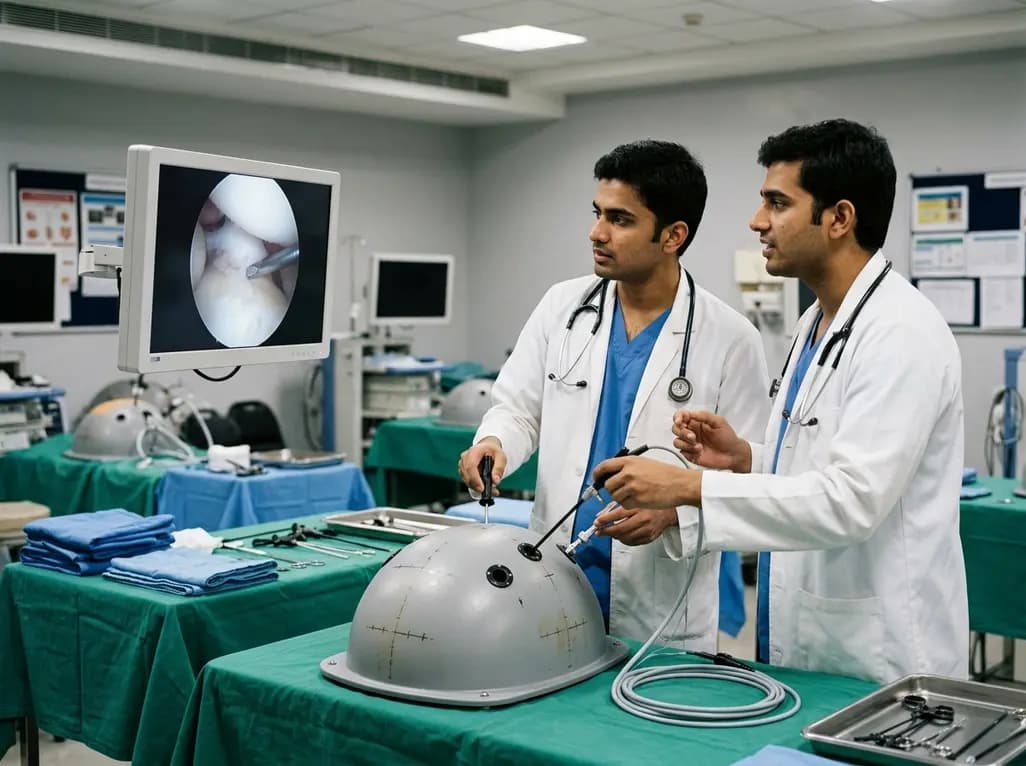
A multicenter trial reveals that the Fundamentals of Arthroscopic Surgery Training (FAST) program demonstrates transfer validity. Trainees showed marked improvement on the ASSET score, proving simulation enhances operating room performance and technical surgical skills.
Today

The SED-SEEN-SEEP consensus outlines an Integrated Care Pathway for preclinical type 1 diabetes, structuring target screening, autoantibody confirmation, metabolic staging, and proactive follow-up to prevent diabetic ketoacidosis and facilitate early disease-modifying intervention.
Yesterday
