.webp&w=384&q=75)

Loading, please wait...
Loading, please wait...
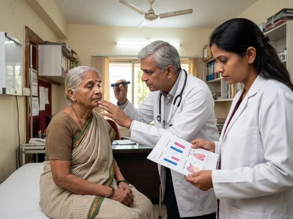
Myasthenia gravis (MG) has traditionally been viewed as a disease of young women or older men. However, rising global life expectancy has shifted the epidemiological landscape. Consequently, clinicians now frequently encounter very late-onset myasthenia gravis (vloMG), defined as onset at or after age 65. This cohort presents unique diagnostic and therapeutic challenges that differ significantly from younger patients. Understanding the nuances of vloMG is essential for geriatricians and neurologists, as misdiagnosis is common due to age-related comorbidities. Research indicates that the clinical phenotype of very late-onset myasthenia gravis often involves specific autoantibody profiles and distinct thymic pathology. Furthermore, the sex distribution in this demographic does not follow the classic female-to-male ratios seen in early-onset cases. By analyzing these variables through meta-analysis, we can better appreciate the heterogeneous nature of the disease across the lifespan. This article explores the specific characteristics of vloMG, contrasting them with early-onset (EOMG) and late-onset myasthenia gravis (LOMG) to provide a comprehensive overview for medical professionals.
In younger populations, myasthenia gravis exhibits a clear female predominance, typically peaking in the second and third decades. However, very late-onset myasthenia gravis demonstrates a striking reversal or equalization of this trend. Epidemiological data suggests that as the age of onset increases, the proportion of male patients rises significantly. In many cohorts of vloMG, men are affected more frequently than women, or the ratio remains nearly equal. This shift is likely driven by various factors, including immunosenescence and hormonal alterations. Unlike EOMG, which is often associated with specific HLA types, vloMG appears less tied to these genetic markers. The rising incidence of MG in elderly males has major implications for clinical practice, particularly in India. Doctors must maintain a high index of suspicion for MG in older men presenting with ptosis, rather than assuming symptoms are merely "age-related." Recognizing this sex-specific trend in very late-onset myasthenia gravis helps in early diagnosis and prevents the delay of life-improving treatments.
The role of the thymus in myasthenia gravis pathogenesis is well-documented, yet its relevance varies by age. In early-onset patients, thymic follicular hyperplasia is common, often justifying thymectomy. In contrast, very late-onset myasthenia gravis is typically associated with thymic involution. As individuals age, thymic tissue is replaced by fat, leading to decreased T-cell production. Despite this physiological decline, thymomas can still occur in the elderly, although they are generally less frequent in the vloMG group compared to the LOMG group. Interestingly, meta-analyses suggest that the incidence of thymoma in patients over 65 is lower than in those between ages 50 and 64. For clinicians, this means that while chest imaging remains mandatory for all new MG diagnoses, the likelihood of finding active hyperplasia is minimal in very late-onset cases. Consequently, the role of thymectomy in vloMG is controversial and usually reserved for confirmed thymoma cases. Understanding these pathological differences is crucial for avoiding unnecessary surgical interventions in older adults with multiple surgical risks.
The immunological signature of very late-onset myasthenia gravis is predominantly characterized by acetylcholine receptor (AChR) antibodies. Most studies show that the vast majority of vloMG patients are seropositive for AChR antibodies, with lower frequencies of MuSK or LRP4 antibodies compared to younger cohorts. This high seropositivity rate is helpful for diagnosis, as the AChR antibody test is highly specific. However, a small subset of elderly patients may remain seronegative, requiring more advanced testing like repetitive nerve stimulation. The presence of these antibodies in very late-onset myasthenia gravis often correlates with a generalized clinical presentation, though ocular-onset is also frequent. Moreover, the titers of AChR antibodies in the elderly do not necessarily correlate with disease severity, which is a common point of confusion. Focusing on the primary autoantibody profile allows for a more streamlined diagnostic process, ensuring that very late-onset myasthenia gravis is identified promptly before the onset of respiratory complications. This focus on serology helps differentiate MG from other geriatric muscle disorders.
Diagnosing very late-onset myasthenia gravis is notoriously difficult because symptoms often mimic other age-related conditions. For instance, ptosis and diplopia may be mistaken for senile blepharochalasis or cranial nerve palsies due to microvascular disease. Similarly, generalized weakness is frequently attributed to frailty or heart failure. Consequently, the diagnostic lag—the time between symptom onset and a correct diagnosis—is often longer in the vloMG group. Clinicians must perform a thorough neurological examination, specifically looking for "fatigability," the hallmark of MG. In India, where healthcare access varies, elderly patients might not report mild symptoms until severe bulbar or respiratory issues emerge. Furthermore, multiple comorbidities complicate the use of certain diagnostic tests. Therefore, integrating clinical findings with antibody testing is the most reliable approach for confirming very late-onset myasthenia gravis. Education regarding these "masqueraders" is vital for primary care physicians to ensure timely referral to neurologists and improve prognosis. Recognizing these nuances reduces the burden of undiagnosed autoimmune disease in the aging population.
Managing very late-onset myasthenia gravis requires a delicate balance between controlling autoimmunity and minimizing side effects. Elderly patients are more susceptible to corticosteroid adverse effects, such as osteoporosis and hyperglycemia. Non-steroidal immunosuppressants also require close monitoring for bone marrow suppression and hepatotoxicity. Polypharmacy is a significant concern; many medications for hypertension can exacerbate MG symptoms. Beta-blockers and certain antibiotics should be used with extreme caution. Despite these challenges, the prognosis for vloMG is generally favorable. Many patients respond well to low-dose pyridostigmine and cautious immunosuppression. Intravenous immunoglobulin (IVIg) is often better tolerated than long-term high-dose steroids for acute exacerbations. Ultimately, the goal of treating very late-onset myasthenia gravis is to maintain quality of life and functional independence while avoiding treatment-related morbidity. Personalized medicine, considering biological age and comorbidities, remains the gold standard. As our understanding of vloMG evolves, clinicians can offer more targeted therapies that respect the physiological limitations of the elderly.
Very late-onset myasthenia gravis (vloMG) is clinically defined as the onset of symptoms at or after the age of 65 years. This classification distinguishes it from early-onset cases (before 50) and late-onset cases (50 to 64), helping clinicians anticipate specific disease patterns and co-existing geriatric health issues.
Unlike early-onset myasthenia gravis, which shows a strong female predominance, very late-onset myasthenia gravis often shows a male predominance or an equal sex ratio. As the age of onset increases, the clinical demographic shifts toward older men, necessitating a high index of clinical suspicion regardless of patient gender.
Thymic pathology in very late-onset myasthenia gravis is typically characterized by thymic atrophy rather than the hyperplasia seen in younger patients. While the incidence of thymoma is generally lower in the vloMG group compared to late-onset cases, chest imaging is still required to rule out neoplastic changes.
Disclaimer: This content is for informational and educational purposes only. It is not intended as a substitute for professional medical advice, diagnosis, or treatment. Always seek the advice of your physician or other qualified health provider with any questions you may have regarding a medical condition. Refer to the latest local and national guidelines for clinical practice.
References
Liampas I et al. Very late-onset myasthenia gravis: a systematic review and meta-analysis of sex distribution, thymic pathology and autoantibodies. J Neurol. 2026 Jul 16. doi: 10.1007/s00415-026-14014-6. PMID: 42461294.
Gilhus NE. Myasthenia Gravis. N Engl J Med. 2016;375(26):2570-2581.
Bubuioc AM, et al. Myasthenia Gravis: Diagnostic Errors and Delay. Front Neurol. 2021;12:635409.

Read summarized clinical updates, watch expert medical content, and earn CME certifications right from your smartphone.
Open your smartphone camera to scan and install.





A comprehensive review of very late-onset myasthenia gravis (onset ≥65), focusing on sex distribution, autoantibodies, and thymic pathology to improve geriatric diagnosis and management.
3 months ago

As monsoon seasons elevate arboviral disease threats, Tamil Nadu has initiated an ICMR-monitored evaluation of the tetravalent dengue vaccine in Tenkasi district. Clinicians emphasize clinical monitoring, public prevention, and high-quality early pediatric care to reduce disease morbidity across endemic regions.
Today

Leucine Rich Bio has introduced South Asia's first comprehensive vaginal microbiome test, BugSpeaks. By profiling microbial dysbiosis, pathogens, and antimicrobial resistance, this next-generation diagnostic platform provides clinicians with deeper insights into recurrent infections, fertility, and reproductive health.
Today

Review how tailored exercise programs benefit frail older patients with heart failure. Explore pathophysiological mechanisms, individualized training prescriptions, and strategies to overcome adherence barriers in clinical practice.
Today

This comparative study evaluates posterior labral tears with and without glenohumeral instability, highlighting distinct demographic patterns, MRI underestimation in stable shoulders, and successful arthroscopic repair outcomes.
Today

A clinical study demonstrates that early intervention with tocilizumab, either as monotherapy or bridged with corticosteroids, achieves significant proptosis reduction and resolves active inflammation in treatment-naive thyroid eye disease.
Today
