.webp&w=384&q=75)

Loading, please wait...
Loading, please wait...
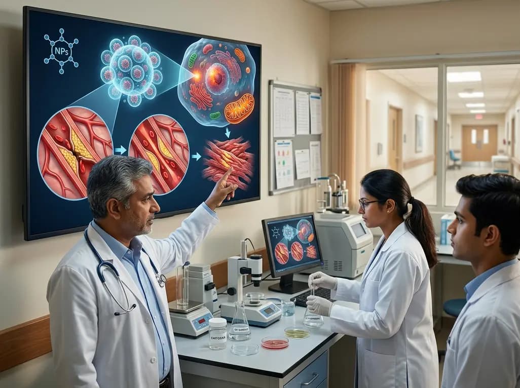
Ischemia-reperfusion injury presents a formidable clinical paradox where the restoration of vital blood flow unexpectedly exacerbates acute cellular destruction across ischemic organs. This complex pathophysiological cascade frequently complicates emergency revascularization, major organ transplantation, severe vascular trauma, acute myocardial infarction, and ischemic stroke. Although rapid reperfusion remains indispensable to salvage ischemic tissues, sudden reoxygenation triggers catastrophic metabolic cascades. Consequently, clinicians encounter severe microvascular dysfunction, oxidative bursts, and uncontrolled tissue necrosis that standard pharmacological regimens cannot adequately mitigate. To resolve this therapeutic bottleneck, innovative drug delivery systems are actively investigated. Among these technologies, chitosan nanoparticles for ischemia-reperfusion injury have emerged as a highly versatile nanomedicine platform. These biocompatible carriers enable targeted molecular interventions directly within damaged vascular beds.
The progression of reperfusion injury involves a dynamic cascade of interconnected cellular and biochemical derangements. Initially, prolonged arterial occlusion depletes intracellular adenosine triphosphate, halts oxidative phosphorylation, and destabilizes membrane transport proteins. Consequently, calcium ions accumulate rapidly within the cytosol and mitochondrial matrix during early ischemia. When oxygenated blood flow is restored, this calcium overload drives the abrupt opening of mitochondrial permeability transition pores. As a direct result, mitochondrial membrane potential collapses, and cells release pro-apoptotic cytochrome c into the cytoplasm. Furthermore, reoxygenation provokes immediate bursts of reactive oxygen species from xanthine oxidase and dysfunctional respiratory complexes. These free radicals trigger widespread lipid peroxidation, break down cellular membranes, and induce direct DNA strand damage. Simultaneously, severe endothelial dysfunction diminishes protective nitric oxide levels and promotes profound microvascular plugging. Activated vascular endothelial cells then upregulate adhesion molecules, recruiting circulating neutrophils and macrophages directly into vulnerable parenchyma. Therefore, severe inflammation, microvascular thrombosis, and regulated necrotic pathways converge to accelerate widespread secondary tissue death.
Traditional pharmacological agents frequently fail in reperfusion scenarios due to poor bioavailability, rapid renal clearance, and negligible accumulation in injured microvasculature. In contrast, natural biopolymer nanocarriers offer exceptional physicochemical versatility to overcome these pharmacokinetic limitations. Chitosan, derived from the deacetylation of natural chitin, possesses superior biocompatibility, minimal systemic toxicity, and complete biodegradability. In addition, its primary amine groups confer a distinctive positive surface charge under physiological conditions. This cationic profile enables efficient electrostatic interaction with negatively charged biological membranes, endothelial surfaces, and therapeutic cargo. Consequently, researchers can functionalize chitosan nanocarriers to encapsulate diverse compounds, including hydrophobic antioxidants, anti-inflammatory molecules, peptide therapeutics, and nucleic acids. Moreover, surface chemical modifications allow for passive accumulation through increased endothelial permeability at the injury site or active molecular targeting toward specific vascular receptors. Furthermore, bioengineers can tailor these nanocarriers to display stimuli-responsive drug release triggered by tissue acidosis, elevated local reactive oxygen species, or specific enzymatic cascades. Thus, chitosan nanoparticles provide controlled, sustained therapeutic concentrations precisely when and where ischemic tissues require protection.
Chitosan-based nanomedicines exert multi-targeted cytoprotective actions by interrupting primary molecular pathways of reperfusion damage. First, these formulations effectively neutralize excessive oxidative stress. When loaded with potent radical scavengers like curcumin, coenzyme Q10, or catalase, nanoparticles deliver concentrated antioxidant capacity directly to injured cells. Therefore, they significantly reduce lipid peroxidation biomarkers, restore endogenous glutathione reserves, and normalize superoxide dismutase activity. Second, nanocarriers substantially suppress sterile inflammatory cascades. By silencing nuclear factor kappa B signaling and inhibiting downstream cytokine expression, chitosan formulations prevent aggressive neutrophil infiltration and microvascular plugging. Third, these therapeutic platforms preserve vital mitochondrial integrity. By stabilizing mitochondrial outer membranes, preventing calcium hyperaccumulation, and inhibiting transition pore opening, they sustain baseline cellular bioenergetics. Subsequently, cells avert pro-apoptotic caspase cascades and preserve functional cellular architecture. In addition, localized drug release protects the delicate microvascular endothelium, stimulating endothelial nitric oxide synthase and preserving tissue perfusion. Overall, these concurrent protective mechanisms significantly attenuate tissue necrosis and enhance intrinsic organ recovery.
Substantial preclinical research validates the efficacy of engineered chitosan nanoparticles across various vital organ models. In acute myocardial infarction models, functionalized chitosan nanocarriers successfully target infarcted myocardium, dramatically decreasing total myocardial infarct size and preventing adverse ventricular remodeling. Similarly, in experimental ischemic stroke studies, chitosan nanoparticles successfully cross compromised blood-brain barriers to deliver neuroprotective neurotrophins and antioxidants. Consequently, treated subjects demonstrate remarkable reductions in cerebral edema, decreased neuronal apoptosis, and significantly improved neurological functional scores. Furthermore, renal ischemia-reperfusion models demonstrate that chitosan nanocarriers preferentially accumulate within injured proximal renal tubular cells. As a result, these nanoparticles markedly attenuate acute kidney injury, reduce serum creatinine levels, and suppress tubular necrosis. In hepatic and intestinal ischemia models, chitosan formulations mitigate microvascular thrombosis and suppress systemic inflammatory release, preserving essential parenchymal architecture and mucosal barrier integrity. In summary, reproducible preclinical findings consistently highlight the broad therapeutic utility of these nanocarriers across diverse clinical organ beds.
Despite compelling experimental successes, translating chitosan-based nanotherapies into human clinical practice presents several challenges. First, standardizing the molecular weight and deacetylation degree of natural chitosan remains technically demanding, which can introduce batch-to-batch variation in drug loading and release kinetics. Therefore, establishing reproducible, scalable manufacturing processes under good manufacturing practice conditions is imperative. Second, comprehensive pharmacokinetic and immunological profiling in large animal models is essential to guarantee absolute long-term biocompatibility and exclude unexpected hypersensitivity reactions. In addition, determining optimal therapeutic timing remains crucial because interventions must be administered promptly during the narrow window of acute revascularization. Looking forward, the integration of targeted peptide ligands and advanced stimuli-responsive polymers promises to enhance tissue specificity further. Moreover, combining chitosan nanocarriers with cutting-edge gene-editing machinery, small interfering RNA, or stem-cell-derived secretomes may unlock unprecedented regenerative possibilities for clinical practice.
Chitosan exhibits exceptional biocompatibility, natural biodegradability, and low cytotoxicity in biological systems. Furthermore, its cationic surface charge enables efficient electrostatic binding with negatively charged cellular membranes and therapeutic cargo. Consequently, clinicians and researchers can easily functionalize chitosan nanocarriers to achieve controlled, stimuli-responsive drug release directly within inflamed, ischemic tissues.
During acute ischemia-reperfusion, endothelial integrity becomes compromised, which enhances vascular permeability at the specific injury site. Therefore, chitosan nanoparticles leverage this increased permeability for passive accumulation within damaged tissues. Additionally, surface modifications with specific targeting ligands facilitate receptor-mediated endocytosis, allowing therapeutic payloads to effectively cross cellular and microvascular barriers.
Key translation hurdles include maintaining rigorous batch-to-batch consistency regarding chitosan polymer length and deacetylation degree. Moreover, developers must establish scalable, sterile manufacturing protocols under strict regulatory standards. Comprehensive long-term biosafety, systemic clearance, and immunogenicity evaluations in large animal models are also essential before initiating human clinical trials.
Disclaimer: This content is for informational and educational purposes only and is not intended as medical advice. Healthcare professionals should exercise their independent clinical judgment. Refer to the latest local and national guidelines for clinical practice.
References
1. Dong Y et al. Chitosan Nanoparticles for the Treatment of Ischemia-Reperfusion Injury: Mechanisms, Preclinical Evidence, Challenges, and Future Perspectives. J Drug Target. 2026 Aug 22. doi: 10.1080/1061186X.2026.2723295. PMID: 42630133.
2. Wu MY, Yiang GT, Liao WT, et al. Current Mechanistic Concepts in Ischemia and Reperfusion Injury. Cell Physiol Biochem. 2018;46(4):1650-1667. doi:10.1159/000489241.
3. Mohammed MA, Syeda JTM, Wasan KM, Wasan EK. An Overview of Chitosan Nanoparticles and Its Applications in Non-Parenteral Drug Delivery. Pharmaceutics. 2017;9(4):53. doi:10.3390/pharmaceutics9040053.

Read summarized clinical updates, watch expert medical content, and earn CME certifications right from your smartphone.
Open your smartphone camera to scan and install.





Chitosan nanoparticles offer a breakthrough nanomedicine platform to mitigate ischemia-reperfusion injury across cardiac, cerebral, renal, and hepatic tissues by targeting oxidative stress, mitochondrial collapse, and acute inflammation.
last month

A recent case shows that exertional rhabdomyolysis can trigger marked transaminase spikes despite moderate creatine kinase elevation. Preserved hepatic synthetic function confirms muscle injury rather than liver failure, guiding conservative hydration and preventing unnecessary invasive diagnostics.
Today

A pharmacometric and machine learning study reveals that specific vaginal bacteria alter cervical antiretroviral exposure, highlighting the need for precision HIV PrEP strategies.
Today

Abdominal aortic aneurysm rupture remains a catastrophic vascular emergency. Groundbreaking research reveals that USP53 accelerates aneurysm progression by reprogramming smooth muscle cell metabolism toward aerobic glycolysis, highlighting a promising target beyond conventional diameter-based surveillance.
Today

A breakthrough study leverages natural language processing to accurately identify incident medication-related osteonecrosis of the jaw and track antiresorptive drug holidays from clinical narratives in electronic health records, advancing osteoporosis pharmacovigilance and dental safety.
Today

Recent research defines ferro-aging as a conserved iron-lipid axis driving organ decline across primates. Age-related iron dyshomeostasis stimulates ACSL4-mediated lipid peroxidation, promoting cellular senescence. Notably, vitamin C directly inhibits ACSL4, suppressing phospholipid oxidation and multi-organ decay.
Today
