.webp&w=384&q=75)

Loading, please wait...
Loading, please wait...
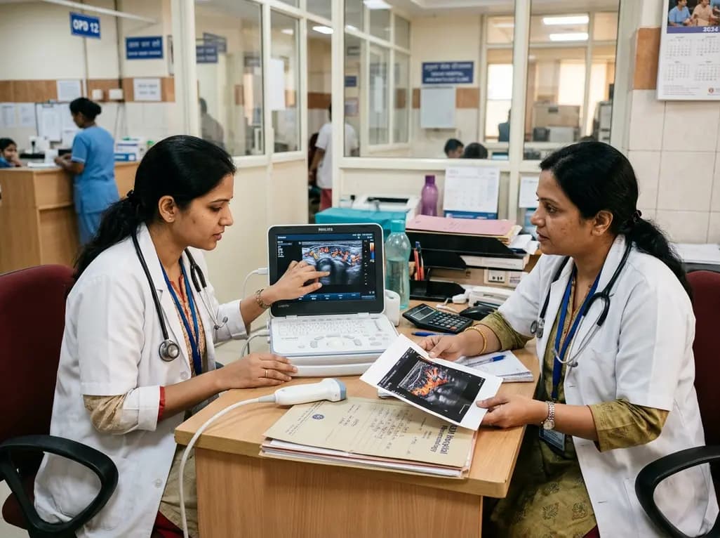
Accurate assessment of joint inflammation remains a cornerstone in managing refractory inflammatory joint disease. Rheumatologists frequently encounter difficulty when monitoring therapeutic responsiveness in patients failing targeted disease-modifying agents. In this setting, the clinical application of CEUS in rheumatoid arthritis provides clinicians with unmatched microvascular resolution. Active synovial proliferation leads to hypervascularity, which reflects ongoing inflammation and heralds joint destruction. While clinical scores like DAS28-CRP offer a systemic snapshot, objective imaging helps confirm true tissue-level activity. Consequently, advanced sonographic modalities provide essential objective data to guide therapeutic switching.
Rheumatoid arthritis management relies on timely treatment intensification when patients fail initial biological agents. However, persistent joint pain often confuses clinicians because non-inflammatory causes can mimic active disease. For instance, secondary fibromyalgia, osteoarthritis, or irreversible structural joint damage may elevate subjective joint tenderness counts significantly. Therefore, rheumatologists require precise, objective tools to differentiate active inflammation from chronic mechanical discomfort.
Synovial neoangiogenesis directly drives disease progression and irreversible radiographic erosions in active rheumatoid arthritis. Traditional physical examination and serological inflammatory markers frequently fail to depict localized microvascular changes accurately. Furthermore, conventional grayscale ultrasound cannot reliably distinguish fibrous pannus from actively inflamed vascularized tissue. Doppler imaging enhances evaluation, yet it struggles to detect ultra-low velocity blood flow in tiny synovial capillary loops.
When patients show resistance to second-line biological disease-modifying antirheumatic drugs (bDMARDs), treatment selection becomes extremely difficult. Switching to third-line biologics or targeted synthetic molecules carries substantial clinical and economic consequences. As a result, clinicians need highly sensitive imaging to confirm ongoing vascularized synovitis before altering therapy. Advanced sonographic techniques have emerged as valuable solutions to resolve these diagnostic ambiguities effectively in daily practice.
Contrast-enhanced ultrasound utilizes intravenous microbubble contrast agents to capture microvascular architecture with exceptional sensitivity. These gas-filled microbubbles act as strictly intravascular tracers, reflecting acoustic signals directly from microscopic capillary beds. Consequently, the clinical application of CEUS in rheumatoid arthritis reveals microvascular perfusion that conventional Doppler modalities often overlook.
In clinical evaluation, microbubble perfusion kinetics provide objective semi-quantitative and quantitative parameters of synovial vascularity. The technique clearly delineates hypervascular synovial thickening from inactive joint effusions, joint debris, or avascular scar tissue. Moreover, recent clinical research demonstrates that contrast enhancement exhibits a significant correlation with composite disease indices, such as DAS28-CRP. Nonparametric bootstrap analyses confirm that contrast imaging achieves fair to substantial agreement with systemic disease severity scores.
Additionally, this modality delivers crucial diagnostic confidence in complex joints, particularly the radiocarpal and midcarpal regions. By highlighting active vascular signals, sonographers can accurately grade local inflammatory burden and monitor therapeutic response. This direct visualization helps clinicians identify residual subclinical disease in patients presenting with borderline clinical findings. Therefore, contrast imaging serves as an indispensable diagnostic benchmark for synovial hypervascularity during therapy re-evaluation.
Although contrast agents deliver excellent clarity, clinicians actively seek non-invasive diagnostic options for routine clinical outpatient workflows. Superb microvascular imaging represents an advanced Doppler algorithm designed to visualize low-velocity capillary blood flow. Unlike conventional color or power Doppler, this innovative technology applies adaptive wall filters to separate tissue motion artifacts from genuine slow blood flow.
As a result, this non-contrast method captures slow microvascular flow without requiring intravenous cannulation or exogenous contrast media. Studies comparing both imaging modalities show a strong positive correlation in grading synovial vascularity. This high concordance indicates that non-contrast microvascular imaging effectively mirrors the vascular distribution patterns visualized during contrast examinations.
Nevertheless, clinical trials indicate that non-contrast microvascular imaging yields weaker direct agreement with clinical composite activity scores than contrast ultrasound. The technique occasionally picks up subtle physiological flow signals or minor tissue motion artifacts, which may alter grading concordance. Despite these subtle differences, the non-contrast method remains a highly accessible, rapid, and well-tolerated imaging modality. It provides immediate chairside information regarding synovial microcirculation, enhancing routine rheumatologic assessment in busy outpatient clinics.
Assessing disease activity accurately is vital to optimize treat-to-target strategies in biologic-resistant rheumatoid arthritis cohorts. In recent comparative investigations, researchers evaluated both sonographic techniques against DAS28-CRP in patients failing second-line biologics. Nonparametric statistical analyses revealed that contrast-enhanced ultrasound achieved superior statistical agreement with DAS28-CRP compared to non-contrast microvascular imaging.
Specifically, contrast imaging exhibited statistically significant fair agreement with DAS28-CRP classifications, achieving a kappa coefficient of 0.38. In contrast, non-contrast microvascular imaging demonstrated a lower kappa coefficient of 0.13, indicating weaker alignment with composite disease indices. However, the direct correlation between contrast imaging and microvascular imaging remained strong, with a Spearman coefficient of 0.82.
These findings suggest that while both modalities detect synovial microvessels, contrast imaging aligns more closely with systemic inflammatory burden. The heightened sensitivity of intravascular microbubbles ensures precise identification of active inflammatory hyperperfusion. Consequently, when evaluating therapy failure, contrast enhancement offers greater reliability in confirming whether elevated composite scores truly stem from active synovitis. Clinicians can confidently rely on these objective vascular metrics to substantiate major therapeutic transitions.
In Indian healthcare settings, managing biologic-resistant rheumatoid arthritis presents unique clinical and socioeconomic challenges. Biologic therapies, such as TNF inhibitors, interleukin-6 blockers, and targeted JAK inhibitors, involve substantial out-of-pocket healthcare expenses for most families. Therefore, verifying true biological non-responsiveness is critical before switching patients to costlier third-line interventions. Misinterpreting non-inflammatory pain as active disease risks unnecessary and expensive escalation of immunosuppressive medications.
Integrating advanced sonographic techniques into Indian rheumatology practice significantly improves diagnostic precision. High-end ultrasound platforms equipped with microvascular imaging algorithms are increasingly accessible across tertiary medical centers in India. While contrast agents incur additional direct costs, their selective use in ambiguous biologic failure cases prevents expensive therapeutic missteps.
Furthermore, objective vascular assessment protects patients from undue exposure to advanced immunosuppressive regimens and associated infection risks, such as latent tuberculosis reactivation. Routine non-contrast microvascular scans can serve as rapid frontline screening tools during regular outpatient visits. Meanwhile, contrast-enhanced examinations can remain reserved for complex, treatment-refractory scenarios requiring definitive validation. Ultimately, adopting these advanced imaging protocols optimizes therapeutic algorithms, enhances patient outcomes, and ensures judicious utilization of limited healthcare resources across Indian clinical centers.
Contrast-enhanced ultrasound provides superior diagnostic alignment with clinical disease activity scores compared to Superb Microvascular Imaging. While both techniques correlate strongly in identifying synovial vascular architecture, contrast microbubbles detect capillary perfusion with greater sensitivity. Consequently, contrast imaging shows stronger agreement with systemic inflammatory markers like DAS28-CRP. This makes it an exceptionally reliable modality for evaluating treatment response in biologic-resistant rheumatoid arthritis patients.
Assessing synovial vascularity is crucial because persistent joint pain does not always indicate active inflammatory disease. Non-inflammatory factors, such as mechanical cartilage wear or fibromyalgia, frequently inflate clinical tenderness scores. Direct visualization of synovial microvessels confirms ongoing inflammatory pannus perfusion. This objective confirmation prevents unnecessary and expensive medication escalations while ensuring that true biological non-responders receive appropriate third-line therapies promptly.
Contrast-enhanced ultrasound is safe, well-tolerated, and avoids ionizing radiation or nephrotoxic risks associated with iodinated contrast agents. The microbubble agents are eliminated entirely through the lungs within minutes. However, the technique requires intravenous access, dedicated contrast-specific ultrasound software, and experienced sonographers. Therefore, many clinicians utilize non-contrast microvascular imaging for initial screening and reserve contrast ultrasound for complex diagnostic dilemmas.
Disclaimer: This content is for informational and educational purposes only. It is not intended to provide medical advice or substitute for professional medical consultation, diagnosis, or treatment. Healthcare providers must exercise independent clinical judgment when diagnosing and treating patients. Refer to the latest local and national guidelines for clinical practice.
References

Read summarized clinical updates, watch expert medical content, and earn CME certifications right from your smartphone.
Open your smartphone camera to scan and install.





A comparative study evaluates CEUS versus Superb Microvascular Imaging (SMI) in assessing synovial vascularity in biologic-resistant rheumatoid arthritis, highlighting CEUS's closer correlation with DAS28-CRP.
last month
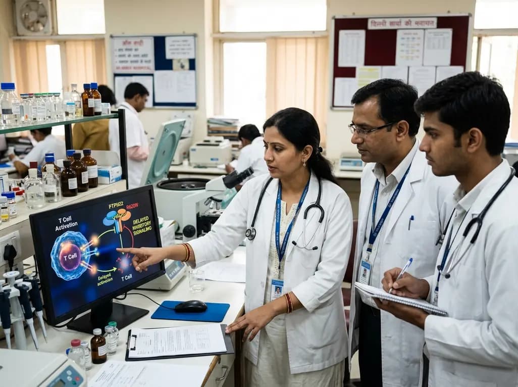
New immunological research reveals how PTPN22 forms a delayed inhibitory signaling complex to restrain T cell activation, explaining how the autoimmune-linked R620W variant disrupts feedback control and promotes persistent inflammation in conditions like rheumatoid arthritis and type 1 diabetes.
Today

A nationwide study covering 3.7 million Swedish births reveals that prenatal exposure to TORCH pathogens significantly elevates long-term risks of intellectual disability and autism. Healthcare providers must recognize clinical indicators, promote prenatal prevention, and track early pediatric neurodevelopment.
Today
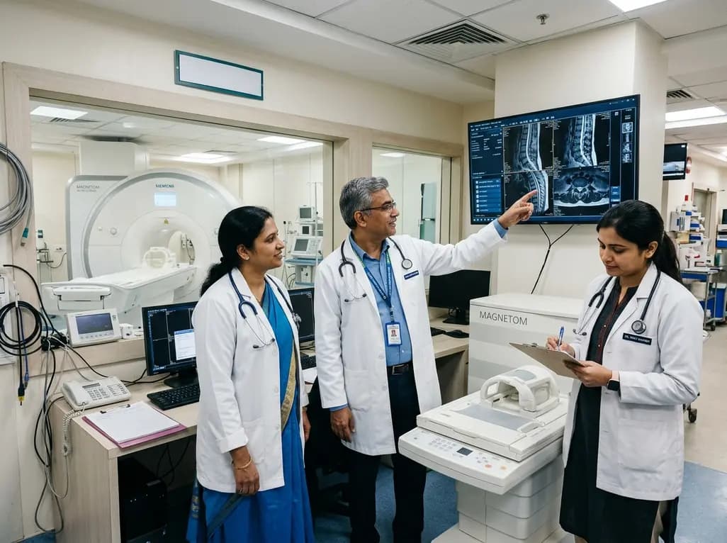
Under India's PLI initiative, domestic production of 57 critical medical devices has commenced, including MRI and CT scanners, cath labs, and heart valves. Global partnerships and local investments are expanding high-end technology, curbing import reliance, and strengthening clinical accessibility nationwide.
Today

A longitudinal study reveals that patients undergoing staged bilateral hip arthroscopy experience progressive contralateral labral deterioration during surgical intervals. However, structural degradation does not compromise 2-year postoperative clinical outcomes or patient-reported satisfaction scores.
Today

SWEDEPAD-1 trial insights show paclitaxel-coated devices do not improve long-term limb salvage in patients with chronic limb-threatening ischemia and tissue loss. While one-year reinterventions decreased, the devices were linked to a higher risk of major amputation at three months.
Yesterday
