.webp&w=384&q=75)

Loading, please wait...
Loading, please wait...
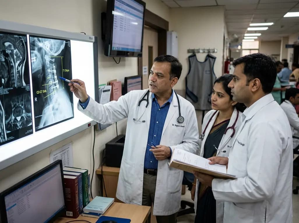
Managing degenerative cervical myelopathy alongside sagittal malalignment presents complex biomechanical hurdles for spine surgeons. When posterior decompressive procedures are performed, cervical kyphosis progression remains a formidable postoperative complication that threatens spinal stability. Posterior surgical approaches effectively relieve neural compression across multiple stenotic levels. However, altering the posterior tension band and facet complexes can accelerate sagittal collapse in predisposed spines. Clinicians must identify preoperative risk factors to distinguish patients who will maintain structural stability from those at high risk of progressive deformity. Recent clinical investigations provide vital quantitative parameters that help surgeons anticipate postoperative sagittal collapse and personalize operative approaches accordingly.
Degenerative cervical myelopathy and canal stenosis frequently coexist with loss of normal cervical lordosis. Posterior decompressive techniques, including laminoplasty and laminectomy with instrumented fusion, provide substantial canal expansion. However, treating patients who already possess baseline kyphotic alignment requires delicate surgical judgment. In these patients, posterior decompression can compromise the posterior musculoligamentous complex. Consequently, this biomechanical alteration elevates the mechanical load placed on the anterior column.
When anterior vertebral support fails under altered load distribution, progressive angular deformity often follows. Although posterior decompression relieves direct neurological compression, uncorrected or worsening sagittal malalignment may jeopardize long-term functional recovery. Furthermore, progressive deformity increases mechanical strain across adjacent segments, elevating the risk of revision surgeries. Surgeons historically debated whether mild or moderate baseline kyphosis absolute contraindicates posterior decompression. Clinical evidence now indicates that baseline alignment alone does not dictate failure. Instead, dynamic flexibility and global sagittal harmony play decisive roles in maintaining postoperative alignment. Therefore, surgical teams must evaluate composite biomechanical parameters before finalizing their operative strategy.
Recent clinical studies have clarified specific radiographic variables that directly influence cervical kyphosis progression after posterior decompressive surgery. Interestingly, preoperative baseline C2-C7 Cobb angles often show similar baseline values between patients who maintain alignment and those who deteriorate. For example, baseline alignment may hover near mild kyphosis in both cohorts. Yet, postoperative trajectories diverge drastically over time.
Patients who experience progressive deformity exhibit a sharp post-surgical drop in lordotic alignment, accompanied by worsening sagittal vertical axis parameters. Conversely, patients who maintain stability often achieve spontaneous lordotic remodeling or preserve neutral alignment. Additionally, radiographic analysis highlights significant changes in T1 slope and cervicothoracic junction balance among progressing cohorts. When the C2-C7 sagittal vertical axis increases significantly, the head shifts anteriorly relative to the thoracic cage. This anterior translation exponentially multiplies the flexion bending moment exerted on the cervical spine. Therefore, surgeons must look beyond static baseline Cobb angles. Evaluating dynamic spinopelvic and cervicothoracic parameters on standing dynamic radiographs provides essential predictive value before performing posterior decompression.
Among all preoperative radiographic parameters, dynamic cervical range of motion serves as an indispensable indicator of structural resilience. Specifically, preoperative extension range of motion demonstrates strong predictive capability for postoperative stability. Patients with preserved cervical extension retain dynamic muscular and ligamentous capacity to counteract anterior gravitational forces. In contrast, patients with restricted extension mobility lack the biomechanical flexibility needed to support the reconstructed cervical column.
When surgeons disrupt the posterior tension band during decompression, the spine must rely on residual dynamic stabilizers to preserve sagittal posture. If preoperative extension capacity is severely diminished, the cervical spine cannot dynamically compensate for the surgical destabilization. Consequently, the cervical column steadily drifts into progressive kyphosis. Therefore, routine preoperative workups must incorporate standing flexion-extension radiographs to quantify dynamic extension reserves accurately. Identifying patients with limited dynamic extension helps surgeons recognize stiff, high-risk deformities that demand rigid anterior reconstruction or multi-column stabilization rather than isolated posterior decompression.
Cervical sagittal alignment functions as an integrated component of global spinal balance. In particular, T1 slope serves as a fundamental foundation for subaxial cervical lordosis. A lower T1 slope demands less cervical lordosis to maintain horizontal gaze, whereas a steep T1 slope requires substantial lordosis. However, when a low T1 slope couples with an increased C2-C7 sagittal vertical axis, biomechanical instability escalates rapidly.
Recent findings reveal that patients experiencing postoperative kyphotic collapse often present with significantly lower T1 slopes alongside increased anterior sagittal vertical axis translation. Under these conditions, the subaxial spine struggles to maintain equilibrium over the thoracic inlet. As a result, the posterior cervical musculature experiences chronic fatigue and mechanical failure. This imbalance accelerates anterior tilt and destabilizes decompressed segments. Moreover, severe mismatch between T1 slope and cervical lordosis correlates with persistent neck pain and functional disability. Thus, comprehensive preoperative planning must incorporate full cervicothoracic junction evaluation to ensure that posterior constructs provide adequate mechanical alignment relative to the patient's individual thoracic anatomy.
Selecting the optimal surgical approach requires balancing neural decompression goals against deformity progression risks. While posterior approaches offer expansive multi-level decompression with lower dysphagia rates, their application in kyphotic spines requires stringent patient selection. Surgeons must avoid isolated posterior decompression in patients exhibiting poor dynamic extension, marked sagittal translation, or low baseline T1 slope.
Instead, high-risk candidates benefit substantially from anterior cervical discectomy and fusion or combined anterior-posterior circumferential stabilization. Anterior reconstruction directly restores disc space height, removes anterior osteophytes, and establishes rigid structural lordosis. Alternatively, if a posterior approach is selected, incorporating multi-level instrumented fusion with rigid lateral mass or pedicle screws can mitigate the risk of sagittal collapse. Furthermore, preserving the C2 muscular attachments and minimizing facet capsule violation during posterior exposure protects key biomechanical tension bands. By matching surgical technique to patient-specific biomechanical parameters, surgical teams can achieve complete neural decompression while effectively preventing severe postoperative structural failure.
Preserving cervical sagittal alignment directly influences long-term patient-reported outcomes and neurological recovery. Clinical studies demonstrate that both alignment-maintained and kyphosis-progressed cohorts achieve initial neurological improvements, as measured by modified Japanese Orthopedic Association scores. However, patients who suffer progressive kyphosis often endure persistent mechanical axial neck pain and functional limitations.
Over time, progressive anterior curvature stretches the spinal cord across ventral vertebral bodies. This continuous mechanical tethering induces cord ischemia and can trigger delayed neurological deterioration. Additionally, uncorrected sagittal malalignment impairs horizontal gaze, alters forward visual fields, and diminishes overall quality of life scores. Compensatory hyperlordosis in the subaxial or cranial segments may also cause rapid adjacent segment degeneration. Therefore, achieving robust neural decompression without securing durable sagittal alignment represents an incomplete surgical victory. Spine specialists must prioritize both neurological decompression and sagittal preservation to optimize long-term clinical success and patient satisfaction.
Posterior decompression can disrupt the posterior ligamentous complex, paraspinal musculature, and facet joints that maintain spinal balance. When patients lack adequate preoperative extension mobility or possess unfavorable cervicothoracic alignment, the biomechanical load shifts anteriorly. This increased stress leads to structural collapse and progressive kyphotic deformity over time.
Preoperative extension range of motion reflects the dynamic flexibility and muscular strength of the cervical spine. Patients with preserved extension mobility can dynamically compensate for posterior structural alterations. Conversely, limited extension mobility indicates stiff, compromised stabilizing tissues, significantly increasing the risk of postoperative sagittal collapse following posterior decompression.
Surgeons should avoid isolated posterior decompression when patients exhibit rigid kyphosis, severely restricted dynamic extension range of motion, high sagittal vertical axis translation, or significant anterior cord compression. In such cases, anterior reconstruction or circumferential instrumented fusion provides superior sagittal realignment, structural stability, and durable neurological protection.
Disclaimer: This content is for informational and educational purposes only and is not intended as medical advice. It is not a substitute for professional medical judgment, diagnosis, or treatment. Always seek the advice of a qualified healthcare provider with any questions you may have regarding a medical condition. Consideration of the individual patient's clinical circumstances is essential in clinical decision-making. Refer to the latest local and national guidelines for clinical practice.
References

Read summarized clinical updates, watch expert medical content, and earn CME certifications right from your smartphone.
Open your smartphone camera to scan and install.





A recent retrospective study evaluates preoperative and intraoperative determinants of cervical kyphosis progression following posterior decompression in patients with degenerative kyphosis and stenosis, highlighting the predictive value of extension range of motion, T1 slope, and C2-C7 sagittal vertical axis.
4 weeks back

A systematic review of 41 preclinical studies reveals that baicalin dampens systemic inflammation, inhibits NF-κB and NLRP3 pathways, and improves organ preservation in sepsis models. However, substantial study heterogeneity and a complete lack of human clinical trials highlight the need for further translational research.
Today
Discover the crucial distinctions between mesonephric and mesonephric-like proliferations of the female genital tract. Learn the key histomorphologic, immunohistochemical, and molecular differences essential for accurate diagnosis and clinical management.
Today

A large UK Biobank study reveals that plasma proteomic signatures capture preclinical organ damage and improve multiorgan risk prediction across early CKM syndrome stages 0 to 2 beyond traditional PREVENT clinical models.
Today
A narrative review investigates whether the articularis genus muscle functions as an independent anatomical entity or blends with the vastus intermedius. We evaluate its morphology, role in retracting the suprapatellar bursa, and direct clinical significance in anterior knee pain and arthroplasty.
Today

A Bayesian multilevel meta-analysis reveals that aerobic training combined with moderate carbohydrate restriction modestly lowers HbA1c in type 2 diabetes. However, sparse data and very low certainty leave incremental benefits over exercise or diet alone unproven, highlighting the need for individualized care.
Today
