.webp&w=384&q=75)

Loading, please wait...
Loading, please wait...
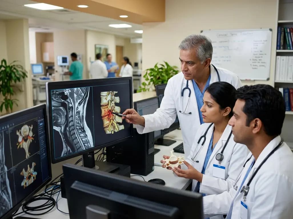
Cervical foraminal stenosis morphology serves as a critical focal point for radiologists and orthopedic surgeons managing patients with persistent radicular pain. While advanced imaging techniques provide exceptionally detailed anatomical views, the correlation between these images and clinical symptoms remains a subject of intense debate in the medical community. For many clinicians, a significant challenge involves determining whether the severity of narrowing seen on a magnetic resonance imaging (MRI) scan accurately reflects the level of disability a patient experiences. Therefore, researchers have sought to identify specific morphological markers that might predict functional recovery after surgical intervention. Despite the sophisticated nature of modern diagnostics, the relationship between the physical structure of the neuroforamen and the subjective experience of pain is often surprisingly complex. Furthermore, the variability in how different observers interpret these scans can lead to inconsistencies in treatment planning. Consequently, understanding the nuances of neuroforaminal canal diameter and nerve root compression is essential for improving patient care. Practitioners in India, where the burden of degenerative spine disease is high, frequently encounter these diagnostic dilemmas. By examining the structural features of the foramen, such as the distance of maximum compression from the ligamentum flavum, doctors hope to refine their surgical indications. Nevertheless, the disconnect between radiological findings and clinical scores like the Neck Disability Index (NDI) suggests that morphology is only one part of the diagnostic puzzle.
To standardize the evaluation of neuroforaminal narrowing, several grading systems have been developed, including the Kim, modified Kim, and Siller methods. Specifically, the Kim classification utilizes axial T2-weighted MRI to categorize the degree of stenosis based on the obliteration of the fat surrounding the nerve root. Additionally, the modified Kim method provides a more detailed assessment of the relationship between the nerve and the surrounding bony and ligamentous structures. In contrast, the Siller method focuses on the narrowest part of the neuroforaminal canal. Each of these systems aims to provide a reproducible score that reflects the biological reality of nerve impingement. Moreover, these radiological measurements include the uncompressed and maximal compressed nerve root diameters, which offer a quantitative view of the pathology. However, the inter-rater reliability of these measurements can vary significantly, even among experienced neuroradiologists. Because of this, some experts argue that a single radiological grade may not capture the dynamic nature of cervical radiculopathy. Furthermore, the length of the neuroforaminal canal where the diameter is restricted also plays a role in the overall morphological picture. These metrics provide a framework for surgeons to communicate the severity of disease. Yet, as the study indicates, these grades often fail to correlate with the functional disability reported by the patient pre-operatively. This discrepancy highlights the need for a more holistic approach to spine assessment that integrates imaging with clinical history.
When conservative management fails to provide relief for cervical radiculopathy, surgical decompression often becomes necessary. The two primary techniques utilized are Anterior Cervical Discectomy (ACD) and Posterior Cervical Foraminotomy (PCF). Each procedure offers distinct advantages depending on the location and nature of the compression. For instance, ACD is typically preferred when the pathology is located anteriorly, such as a herniated disc or an osteophyte. Conversely, PCF allows for a direct approach to posterior compression without the need for fusion. Notably, the choice of surgical technique is often guided by the morphological features identified on pre-operative imaging. Surgeons carefully analyze the cervical foraminal stenosis morphology to determine which path offers the safest and most effective route to decompression. Additionally, the goal of both procedures is to restore the functional capacity of the patient and reduce pain scores significantly. Recent data shows that both ACD and PCF lead to substantial improvements in the Neck Disability Index (NDI) following surgery. Specifically, the change in NDI scores represents the primary measure of surgical success. Interestingly, the study found that neither pre-operative NDI nor the magnitude of functional improvement was statistically different between the two surgical cohorts. This suggests that for many patients, the choice of approach may be less critical than the achievement of adequate decompression. Thus, the surgeon's expertise and the specific anatomical constraints of the patient often dictate the procedural choice more than radiological grades alone.
A primary objective of many clinical studies is to determine if specific radiological markers can predict how well a patient will recover after surgery. In the case of cervical radiculopathy, researchers investigated whether features like the length of the canal or the degree of nerve compression correlated with post-operative NDI changes. Surprisingly, the results indicated that no specific morphological factor was significantly associated with pre-operative disability. Furthermore, none of the radiological grades, including Kim or Siller, served as reliable predictors of the functional outcome six weeks after the operation. This lack of correlation suggests that the degree of anatomical narrowing is not the sole driver of the patient's symptoms. Instead, factors such as nerve sensitivity, the duration of symptoms, and individual pain thresholds may play more prominent roles. Moreover, the pre-operative NDI score itself was found to be the strongest predictor of the post-operative NDI score. Patients who started with higher levels of disability often remained more disabled compared to those with lower initial scores, even after successful surgery. This finding underscores the importance of managing patient expectations regarding recovery. While surgery effectively reduces pain, it may not completely eliminate the functional limitations associated with chronic nerve compression. Consequently, clinicians should emphasize that radiological severity does not always dictate the path of recovery. Therefore, the focus should remain on the clinical presentation and the patient's subjective experience rather than relying exclusively on MRI findings to gauge potential success.
An interesting finding in the study of cervical radiculopathy involves the significant difference in pre-operative disability scores between male and female patients. Specifically, female patients reported a mean NDI that was notably higher than their male counterparts. This trend persisted despite no significant differences in the radiological severity of their conditions. Such a disparity suggests that gender-related factors may influence how patients perceive and report their functional limitations. Furthermore, psychological and sociocultural variables often interact with physical pathology to shape the experience of chronic pain. For example, some studies suggest that women may experience higher levels of central sensitization or have different coping mechanisms for spinal pain. Additionally, clinicians must account for these differences when evaluating a patient's readiness for surgery and their likely post-operative trajectory. Although the radiological cervical foraminal stenosis morphology was similar across genders, the clinical impact varied. This observation reinforces the idea that imaging provides only a narrow view of the patient's overall health status. Moreover, it highlights the necessity of using validated tools like the NDI to capture the patient's perspective. By recognizing these demographic nuances, doctors in India can provide more personalized care and better counsel patients on what to expect during the healing process. Ultimately, the successful management of cervical radiculopathy requires a deep understanding of both the anatomical structures and the unique characteristics of the individual patient.
For practitioners in India, the findings of this study offer valuable insights into the daily management of degenerative spine disease. First and foremost, it is clear that while MRI is indispensable for localization, its ability to predict functional outcomes is limited. Therefore, surgeons should avoid making definitive prognostic statements based solely on the severity of foraminal narrowing. Instead, a comprehensive clinical examination remains the cornerstone of diagnosis. Furthermore, the research validates both ACD and PCF as effective treatments for radiculopathy, regardless of the anterior or posterior nature of the compression. Consequently, the choice between these techniques should be based on the surgeon's comfort and the specific surgical goals for each patient. Additionally, the significant improvement in NDI scores post-operatively provides strong evidence for the efficacy of surgical intervention in symptomatic cases. However, clinicians should also be mindful of the gender differences in reported disability. By acknowledging that female patients may present with higher NDI scores, doctors can better tailor their pre-operative counseling. Moreover, the lack of correlation between radiological grades and outcomes suggests that grading systems like Kim or Siller should be used primarily for descriptive purposes rather than for decision-making. Specifically, the morphological features of the foramen do not appear to be the primary drivers of surgical success. In conclusion, while imaging remains a vital tool, the patient's clinical symptoms and subjective disability must always take precedence in the management of cervical foraminal stenosis.
While the Kim, modified Kim, and Siller methods are all utilized to assess stenosis, research suggests that none are superior at predicting functional outcomes. These systems are useful for anatomical description and inter-clinician communication but do not consistently correlate with a patient's pre-operative disability or their post-operative recovery speed.
Current research indicates that the radiological severity of stenosis does not reliably predict the change in a patient's Neck Disability Index after surgery. Surgical success is generally achieved regardless of the initial radiological grade, provided that adequate neural decompression is performed during the procedure, either through an anterior or posterior approach.
Studies show that female patients often report higher pre-operative Neck Disability Index (NDI) scores compared to males, despite having similar radiological findings. This suggests that gender-specific factors, including pain perception, central sensitization, and sociocultural influences, may play a significant role in how disability is experienced and reported by patients.
Disclaimer: This content is for informational and educational purposes only. It is not intended as medical advice or to replace the professional judgment of a healthcare provider. Always seek the advice of a physician or other qualified health provider with any questions regarding a medical condition. Refer to the latest local and national guidelines for clinical practice.
References
Meacock J et al. Radiological cervical foraminal stenosis severity and morphology as a predictor of pre-operative function and functional surgical outcome. Br J Neurosurg. 2025 Oct. doi: 10.1080/02688697.2024.2376647. PMID: 39001642.
Park MS, et al. Clinical significance of the Kim's classification of cervical foraminal stenosis. Spine (Phila Pa 1976). 2017;42(11):E631-E637.
Lee JE, et al. Interreader reliability and clinical validity of a magnetic resonance imaging grading system for cervical foraminal stenosis. J Comput Assist Tomogr. 2017;41(6):926-930.
"
Read summarized clinical updates, watch expert medical content, and earn CME certifications right from your smartphone.
Open your smartphone camera to scan and install.





This article examines the relationship between cervical foraminal stenosis morphology and surgical outcomes. While MRI grading systems like Kim and Siller provide detailed anatomical data, the study reveals a surprising lack of correlation between radiological severity and pre-operative disability or recovery.
3 months ago

A retrospective cohort study of 7,716 ICU patients from the MIMIC-IV database demonstrates that Gradient Boosting Decision Trees significantly outperform traditional SOFA scores in predicting in-hospital mortality among patients with sepsis and diabetes, with SHAP analysis providing clinical interpretability.
Today

A prenatal case highlights severe glutaric acidemia type II presenting as bilateral hyperechoic cystic kidneys at 18 weeks. Whole-exome sequencing revealed a novel homozygous ETFDH frameshift variant, demonstrating the power of rapid genetic testing in fetal renal anomalies.
Today
This article reviews the forensic and clinical complexities of acute myocarditis arising on left ventricular hypertrophy in children, highlighting early diagnostic red flags and multidisciplinary postmortem autopsy protocols.
Today

This comparative study evaluates posterior labral tears with and without glenohumeral instability, highlighting distinct demographic patterns, MRI underestimation in stable shoulders, and successful arthroscopic repair outcomes.
Yesterday

A clinical study demonstrates that early intervention with tocilizumab, either as monotherapy or bridged with corticosteroids, achieves significant proptosis reduction and resolves active inflammation in treatment-naive thyroid eye disease.
Yesterday
