.webp&w=384&q=75)

Loading, please wait...
Loading, please wait...

Cerebrospinal fluid overdrainage represents a hazardous yet treatable complication of neurosurgical diversion procedures. Clinicians frequently encounter overdrainage in patients with ventriculoperitoneal or lumboperitoneal shunts. However, cervical epidural venous engorgement remains an under-recognized consequence that can cause spinal cord compression. When excessive cerebrospinal fluid drainage occurs, the loss of fluid volume causes secondary spinal hypovolemia. Consequently, thin-walled epidural veins must expand rapidly to fill the extradural space. This mechanical vascular expansion directly impinges upon the adjacent cervical cord and neural roots.
Patients frequently develop progressive cervical myelopathy rather than classic signs of intracranial hypotension. Indeed, standard intracranial symptoms such as low-pressure orthostatic headaches are noticeably absent in many chronic cases. Therefore, attending clinicians frequently attribute the resulting neurological deterioration to degenerative cervical spondylosis. Misinterpreting these secondary compressive vascular masses can lead to unnecessary, high-risk surgical laminectomies. Conversely, early recognition of cervical epidural venous engorgement allows teams to prioritize non-invasive shunt recalibration. Timely intervention successfully restores balanced fluid dynamics and prevents permanent neurological disability.
The fundamental mechanism behind this vascular syndrome relies upon the Monro-Kellie doctrine applied across the craniospinal axis. Cerebrospinal fluid diversion creates negative hydrostatic pressure gradients, particularly when patients maintain upright postures. Because the rigid bony spinal canal maintains a constant total volume, any reduction in fluid volume forces immediate vascular compensation. Consequently, low-resistance epidural venous plexuses experience compensatory engorgement to preserve physical equilibrium. Furthermore, the avalvular nature of Batson's vertebral venous plexus facilitates rapid vascular dilation under negative intraspinal forces.
Moreover, persistent venous hypertension within the epidural canal can trigger secondary microvascular breakdown. In severe cases, fragile engorged venous channels can rupture spontaneously, producing an acute epidural hematoma. Such hematomas rapidly exacerbate cord compression at the cervicomedullary junction and upper cervical levels. In addition, chronic venous stasis can impair local venous drainage from the spinal cord parenchyma. This localized congestion induces parenchymal ischemia, spinal cord edema, and persistent myelomalacia. Thus, mechanical compression and venous congestion combine to drive progressive functional deficits.
The clinical manifestations of this condition present distinctive diagnostic pitfalls for neurosurgeons and neurologists alike. Systematic evidence demonstrates that symptoms emerge after a remarkably prolonged latency period. Specifically, the median duration from initial shunt implantation to symptom manifestation reaches nearly fifteen years. Nevertheless, symptoms can also emerge acutely within days following shunt insertion or valve adjustment. Therefore, clinicians must maintain high clinical suspicion regardless of whether the shunt operation was recent or remote.
Furthermore, the predominant clinical presentation involves progressive cervical myelopathy characterized by spastic quadriparesis, gait ataxia, and sensory loss. Patients also report upper extremity clumsiness, hyperreflexia, and occasional radicular pain. Remarkably, most patients present without typical orthostatic headaches, nausea, or cranial nerve palsies. Because typical intracranial hypotension clues are absent, physicians often overlook the ventricular shunt as the primary etiologic culprit. Consequently, patients frequently experience substantial diagnostic delays before obtaining proper neuroimaging. Rapid clinical identification remains vital because early treatment completely reverses neurological deficits.
Magnetic resonance imaging represents the indispensable gold standard for confirming shunt-induced epidural venous dilation. Spine magnetic resonance imaging characteristically reveals extensive, circumferential extradural soft tissue masses compressing the upper cervical spinal cord. On T2-weighted sequences, these engorged venous structures display prominent flow voids or intermediate signal intensity. In addition, T1-weighted contrast-enhanced images show intense, homogeneous enhancement of the dilated epidural plexus. Clinicians also observe associated cord parenchymal hyperintensity on T2-weighted images, which reflects secondary compressive edema.
However, radiologists must carefully distinguish these vascular structures from epidural abscesses, metastases, and primary epidural hematomas. Brain magnetic resonance imaging provides vital diagnostic adjuncts by searching for concurrent intracranial hypotension signs. These intracranial features include diffuse pachymeningeal enhancement, sagging of the midbrain, and engorged dural sinuses. Nevertheless, spine specialists must remember that intracranial imaging can occasionally appear completely normal. Therefore, demonstrating prominent, non-thrombosed epidural venous plexuses in a shunted patient strongly confirms over-shunting myelopathy.
Management strategies must center primarily on reversing cerebrospinal fluid overdrainage rather than performing invasive direct spinal decompression. In most reported clinical cases, direct laminectomy fails to resolve the underlying pressure deficit and carries substantial hemorrhage risks. Instead, clinicians should promptly increase the opening pressure in programmable shunt valves. Non-invasive magnetic valve reprogramming immediately restores physiological intraspinal pressures and normalizes venous caliber. Consequently, engorged venous networks collapse rapidly once intraspinal hypotension resolves.
Furthermore, patients with fixed-pressure shunt systems generally require formal surgical revision. Surgeons should convert fixed-pressure systems to adjustable programmable valves equipped with gravitational or antisiphon devices. Antisiphon devices effectively eliminate the siphon effect that triggers massive fluid overdrainage whenever the patient stands upright. In rare emergency cases involving acute, large epidural hematomas, localized surgical hematoma evacuation may become necessary alongside shunt modification. Overall, systematic reviews confirm that restoring proper fluid dynamics achieves dramatic functional recovery and radiologic resolution.
This systematic analysis delivers critical practical insights for contemporary neurology, neurosurgery, and spine practice. First, clinicians must always obtain a comprehensive surgical history in patients presenting with unprovoked cervical myelopathy. Even if a ventriculoperitoneal shunt was inserted decades earlier, late-onset hydrodynamic overdrainage remains entirely possible. In addition, clinicians must avoid premature spine decompression when magnetic resonance imaging indicates prominent epidural soft-tissue enhancement. Performing decompressive laminectomy in the presence of severe venous engorgement can precipitate torrential, uncontrollable epidural hemorrhage.
Moreover, post-procedure monitoring protocols for hydrocephalus patients should incorporate systematic vigilance for subtle spinal symptoms. Clinicians typically monitor ventricular size through cranial computed tomography; however, normal ventricular dimensions do not rule out spinal hypovolemia. Regular clinical assessments must evaluate gait stability, finger dexterity, and long-tract signs during outpatient shunt surveillance. When myelopathy symptoms appear, multidisciplinary collaboration between spine surgeons and hydrocephalus specialists ensures prompt shunt recalibration. Ultimately, timely identification of this reversible condition prevents permanent quadriplegia.
Shunt overdrainage removes excessive cerebrospinal fluid, creating negative intraspinal hydrostatic pressure. According to the Monro-Kellie doctrine, the rigid spinal canal requires constant volumetric balance. Consequently, avalvular epidural venous plexuses rapidly dilate to replace lost fluid volume. This compensatory vascular engorgement compresses the adjacent cervical spinal cord and nerve roots.
In chronic shunt overdrainage, intracranial compliance gradually adapts over many years, dampening the pain-sensitive dural stretch responsible for orthostatic cephalalgia. Instead, the persistent hydrostatic gradient shifts fluid dynamics downward into the spinal axis. Consequently, venous engorgement predominates in the cervical canal, producing compressive myelopathy without typical postural cranial symptoms.
Yes, non-invasive valve reprogramming effectively reverses this condition in most patients. Increasing valve resistance corrects intraspinal hypotension and restores physiological cerebrospinal fluid pressure. Once normal hydrostatic balance returns, the engorged epidural venous plexus promptly collapses. This decompression relieves pressure on the cervical cord, allowing significant neurological recovery and resolving symptoms.
Disclaimer: This content is for informational and educational purposes only... Refer to the latest local and national guidelines for clinical practice.
References

Read summarized clinical updates, watch expert medical content, and earn CME certifications right from your smartphone.
Open your smartphone camera to scan and install.





Cervical epidural venous engorgement is a rare, reversible complication of CSF overdrainage causing compressive myelopathy. Recognizing this entity prevents unnecessary spine surgery and enables prompt reversal through non-invasive shunt valve reprogramming.
Today

Survival analyses often report hazard ratios, but mathematical non-collapsibility creates crucial differences between conditional and marginal estimates. Clinicians must account for unmeasured confounding and model selection when interpreting trial endpoints and real-world cardiovascular outcomes.
Today

A cross-sectional study of 700 college students highlights significant contraceptive knowledge disparities between sexually active and inactive young adults. The findings demonstrate a critical need for proactive, comprehensive sexual health counseling before sexual debut.
Yesterday
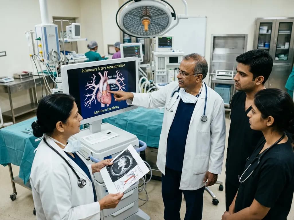
Autologous tissue-engineered vascular graft patches created via in-body tissue architecture show excellent mid-term durability and safety for pulmonary artery reconstruction in congenital heart surgery, particularly for peripheral branch augmentation.
Today

A novel cluster analysis of real-world datasets reveals four distinct CPPD disease phenotypes, validating existing EULAR categories while uncovering previously unrecognized monoarticular and axial presentations to refine patient management.
Today

Recent pharmacokinetic research demonstrates that trace dermal exposure to microgram quantities of trenbolone and metenolone produces detectable urinary metabolites for days to over a week, providing critical objective data for evaluating unintentional contamination claims in sports drug testing.
Today
