.webp&w=384&q=75)

Loading, please wait...
Loading, please wait...

Degenerative cervical spine conditions frequently present diagnostic and therapeutic challenges for spine surgeons and neurologists. Among these disorders, cervical central spinal stenosis remains a leading cause of cervical spondylotic myelopathy and progressive functional impairment. Conventional magnetic resonance imaging predominantly emphasizes canal narrowing and direct spinal cord compression. However, researchers increasingly recognize the importance of the surrounding muscular envelope. The cervical multifidus and rotatores muscles provide essential dynamic stability and segmental control to the subaxial cervical spine. However, how progressive spinal cord compression influences paraspinal muscle health has historically remained poorly understood. A landmark investigation now provides critical insights into the structural degeneration of these stabilizing muscles. Specifically, the study reveals significant associations between canal stenosis severity and deep paraspinal muscle fat infiltration.
Degenerative changes in the cervical spine involve complex interplay among intervertebral discs, facet joints, and supporting soft tissues. As canal diameter narrows in cervical central spinal stenosis, compression of neural elements disrupts physiological signaling to deep stabilizing muscles. Historically, spine clinicians attributed paraspinal muscle deterioration primarily to localized mechanical disuse or general aging. However, recent evidence suggests that neural compromise plays a far more direct, pathogenic role in structural muscle degeneration.
The subaxial cervical paraspinal musculature, particularly the deep multifidus and rotatores groups, ensures segmental cervical lordosis. Additionally, these muscles resist shear forces during cervical motion. When severe central canal compromise occurs, denervation pathways may trigger muscular fatty infiltration. This pathologic replacement of active muscle fibers by non-contractile adipose tissue diminishes muscular functional capacity. Consequently, patients experience diminished mechanical stabilization across subaxial motion segments. Furthermore, severe fat infiltration can alter postoperative biomechanics, possibly exacerbating neck disability and accelerating adjacent segment degeneration. Recognizing the connection between central cord compression and paraspinal fatty degeneration helps clinicians evaluate patient outcomes. Furthermore, this insight explains why functional recovery varies widely after surgical decompression.
To clarify the relationship between central canal compromise and paraspinal composition, researchers conducted a rigorous retrospective Level III study. The investigation evaluated one hundred fifty-six consecutive patients who underwent anterior cervical discectomy and fusion for cervical pathology. All participants completed standardized preoperative cervical magnetic resonance imaging, which allowed detailed morphometric analysis of the cervical spine.
The investigators segmented the deep cervical multifidus and rotatores musculature bilaterally from the C3 level down to C7. Using custom-written MATLAB software, the team quantitatively measured the percentage of fat infiltration within these deep muscle compartments. Additionally, observers graded the severity of cervical central stenosis at each subaxial level using an established radiological classification scheme. The researchers designated Grade 3 stenosis as the primary reference comparator. This severe grade represents cerebrospinal fluid space loss and spinal cord deformation exceeding twenty-five percent. Multivariable linear regression models systematically adjusted for key confounding patient variables, including age, sex, and body mass index. This rigorous quantitative methodology minimized measurement bias. Consequently, the authors isolated the independent predictive impact of spinal cord compression on paraspinal fatty changes across individual subaxial levels.
The study demonstrated highly significant, level-specific correlations between the magnitude of central cord compression and multifidus fat infiltration. Most importantly, spinal cord compression at a designated level was consistently associated with increased fat accumulation in paraspinal muscles below that level. This caudal pattern provides critical mechanistic clues regarding the underlying pathophysiology.
Specifically, multivariable linear regression models showed that cord compression at C3/4 and C4/5 significantly correlated with greater multifidus fat infiltration. These fatty changes extended from C3 to C6 and C5 to C7, respectively. Furthermore, cord compression located at C5/6 or C6/7 was significantly linked to elevated fatty infiltration at the C7 level. The cervical multifidus and rotatores receive segmental motor innervation from the medial branches of the cervical dorsal rami. Therefore, this caudal progression strongly indicates neurogenic muscle atrophy rather than generalized disuse. Anterior horn cell compression or disrupted descending spinal motor pathways impair normal motor unit trophic support. Therefore, denervated muscle fibers progressively degenerate and undergo fibrofatty substitution below the site of spinal cord compression. These distinctive level-specific correlations highlight an active neurogenic mechanism driven by mechanical cord compromise.
Anterior cervical discectomy and fusion remains the gold standard surgical intervention for symptomatic central cervical stenosis and myelopathy. The anterior surgical approach traverses anterior soft tissue planes and spares posterior cervical musculature. Nevertheless, preoperative posterior muscle quality remains crucial for clinical success. Paraspinal muscles maintain cervical sagittal balance and absorb physiological loads following anterior stabilization.
When patients present with substantial preoperative fat infiltration, the loss of active posterior muscular support compromises post-surgical spinal biomechanics. Consequently, these individuals face a heightened risk of persistent axial neck pain and loss of cervical lordosis. In addition, accelerated mechanical stress can compromise adjacent motion segments. Furthermore, neurogenic muscle atrophy may not spontaneously reverse following successful anterior decompression. Therefore, preoperative magnetic resonance imaging assessments should extend beyond canal dimensions to evaluate posterior musculature health. Identifying profound multifidus fatty infiltration enables surgical teams to anticipate postoperative challenges and refine surgical planning. For instance, clinicians may tailor fixation constructs based on the structural integrity of the paraspinal muscular sleeve. They can also adjust postoperative immobilization protocols accordingly.
These findings establish that paraspinal muscle parameters offer valuable objective biomarkers of chronic neurologic impairment in cervical stenosis. For orthopedic surgeons, neurosurgeons, and physiatrists, integrating muscular quality assessments into routine preoperative evaluations provides comprehensive prognostic perspectives. In routine clinical practice, clinicians frequently encounter patients whose persistent postoperative disability seems disproportionate to radiological canal decompression. Underlying neurogenic muscle degeneration provides an anatomical explanation for this common clinical scenario.
Moreover, these discoveries emphasize the vital necessity of early, proactive rehabilitation programs. Because denervated and fat-infiltrated muscles suffer from diminished endurance and altered recruitment patterns, generalized neck exercises may prove insufficient. Instead, rehabilitation specialists must design targeted neuromuscular re-education protocols that selectively recondition the deep multifidus and rotatores. Initiating supervised muscular conditioning both preoperatively and throughout postoperative recovery may optimize cervical dynamic stabilization. Additionally, timely surgical decompression before advanced cord compression occurs may prevent irreversible caudal muscle fatty degeneration. Ultimately, combining precise surgical decompression with focused neuromuscular rehabilitation optimizes functional recovery and preserves long-term spinal health.
Cervical central spinal stenosis causes mechanical compression of the spinal cord and anterior horn cells, disrupting descending and segmental motor pathways. Because paraspinal muscles depend on continuous neural trophic signaling, chronic denervation triggers progressive muscle fiber atrophy and subsequent fibrofatty tissue replacement below the compressed segment.
Evaluating multifidus muscle quality before anterior cervical discectomy and fusion provides vital prognostic information regarding spinal dynamic stability. Patients with extensive fatty infiltration face an increased likelihood of persistent axial neck discomfort, loss of cervical lordosis, and heightened biomechanical stress on adjacent spinal motion segments postoperatively.
While advanced fatty substitution may not fully reverse, targeted neuromuscular rehabilitation can significantly improve functional capacity. Specific exercises activate residual muscle fibers, enhance dynamic stability, and compensate for segmental weakness. Initiating tailored motor control training early in recovery optimizes biomechanical support and functional outcomes following surgical decompression.
Disclaimer: This content is for informational and educational purposes only, and does not substitute professional medical advice, diagnosis, or treatment. Refer to the latest local and national guidelines for clinical practice.
References

Read summarized clinical updates, watch expert medical content, and earn CME certifications right from your smartphone.
Open your smartphone camera to scan and install.





A retrospective Level III study reveals that severe cervical central spinal stenosis correlates with level-specific paraspinal multifidus fat infiltration below the compression site, indicating neurogenic atrophy and highlighting key prognostic factors for ACDF surgery.
Today

A recent Journal of Clinical Oncology study demonstrates that industry payments to oncologists directly sway prescribing behavior. Oncologists who received marketing transfers favored promoted therapies over non-promoted alternatives, highlighting critical ethical concerns for evidence-based cancer practice.
Today

A recent qualitative study examines how college students seek digital sexual health information under persistent uncertainty. Discover the concept of functional trust, the clinical hazards of search abandonment, and evidence-based approaches clinicians can adopt to guide youth toward verified reproductive healthcare.
Today

A randomized controlled trial demonstrates that curated FOAMed videos and podcasts produce superior gains in high-risk ECG interpretation and acute coronary syndrome clinical decision-making compared with print-based materials among interprofessional emergency care teams.
Today
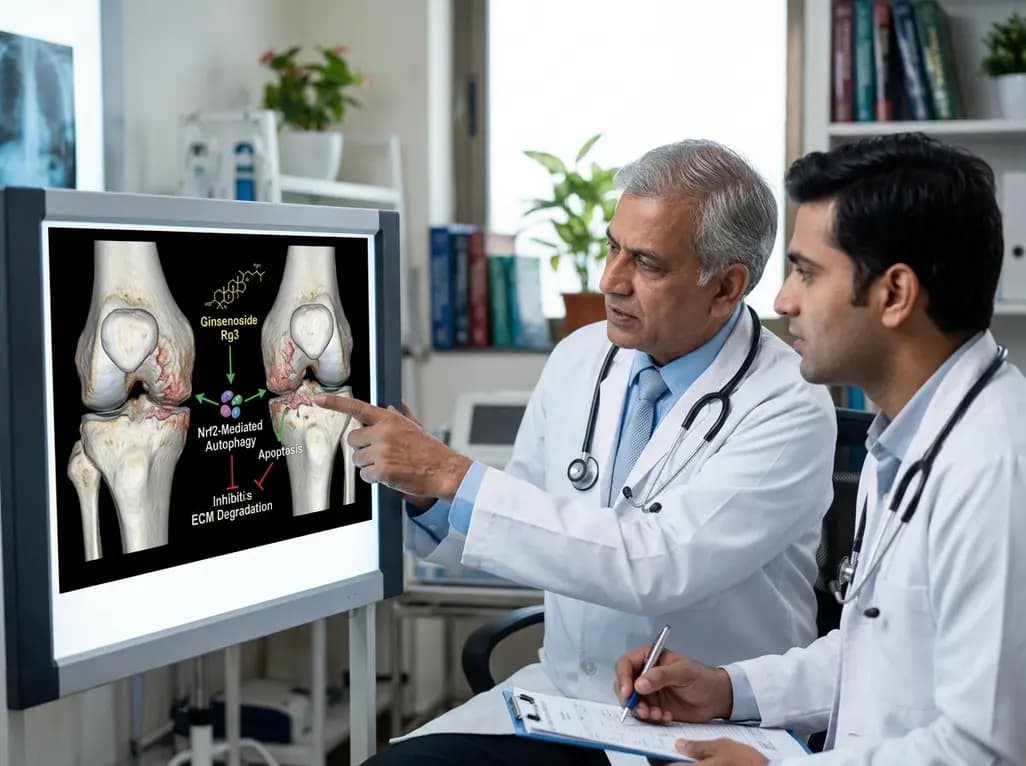
A study reveals that Ginsenoside Rg3 attenuates osteoarthritis progression by modulating the Nrf2-mediated autophagy pathway, reducing chondrocyte apoptosis, and curbing extracellular matrix degradation, offering promising chondroprotective potential.
Today

A cross-sectional study demonstrates that lower pediatric motor competence strongly correlates with adverse metabolic syndrome markers, including central adiposity, elevated triglycerides, and low HDL cholesterol, emphasizing early neuromuscular interventions in youth with overweight and obesity.
Today
