.webp&w=384&q=75)

Loading, please wait...
Loading, please wait...
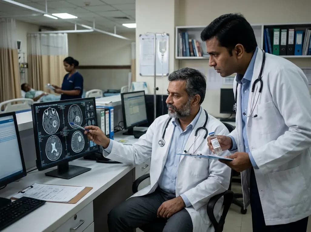
Diffuse gliomas present significant diagnostic and therapeutic monitoring hurdles in modern clinical practice. Traditional tissue biopsy carries surgical morbidity, and neuroimaging often fails to distinguish true tumor progression from treatment-induced pseudoprogression or radiation necrosis. Consequently, cerebrospinal fluid biomarkers have emerged as an invaluable liquid biopsy modality in neuro-oncology. Cerebrospinal fluid flows through the ventricular system and subarachnoid spaces in direct contact with the central nervous system. Because it circulates in immediate proximity to primary brain tumors, this fluid captures tumor-derived circulating cell-free DNA, extracellular vesicles, oncometabolites, and proteomic signatures that reflect disease burden with high biological fidelity.
Primary central nervous system malignancies shed molecular debris directly into surrounding fluid pathways. The blood-brain barrier substantially restricts the systemic extravasation of these tumor components into peripheral blood. Therefore, standard plasma liquid biopsies frequently demonstrate low sensitivity for intracranial gliomas. In contrast, cerebrospinal fluid biomarkers capture high concentrations of tumor-specific genetic mutations, including IDH1/2 alterations, TERT promoter variants, EGFR amplifications, and H3 histone mutations. Additionally, metabolic products such as D-2-hydroxyglutarate provide quantifiable markers of disease activity. However, clinicians must interpret these liquid analytes within the context of dynamic physiological flow, anatomical compartmentalization, and clearance kinetics within the craniospinal axis.
The anatomical location of sampling represents a critical determinant of analyte yield and diagnostic accuracy. Cerebrospinal fluid circulates continuously, creating marked concentration gradients between distinct compartments. Specifically, tumor-derived analytes exhibit markedly higher concentrations in intracranial subarachnoid and ventricular compartments compared to standard lumbar cistern samples. For instance, high-grade supratentorial gliomas shed nucleic acids locally, which rapidly dilute as fluid descends through spinal pathways. Consequently, relying exclusively on lumbar puncture may yield false-negative results or misrepresent molecular burden. Clinicians and researchers must carefully select the sampling site based on tumor proximity, hydrodynamics, and intended downstream analytical thresholds.
Neuroimaging provides essential baseline parameters that directly influence biofluid analyte recovery. Radiographic evaluation of tumor contact with the ventricular ependyma or pial subarachnoid space strongly correlates with cell-free DNA concentrations. Specifically, lesions with direct ventricular abutment release significantly higher quantities of genomic material into circulation than deep subcortical tumors. Furthermore, contrast enhancement on magnetic resonance imaging reflects blood-brain barrier disruption. When this vascular interface breaks down, systemic plasma proteins enter the intracranial compartment in large quantities. Consequently, investigators must account for these plasma-derived proteins when analyzing the cerebrospinal fluid proteome, ensuring accurate discrimination of true tumor-derived signals.
Serial monitoring provides real-time insights into therapeutic efficacy, clonal evolution, and acquired resistance. To facilitate repeat intracranial access without recurrent invasive procedures, neurosurgeons increasingly utilize indwelling access devices such as Ommaya reservoirs or modified ventricular catheter ports. These devices enable safe, longitudinal collection of ventricular fluid across multiple therapy cycles. As a result, longitudinal tracking allows early detection of molecular recurrence before radiographic manifestations appear. However, clinicians must implement rigorous aseptic handling protocols to minimize catheter colonization risks. Moreover, physicians must standardize sample collection volumes to maintain reproducible analyte concentrations during serial testing regimens.
Surgical intervention introduces profound disruptions to the local microenvironment and intracranial fluid dynamics. Gross total resection immediately reduces overall tumor burden, which dramatically lowers circulating tumor fractions. Furthermore, the postoperative inflammatory response triggers an influx of non-neoplastic inflammatory proteins and host nucleic acids that persist for several weeks. Consequently, collecting samples too soon after surgery can confound proteomic and genomic analyses. Clinicians must establish standardized post-resection sampling windows to obtain reliable baseline measurements. Additionally, multi-institutional assay harmonization remains necessary to establish diagnostic thresholds, analytical sensitivity standards, and regulatory concordance across clinical neuro-oncology centers.
Systematic biobanking infrastructure is essential for translating liquid biopsy discoveries into routine neuro-oncology workflows. Standardized protocols must govern every pre-analytical phase, including immediate cold-chain handling, rapid centrifugation to separate cellular debris, and storage at ultra-low temperatures. Furthermore, comprehensive clinical annotation—capturing radiographic tumor volume, anatomical sampling coordinates, steroid administration, and systemic chemotherapy status—is mandatory for meaningful data interpretation. By developing rigorous biobanking repositories, neuro-oncology networks can accelerate biomarker validation, improve clinical trial design, and facilitate individualized precision oncology for patients confronting aggressive brain tumors.
Intracranial cerebrospinal fluid resides closer to primary brain tumors, yielding substantially higher concentrations of tumor-derived DNA and proteins. Lumbar fluid often exhibits severe analyte dilution due to craniospinal flow gradients, which substantially increases the risk of false-negative liquid biopsy results.
Surgical resection dramatically decreases tumor-derived analyte abundance while simultaneously increasing surgical inflammatory proteins and host cellular debris. These post-resection alterations can persist for several weeks, requiring carefully timed collection intervals to ensure valid molecular interpretations.
Ommaya reservoirs provide safe, continuous, and minimally invasive access to intracranial cerebrospinal fluid across treatment cycles. This serial access allows clinicians to evaluate molecular treatment response, detect early clonal resistance, and track disease progression before radiographic changes occur.
Disclaimer: This content is for informational and educational purposes only. It does not constitute medical advice, diagnosis, or treatment recommendations. Refer to the latest local and national guidelines for clinical practice.
References

Read summarized clinical updates, watch expert medical content, and earn CME certifications right from your smartphone.
Open your smartphone camera to scan and install.





Cerebrospinal fluid biomarkers offer a transformative liquid biopsy approach for neuro-oncology. This clinical review explores spatial sampling gradients, radiographic correlates, longitudinal monitoring via access devices, and standardized biobanking protocols for optimizing glioma management.
last month

A recent clinical report details an atypical presentation of bilateral proptosis as the primary manifestation of aggressive multiple myeloma. Explore the diagnostic pathway, imaging findings, and critical multidisciplinary management strategies essential for handling extramedullary plasma cell dyscrasias.
Today

Prenatal evaluation of fetal growth restriction requires an integrated diagnostic approach. While chromosomal microarray remains fundamental, exome sequencing offers significant diagnostic yield in isolated and syndromic cases, especially when ultrasound shows skeletal anomalies or normal placental function.
Today

Discover a novel laparoscopic approach for iatrogenic diaphragmatic hernia repair following pedicled omentoplasty. This modified Sugarbaker technique integrates dorsal pedicle lateralization and round ligament reinforcement to secure the diaphragmatic defect while preserving vital omental vascularity.
Today
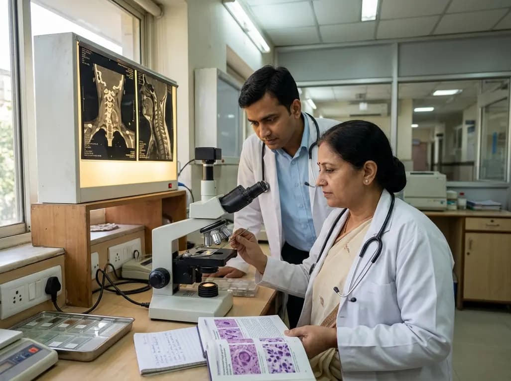
A cervical giant cell tumour presents rare diagnostic hurdles in spinal oncology. Image-guided FNAC provides rapid, reliable evaluation by identifying bland mononuclear stromal cells intimately admixed with osteoclast-type giant cells, aiding timely surgical planning and organ-preserving targeted management.
Today

New research reveals that M1 macrophage-derived exosomes aggravate diabetic nephropathy by transferring WTAP to stabilize S1PR2 mRNA. Silencing WTAP in these vesicles attenuates endothelial injury and renal fibrosis, pointing toward innovative nanomedicine therapies.
Today
