.webp&w=384&q=75)

Loading, please wait...
Loading, please wait...

Endovascular thrombectomy is the established standard of care for acute ischemic stroke with large-vessel occlusion. However, clinical teams frequently encounter futile recanalization, wherein patients fail to regain functional independence despite technically successful mechanical reperfusion. Clinicians traditionally evaluate macrovascular pial collaterals to estimate baseline tissue viability. Nevertheless, conventional arterial grading often overlooks downstream microcirculatory resistance and venous clearance. Recently, investigators established the cerebral collateral cascade model to capture this comprehensive microvascular network. Furthermore, dynamic physiological factors such as the infarct growth rate significantly influence whether salvageable penumbra survives until vessel reopening occurs. Understanding how the cerebral collateral cascade interacts with infarct growth dynamics provides critical insights into preventing futile recanalization after endovascular thrombectomy.
Mechanical thrombectomy routinely restores arterial lumen patency across modern comprehensive stroke centers. However, nearly half of treated patients still face poor functional outcomes at ninety days, a frustrating paradox termed futile recanalization. In standard clinical practice, interventionalists define successful angiographic reperfusion as achieving a modified Thrombolysis in Cerebral Infarction grade of 2b or 3. Yet, macroscopic arterial recanalization does not guarantee microscopic capillary reperfusion. Ischemia-reperfusion injury, microvascular capillary plugging, pericyte constriction, and severe blood-brain barrier disruption frequently cause extensive parenchymal damage. Furthermore, baseline patient variables, including advanced age, chronic comorbidities, admission hyperglycemia, and severe neurological deficits, exacerbate post-ischemic tissue loss. Consequently, arterial reopening cannot salvage penumbra that has already sustained irreversible metabolic injury. Standard digital subtraction angiography provides only a momentary, two-dimensional snapshot of arterial transit. It cannot determine whether circulating blood effectively irrigates capillary beds or drains freely through cerebral veins. Therefore, clinicians urgently need reliable baseline imaging biomarkers to distinguish patients destined for recovery from those at high risk of futile recanalization. Identifying these vulnerable individuals enables neurocritical care teams to anticipate secondary neurological decline and tailor aggressive supportive therapies promptly.
For decades, neurovascular specialists graded collateral circulation solely by evaluating retrograde pial arterial filling on computed tomography angiography. However, this conventional approach overlooks crucial physiological steps governing downstream cerebral hemodynamics. To address this limitation, stroke researchers introduced the comprehensive cerebral collateral cascade framework. This novel paradigm integrates three distinct, interconnected vascular levels: pial arterial collaterals, tissue-level microvascular perfusion, and cortical venous outflow. Leptomeningeal arterial vessels provide the primary retrograde bypass route around an occluded intracranial trunk. Subsequently, microvascular capillary beds determine whether oxygenated blood successfully permeates ischemic brain parenchyma. Finally, robust cortical and deep venous channels clear microvascular flow, preventing capillary congestion and elevated local tissue pressure. Thus, a patient with robust pial collateral vessels may still experience tissue death if microvascular beds exhibit capillary no-reflow or venous stagnation. In contrast, patients with complete cascade preservation maintain penumbral viability far longer across ischemic territory. Furthermore, comprehensive collateral evaluation correlates strongly with final tissue infarct volume. Consequently, analyzing the complete cascade equips stroke clinicians with an accurate view of microvascular integrity before catheter intervention begins.
Ischemic stroke progression does not follow a uniform, clockwork timeline in every patient. Instead, stroke pathophysiology evolves dynamically, varying based on individual cerebral autoregulation and metabolic demands. Clinicians quantify this biological trajectory using the infarct growth rate, calculated by dividing baseline ischemic core volume by the elapsed time from symptom onset to initial imaging. Consequently, this velocity metric categorizes stroke patients into distinct phenotypes: fast progressors and slow progressors. Fast progressors demonstrate rapid core expansion, exhausting ischemic penumbra within a few hours of arterial occlusion. In contrast, slow progressors sustain viable penumbral tissue for extended periods, occasionally preserving brain tissue well beyond standard therapeutic time windows. However, absolute ischemic core volume alone cannot reflect this biological pace without factoring in onset time. For instance, a thirty-milliliter core volume represents a much more dangerous trajectory at ninety minutes than at six hours post-onset. Furthermore, rapid infarct growth signals early microvascular breakdown and impaired cellular survival pathways. Therefore, infarct growth rate provides indispensable prognostic information regarding tissue fate after mechanical thrombectomy. Incorporating this dynamic metric refines acute clinical triage and prognostic stratification.
A multicenter retrospective cohort study investigated 157 consecutive patients with acute anterior circulation large-vessel occlusion undergoing thrombectomy within six hours of symptom onset. All enrolled individuals achieved successful angiographic reperfusion, defined as modified Thrombolysis in Cerebral Infarction grades 2b to 3. Remarkably, seventy patients (44.6%) developed futile recanalization, defined as a ninety-day modified Rankin Scale score between 3 and 6. Multivariable logistic regression identified unfavorable cerebral collateral cascades and elevated infarct growth rates as independent predictors of futile recanalization. Crucially, the researchers documented a statistically significant interaction between collateral cascade status and infarct growth velocity. This finding demonstrates that infarct growth rate fundamentally modifies how collateral vessels influence functional recovery. Specifically, stratified analysis revealed that favorable collateral status substantially reduced the risk of futile recanalization among slow progressors. Conversely, favorable collaterals did not protect against futile recanalization in fast progressors. Rapid ischemic injury appears to overwhelm compensatory microcirculatory mechanisms, rendering anatomical collateral pathways functionally ineffective once core expansion accelerates. Consequently, clinicians must interpret collateral adequacy within the dynamic context of tissue growth kinetics.
These clinical findings provide valuable guidance for emergency stroke protocols and neurocritical intensive care management. Contemporary stroke guidelines emphasize that interventionalists should not withhold mechanical thrombectomy based solely on collateral scoring. Nevertheless, evaluating the interaction between collateral cascade integrity and infarct growth rate refines post-recanalization expectations. For example, patients identified as slow progressors with preserved collateral cascades are prime candidates for complete functional independence. In contrast, fast progressors displaying compromised collateral cascades warrant immediate vigilance for post-procedural complications, including hemorrhagic transformation and malignant cerebral edema. In addition, neurocritical specialists can proactively optimize mean arterial pressure, maintain strict normoglycemia, and avoid hyperthermia to protect marginal microcirculatory beds in high-risk patients. Furthermore, future clinical trials evaluating neuroprotective agents and microvascular cytoprotective therapies should incorporate these combined physiological parameters during patient selection. Automated perfusion software will soon enable real-time calculation of both collateral status and growth kinetics upon arrival. Ultimately, moving beyond anatomical arterial images toward dynamic microvascular assessment empowers clinicians to deliver precise, pathophysiologically tailored stroke care in acute centers.
Futile recanalization occurs when acute ischemic stroke patients achieve successful angiographic vessel reopening (mTICI 2b-3) yet remain functionally dependent or die at ninety days (mRS 3-6). Incomplete capillary reperfusion, severe reperfusion injury, baseline comorbidities, and extensive early irreversible tissue necrosis drive this unfortunate clinical outcome.
The cerebral collateral cascade evaluates three distinct vascular compartments: pial arterial collaterals, microvascular tissue-level capillary perfusion, and cortical venous outflow. Rather than assessing arterial filling alone, this integrated framework captures macrovascular bypass pathways, capillary transit, and venous drainage, offering a comprehensive assessment of the complete microvascular circulation during acute ischemic stroke.
Infarct growth rate reflects the velocity of ischemic core expansion. Among slow progressors, favorable collateral cascades successfully preserve penumbral tissue until recanalization occurs. Conversely, in fast progressors, rapid metabolic breakdown and microvascular collapse outpace collateral compensation. Consequently, anatomical collaterals fail to prevent futile recanalization when ischemic core expansion progresses too rapidly.
Disclaimer: This content is for informational and educational purposes only and should not be considered medical advice. It is not intended to replace professional clinical judgment or guidelines. Refer to the latest local and national guidelines for clinical practice.
References

Read summarized clinical updates, watch expert medical content, and earn CME certifications right from your smartphone.
Open your smartphone camera to scan and install.





A retrospective cohort study demonstrates that the interaction between the cerebral collateral cascade and infarct growth rate predicts futile recanalization after thrombectomy. Evaluating both microvascular status and tissue growth kinetics refines stroke prognosis and post-procedural care.
Today

The CARE study protocol introduces a multilingual, RAG-powered conversational interface designed by IIT Bombay to support tobacco cessation in India. Evaluating feasibility and behavioral outcomes across three cessation centers, this digital health initiative aims to enhance quitting support for diverse users.
Today

A large UK cohort study of 1.4 million women shows that pregnancy complications, including gestational diabetes, hypertension, and preterm birth, substantially raise long-term risks for hypertension, chronic kidney disease, type 2 diabetes, and cardiovascular events, highlighting early postpartum intervention needs.
Today
A cross-sectional study reveals that hypertension knowledge, illness perceptions, depression, and substance use significantly predict self-care behaviors. Addressing mental health and cognitive beliefs in primary care is crucial for optimal blood pressure control and cardiovascular disease prevention.
Today
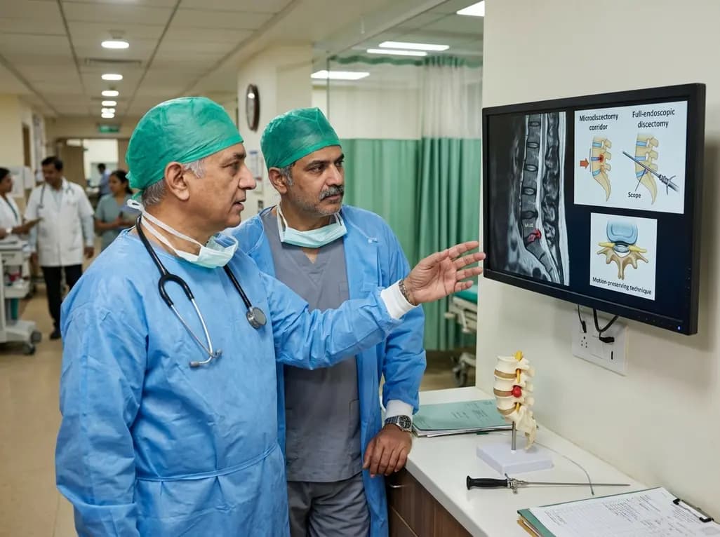
This clinical overview examines recent advancements in lumbar disc herniation surgery, comparing microscopic and full-endoscopic discectomy while highlighting biportal techniques and emerging motion-preserving innovations for spine specialists.
Today

Small interfering RNA (siRNA) therapeutics harness RNA interference to degrade disease-causing messenger RNA. By delivering hepatocyte-targeted, durable gene silencing, agents targeting PCSK9, LPA, and angiotensinogen offer potent cardiovascular risk reduction with infrequent, biannual dosing schedules.
Today
