.webp&w=384&q=75)

Loading, please wait...
Loading, please wait...
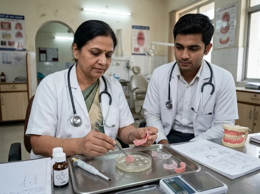
Denture stomatitis represents one of the most common and recurrent mucosal inflammatory conditions among completely and partially edentulous patients. Clinicians frequently prescribe resilient soft lining materials to cushion masticatory loads, absorb functional shocks, and relieve sore atrophic ridges. However, conventional soft liners present persistent clinical drawbacks, including low tear resistance, accelerated material degradation, weak adhesive bonding to polymethyl methacrylate denture bases, and heightened susceptibility to fungal colonization. Recent innovations demonstrate that incorporating cellulose nanofibers in denture liners significantly mitigates fungal colonization while reinforcing key physical parameters. This biomaterial enhancement offers substantial promise for improving patient comfort, tissue preservation, and overall prosthetic longevity in daily practice.
Resilient denture liners provide essential cushioning for geriatric patients presenting with thin, friable oral mucosa and severely resorbed alveolar ridges. In addition, these compliant elastomeric materials distribute occlusal forces uniformly across underlying supportive bone. Nevertheless, standard formulations often suffer from early mechanical deterioration under functional mastication. Repeated chewing cycles induce surface micro-cracking, tearing, and loss of plasticizers, which progressively hardens the elastomeric matrix. Consequently, the material loses its therapeutic resilience within weeks to months of continuous intraoral function.
Furthermore, standard soft liners exhibit weak peel bond strength at the liner-denture acrylic interface. This inadequate interface bond leads to marginal separation, saliva entrapment, and premature prosthesis failure. Clinicians frequently encounter severe material debonding, which necessitates repeated relining appointments and increases chairside time. More critically, the porous nature of deteriorating resilient liners creates ideal microenvironments for microbial colonization. Fungal pathogens adhere avidly to these elastomeric surfaces, converting comfortable prostheses into chronic infective reservoirs. Therefore, prosthodontists require novel reinforcement methods that preserve compliance while arresting fungal adhesion and interfacial breakdown.
Cellulose nanofibers represent bio-derived, high-aspect-ratio nanomaterials known for superior tensile stiffness, non-toxicity, and abundant surface hydroxyl groups. When dental researchers incorporate cellulose nanofibers in denture liners, these nano-scale fibers disperse throughout the elastomer matrix to establish robust structural networks. Fourier Transform Infrared spectroscopy confirms that extensive hydrogen bonding forms between the functional hydroxyl groups of nanofibers and the host polymer chain network. Consequently, this chemical interaction reinforces intermolecular cohesion without compromising overall material flexibility.
Scanning electron microscopy demonstrates that uniform nanofiber dispersion effectively bridges microscopic voids within the polymer. As a result, the reinforced composite matrix displays elevated resistance against crack propagation and internal shear stress. The incorporation of cellulose nanofibers significantly improves tensile strength and Shore A hardness under dynamic masticatory stress. Additionally, the improved molecular cross-linking resists the leaching of plasticizers into saliva. Thus, the modified liner preserves its elastomeric properties over extended clinical durations. By stabilizing the structural framework, cellulose nanofibers protect against the premature hardening and abrasive wear commonly seen in unmodified soft relining systems.
Denture-associated stomatitis occurs primarily due to the opportunistic proliferation of Candida albicans on the intaglio surface of prostheses. Blastoconidia and hyphal forms readily anchor to elastomeric materials, initiating complex biofilms that resist systemic and topical antimicrobials. Conventional prosthodontic principles suggest that increased surface roughness directly increases microbial retention, particularly when roughness exceeds the standard threshold of 0.2 micrometers. However, modified lining materials incorporating cellulose nanofibers demonstrate a remarkable departure from this classic paradigm.
Although the inclusion of cellulose nanofibers slightly increases microscopic surface roughness, quantitative microbial assays confirm a statistically significant decrease in Candida albicans adhesion. Researchers hypothesize that the dense surface distribution of hydroxyl motifs alterations surface free energy, electrical zeta potential, and localized wetting characteristics. Consequently, these physicochemical alterations disrupt the electrostatic attraction and specific adhesin binding mechanisms of candidal blastospores. Furthermore, altered surface topography prevents hyphal penetration into deeper polymer layers. Because the yeast cells cannot establish stable anchor sites, salivary shear forces easily dislodge emerging biofilms. This non-pharmacological antifungal mechanism substantially reduces mucosal erythema without inducing antifungal drug resistance.
Determining the ideal concentration of nano-additives is vital for maximizing clinical outcomes without compromising functional compliance. Recent laboratory evaluations compared control liners against experimental groups modified with 0.5 weight percent and 1.0 weight percent cellulose nanofibers. While both nanofiber concentrations reduced fungal adhesion significantly, the 0.5 weight percent concentration established the most favorable balance of biological and mechanical performance. This optimal loading level reinforced the material structure without causing excessive particle agglomeration.
Specifically, the 0.5 weight percent formulation achieved superior tensile strength and significantly elevated peel bond strength against acrylic denture base resins. Scanning electron microscopy confirmed that higher concentrations, such as 1.0 weight percent, can produce minor localized fiber clustering. These clusters generate microscopic stress concentration points that moderately limit further mechanical gains. Conversely, the 0.5 weight percent group preserved smooth interfacial distribution, maximizing hydrogen bonding with the acrylic polymer. As a result, this specific concentration prevents adhesive delamination while simultaneously curtailing Candida albicans proliferation. Thus, 0.5 weight percent represents the premier candidate concentration for future clinical trials.
Integrating nanotechnology into clinical prosthodontics addresses critical public health demands, particularly for vulnerable geriatric populations. Elderly individuals frequently exhibit reduced salivary flow, compromised immune responses, and diminished manual dexterity, making routine denture hygiene challenging. Incorporating intrinsically bioactive and mechanically durable relining materials provides continuous, passive protection against chronic oral candidiasis. Consequently, patients experience fewer episodes of painful palatal inflammation and enjoy uninterrupted nutritional intake.
Moreover, enhanced interfacial bond strength reduces the frequency of emergency clinical visits caused by liner delamination. Clinicians can perform direct or indirect relining procedures with greater confidence in prosthetic longevity. Dental laboratory technicians can readily incorporate standardized nanofiber masterbatches into commercially available lining polymers without requiring costly equipment changes. Furthermore, the bio-renewable nature of plant-derived cellulose makes this additive exceptionally cost-effective and biocompatible. As translational validation progresses toward human clinical trials, nanocellulose-modified liners promise to redefine quality of life standards for edentulous patients worldwide.
Cellulose nanofibers alter surface free energy, electrical charge, and nanoscale topography across the elastomer. Consequently, these structural modifications disrupt candidal adhesins and impair blastospore binding mechanisms. Although nanoscale roughness increases slightly, the chemical changes prevent stable yeast attachment. Therefore, salivary flow and standard hygiene easily dislodge emerging biofilms, neutralizing the typical microbial risks associated with rougher prosthetic materials.
The 0.5 weight percent loading achieves optimal fiber dispersion and extensive hydrogen bonding with the polymer matrix. In contrast, higher loadings, such as 1.0 weight percent, increase the risk of fiber agglomeration and microscopic stress concentration. Consequently, the 0.5 weight percent formulation delivers maximum tensile strength, superior peel bond resilience, and robust antifungal performance without compromising elasticity.
The addition of cellulose nanofibers produces a modest, controlled increase in Shore A hardness while maintaining essential therapeutic elasticity. Furthermore, the hydrogen-bonded network resists plasticizer loss and mechanical fatigue during functional mastication. As a result, the liner retains its cushioning capability against atrophic ridges over longer periods, preventing premature hardening and ensuring lasting patient comfort.
Disclaimer: This content is for informational and educational purposes only. It is not intended to provide medical advice, clinical diagnosis, or treatment plans. Dental professionals must exercise independent clinical judgment and correlate these findings with specific clinical presentations. Refer to the latest local and national guidelines for clinical practice.
References

Read summarized clinical updates, watch expert medical content, and earn CME certifications right from your smartphone.
Open your smartphone camera to scan and install.





A novel study demonstrates that incorporating 0.5 wt.% cellulose nanofibers into soft denture lining material significantly enhances mechanical properties and reduces Candida albicans adherence, presenting an effective approach to mitigating denture-induced stomatitis in prosthodontic practice.
last month

A multicity study highlights that over 80% of acute respiratory infections remain outside hospitals. Patients frequently choose community pharmacies and digital platforms. Integrating digital health data and retail pharmacy sales can fill critical surveillance gaps and improve early outbreak detection.
Today
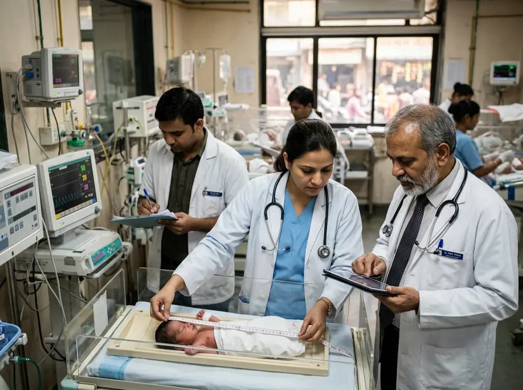
Scandinavian register evidence reveals that neonatal birth length strongly predicts adult health and mortality, whereas birth weight dictates cognitive capacity. Symmetric growth restriction causes far more severe long-term deficits than asymmetric restriction, underscoring the need for comprehensive anthropometry.
Today
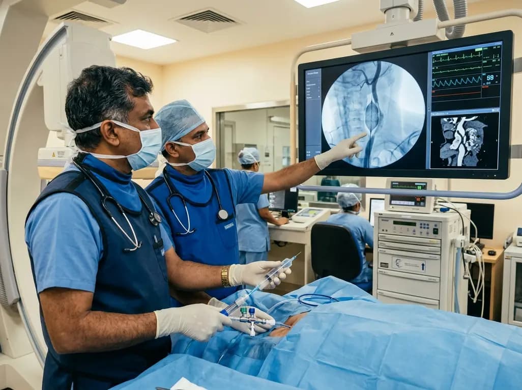
Discover how rescue cutting balloon angioplasty successfully modifies heavily calcified carotid stenosis, enabling stent delivery without baroreceptor instability and ensuring favorable long-term neurologic outcomes.
Today
A breakthrough study reveals that digoxin alleviates osteoarthritis by targeting LSP1 in synovial macrophages, preventing harmful extracellular trap formation and restoring SOD1 activity. This dual-action mechanism protects joint cartilage and offers a promising repurposing strategy for disease modification.
Today
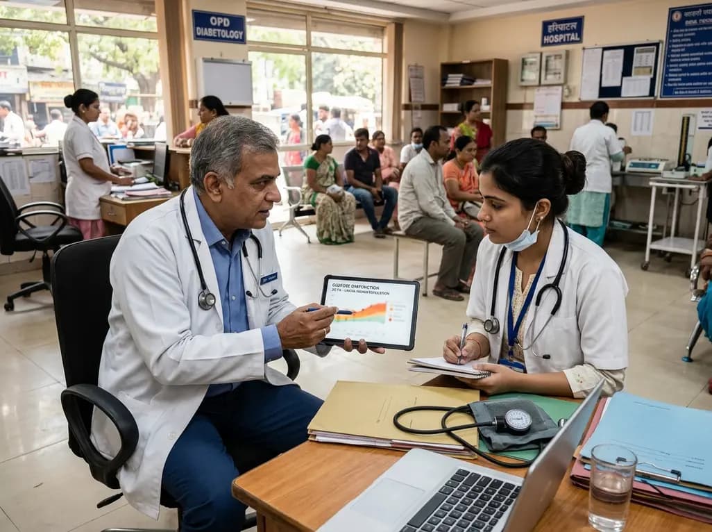
Data from the 2026 Workforce Health Index reveals that 38.7% of urban employees and dependents exhibit abnormal glycemic control. Emerging at increasingly younger ages, this metabolic shift intersects with severe vitamin deficiencies, highlighting the urgent need for early workplace screenings and targeted clinical care.
Today
