

Loading, please wait...
Loading, please wait...

Carpal instability can cause debilitating wrist pain, diminished grip strength, and progressive structural collapse if left unmanaged. Consequently, structured carpal instability rehabilitation serves as a cornerstone of treatment across both non-operative and postoperative care pathways. Clinicians often encounter complex ligament injuries involving scapholunate dissociation or midcarpal laxity in active populations. However, fragmented evidence and heterogeneous clinical protocols frequently complicate therapeutic decision-making. A comprehensive synthesis of current literature clarifies how targeted interventions restore wrist mechanics and protect healing tissues. By integrating active stabilization with precise immobilization, clinicians optimize outcomes for patients recovering from acute trauma or chronic laxity.
Wrist stability depends on an intricate interplay between passive ligamentous constraints, joint geometry, and dynamic muscular vectors. Therefore, clinicians must distinguish between carpal instability dissociative (CID) and carpal instability nondissociative (CIND) before initiating therapy. In dissociative patterns such as scapholunate or lunotriquetral tears, disruptions occur within a single carpal row. Consequently, abnormal kinematic forces alter intercarpal angles and disrupt force transmission during hand loading. Conversely, nondissociative instability involves dysfunction between carpal rows or across the radiocarpal joint without intrinsic interosseous ligament rupture. Patients with CIND often present with painful mechanical clunking and adaptive motor compensations during midcarpal motion. Furthermore, chronic ligamentous laxity can cause secondary cartilage wear and degenerative changes over time. Thus, accurate physical examination and diagnostic imaging must establish whether the instability pattern is static, dynamic, or predynamic. Recognizing these distinct structural patterns guides specific orthotic design and targeted neuromuscular retraining.
Recent evidence syntheses categorize wrist therapy into six essential components that span conservative care and surgical recovery. Specifically, these foundational pillars include immobilization, soft tissue management, joint mobilization, therapeutic exercise, proprioceptive training, and instrumental modalities. During early recovery phases, custom orthoses shield vulnerable ligament complexes from destabilizing torsional shear. In addition, therapists implement meticulous edema control and patient education to mitigate secondary stiffness. As tissue healing progresses, clinicians introduce controlled mobilization to preserve functional arcs of motion. Furthermore, therapeutic exercise forms the primary active intervention across every rehabilitation stage. Clinicians utilize progressive resistance training to build muscular endurance around the carpus. Moreover, adjunctive modalities like electrical stimulation help modulate localized discomfort and facilitate muscle recruitment during early rehabilitation. However, therapists must carefully customize orthotic positioning and exercise dosages according to the specific ligament injury. Standardizing these multimodal interventions ensures a safe transition from initial protection toward unconstrained wrist function.
Active muscular stabilization compensates effectively for deficient passive wrist ligaments. For instance, specific forearm muscles generate rotational vectors that either stabilize or open injured carpal joints. In scapholunate ligament insufficiency, the scaphoid tends to flex abnormally and rotate into pronation. Therefore, rehabilitation prioritizes isometric recruitment of carpal supinators, including the extensor carpi radialis longus and brevis. In addition, the abductor pollicis longus and flexor carpi radialis help seat the scaphoid into its anatomic fossa. Conversely, clinicians deliberately avoid activating muscles that exacerbate scaphoid pronation during early healing phases. Clinicians frequently prescribe the dart thrower's motion arc, moving from radial extension to ulnar flexion. This specialized movement pathway minimizes motion at the scapholunate articulation while maintaining functional midcarpal excursion. Consequently, patients develop muscular coordination without imposing excessive strain on reconstructed or healing ligamentous structures. As a result, systematic motor training reinforces dynamic stabilization while preserving joint congruency.
Carpal ligaments contain rich networks of mechanoreceptors that transmit vital kinesthetic information to the central nervous system. Following acute tears or prolonged postoperative immobilization, patients frequently lose joint position sense and subconscious reflexive protection. As a result, periarticular muscles exhibit dyssynergic contractions and delayed reaction times. Proprioceptive neuromuscular retraining directly addresses these afferent deficits to restore dynamic stability under unexpected perturbations. Therapists often initiate sensorimotor retraining using mirror therapy and conscious joint repositioning drills. Subsequently, patients advance toward unstable surface training, pertubation exercises, and rhythmic stabilization drills. Moreover, introducing visual feedback and dual-task challenges enhances motor cortex reorganization. This progressive neuromuscular re-education trains forearm muscles to react automatically during high-velocity impacts. Ultimately, robust sensorimotor recovery shields healing ligaments and prevents recurrent joint subluxations during demanding manual tasks.
Despite widespread clinical adoption of wrist exercises, notable inconsistencies persist across published rehabilitation literature. In particular, many existing protocols fail to define explicit progression criteria, exercise dosages, or treatment frequency. Most clinical reports emphasize impairment-level outcomes, such as grip strength, passive range of motion, and pain scores. However, few studies evaluate participation-level metrics, including timely return to work, vocational productivity, or athletic performance. Therefore, clinicians must adopt objective functional milestones rather than arbitrary timelines when advancing therapeutic loads. Clinicians should assess dynamic grip stability under varied forearm rotations before clearing patients for unrestricted loading. Furthermore, structured patient education regarding ergonomic modifications protects reconstructed ligaments against repetitive mechanical overload. Developing rigorous, standardized rehabilitation protocols will ultimately bridge current evidence gaps and improve long-term outcomes for complex carpal disorders.
Conservative rehabilitation focuses primarily on dynamic muscular compensation and joint protection for partial ligament tears or mild midcarpal laxity. In contrast, postsurgical protocols follow strict biological healing timelines to safeguard repaired or reconstructed ligaments. Consequently, postoperative programs require initial rigid immobilization, followed by gradual protected motion and progressive loading. Both pathways, however, prioritize targeted neuromuscular re-education, proprioceptive retraining, and activity modification to achieve long-term functional joint stability.
The dart thrower motion guides the wrist along an oblique functional plane from radial extension to ulnar flexion. Biomechanical investigations demonstrate that this specific arc occurs predominantly at the midcarpal joint rather than the radiocarpal level. Therefore, practicing this functional trajectory minimizes rotation and translation between the scaphoid and lunate. Consequently, patients can restore functional mobility and activate dynamic forearm stabilizers without placing excessive stress on healing scapholunate ligaments.
Injured carpal ligaments lose vital sensory mechanoreceptors, which impairs joint position awareness and reflexive muscular guarding. Without prompt intervention, dynamic instability alters intra-articular contact pressures and accelerates degenerative joint wear. Proprioceptive exercises retrain afferent neuromuscular pathways through joint repositioning, balance boards, and rhythmic perturbation drills. Consequently, forearm muscles learn to activate promptly during unexpected loading, maintaining congruent carpal kinematics and protecting articular cartilage against chronic destructive shear forces.
Disclaimer: This content is for informational and educational purposes only and does not substitute professional medical advice, diagnosis, or treatment. Always consult a qualified healthcare provider regarding clinical management, and never disregard medical guidance based on this article. Refer to the latest local and national guidelines for clinical practice.
References
Prato I et al. Rehabilitation strategies for carpal instability: A scoping review of conservative and postsurgical interventions. J Hand Ther. 2026 Nov 12. doi: undefined. PMID: 43020762.
Harwood C, Turner L. Conservative management of midcarpal instability. J Hand Surg Eur Vol. 2016;41(1):102-109.
Esplugas M, Garcia-Elias M, Lluch A, Llusá M. Role of muscles in the stabilization of ligament-deficient wrists. J Hand Ther. 2016;29(2):166-174.

Read summarized clinical updates, watch expert medical content, and earn CME certifications right from your smartphone.
Open your smartphone camera to scan and install.





A comprehensive scoping review synthesizes conservative and postsurgical carpal instability rehabilitation strategies, highlighting immobilization, targeted muscle recruitment, and proprioception to restore wrist stability.
Today
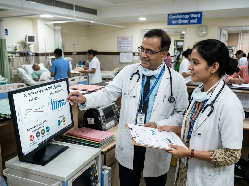
The VOCAL-AHF study reveals that most acute heart failure patients across reduced and non-reduced ejection fraction categories are eligible for in-hospital GDMT initiation. Notably, nearly two-thirds of HFrEF patients qualify for quadruple therapy, while non-reduced EF patients show high eligibility for dual therapy.
Today

Ascertainment bias in cancer case-control studies inflates odds ratios and penetrance estimates when cases are drawn from high-risk clinics. A landmark modeling study demonstrates substantial bias ranging up to 23.72, highlighting the critical need for ascertainment adjustment in genetic risk counseling.
Today
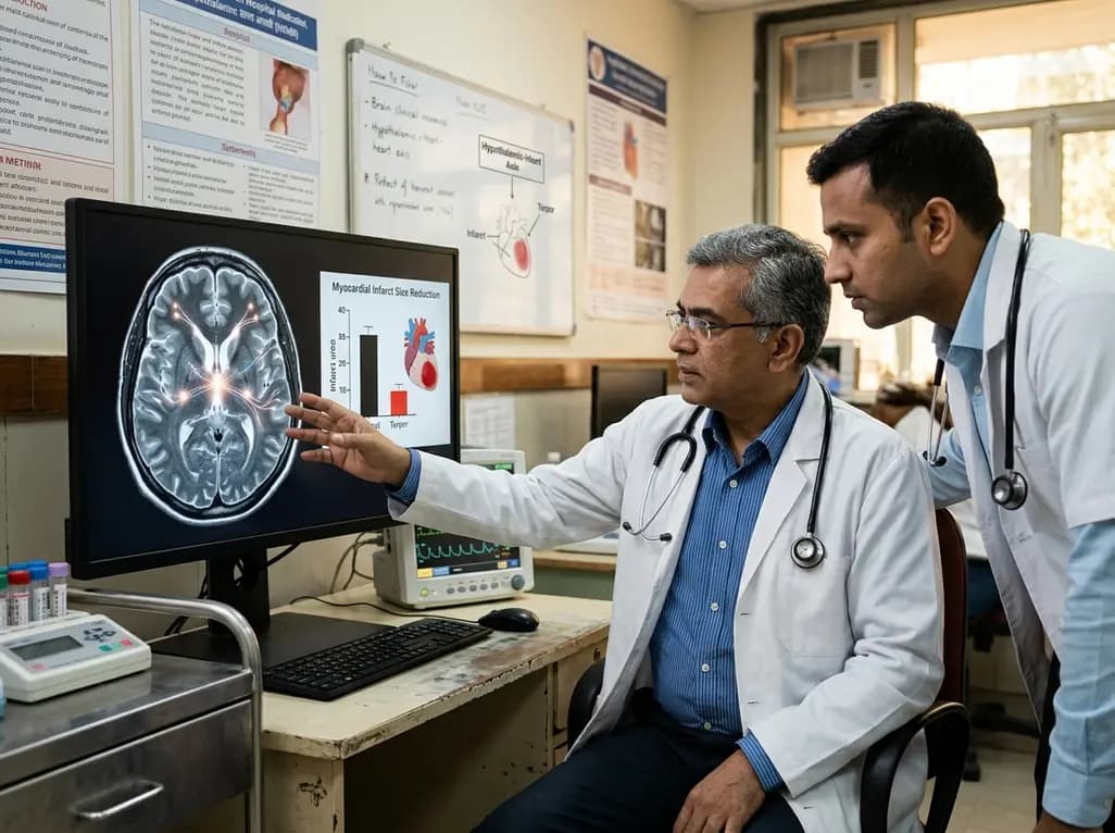
Researchers demonstrate that central induction of synthetic torpor in non-hibernating rats provides striking cardioprotection, reducing myocardial infarct size by ~40% during ischaemia-reperfusion injury through endogenous survival signaling, independent of hypothermia.
Today
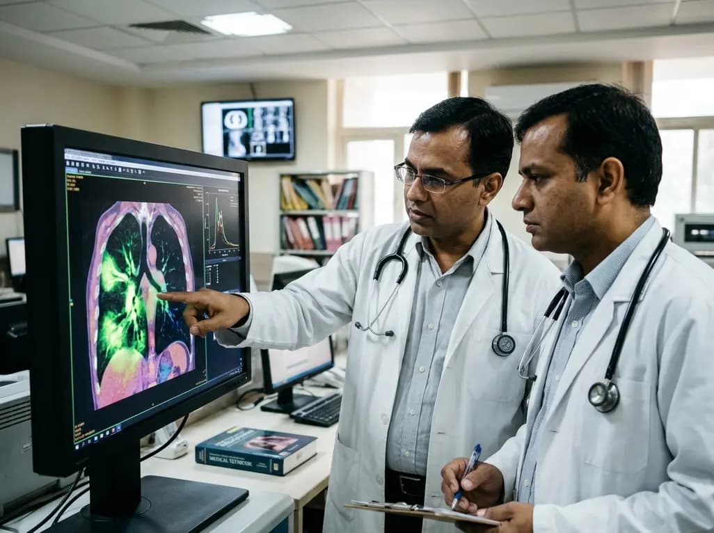
A novel fluorine-functionalized near-infrared copper fluorescent probe selectively illuminates lung tissue, revealing critical links between hyperglycemia, ER stress, and pulmonary malignancy for improved early diagnosis.
Today
