.webp&w=384&q=75)

Loading, please wait...
Loading, please wait...
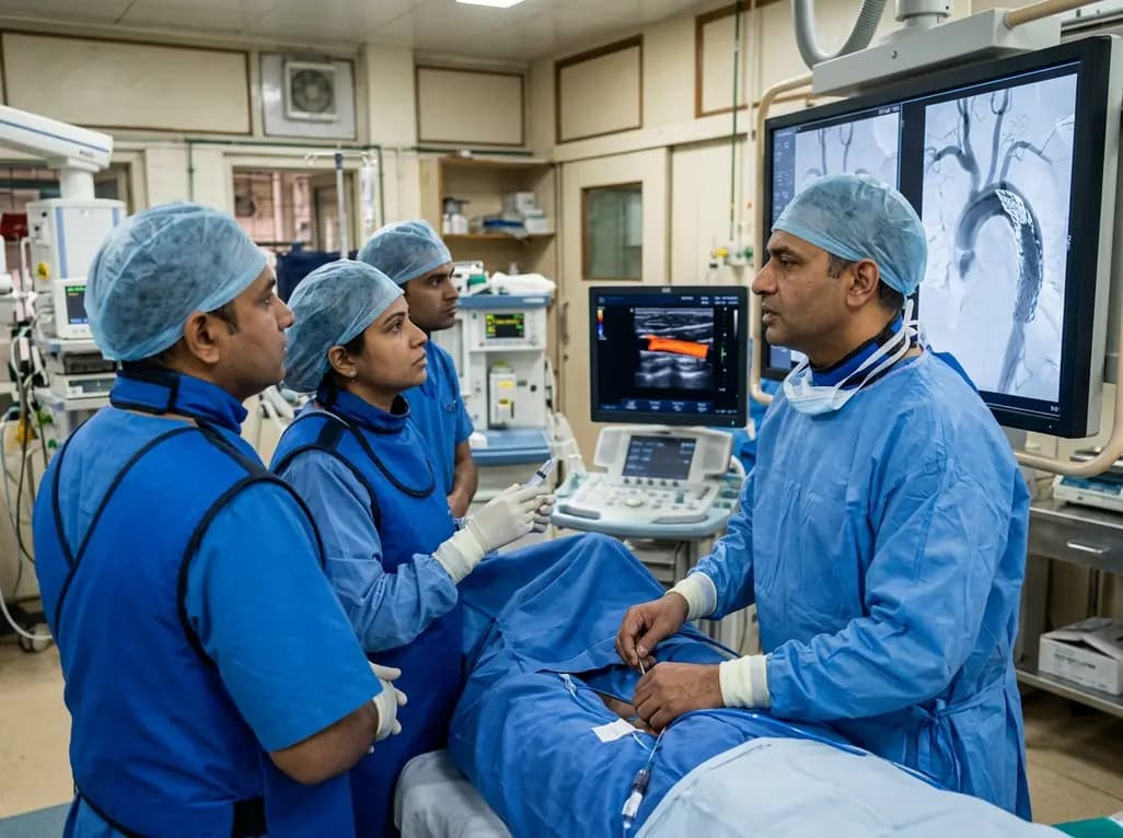
Thoracic endovascular aortic repair has transformed the treatment of complicated Stanford type B aortic dissection and complex thoracic aortic pathologies. However, landing stent grafts near the aortic arch introduces major procedural challenges. Proximal landing zone extension into aortic arch Zone 1 or Zone 2 often poses substantial risks to supra-aortic branch perfusion. Inadvertent coverage or mechanical occlusion of the left common carotid artery constitutes an urgent procedural complication that can precipitate catastrophic ischemic stroke. Therefore, performing rapid carotid revascularization after TEVAR remains imperative when branch compromise occurs. Interventional teams must maintain high vigilance during every arch deployment. Immediate recognition of altered perfusion, followed by prompt endovascular bailout, allows operators to restore antegrade cerebral blood flow before irreversible brain injury develops. This clinical review evaluates the mechanisms of acute carotid occlusion, explores intraoperative monitoring strategies, and outlines emergency revascularization techniques to optimize patient safety.
Achieving an adequate proximal seal in aortic dissection often demands extending the stent graft proximally into the aortic arch. The sharp curvature of the arch and high hemodynamic shear stress create mechanical forces that cause graft migration or misalignment. When deploying a proximal extension to seal a persistent type IA endoleak, the leading edge of the endograft may inadvertently cover the left common carotid artery ostium. Furthermore, balloon molding or radial tension from overlapping stent grafts can induce local arterial dissection, thrombosis, or branch compression. Because the left common carotid artery delivers vital blood flow to the anterior cerebral circulation, sudden occlusion places the ipsilateral cerebral hemisphere at extreme risk. Although the circle of Willis theoretically provides collateral cross-filling from contralateral systems, anatomical variations and incomplete circles occur frequently. Consequently, operators cannot assume collateral flow will prevent cerebral infarction. Understanding these mechanical hazards underscores why surgical teams must meticulously plan landing zones and anticipate emergency bailout options.
Prompt detection of carotid occlusion is the critical first step in preventing irreversible perioperative stroke. During complex thoracic repair, completion angiography must systematically visualize all supra-aortic branches. A sudden absence of contrast opacification in the left common carotid artery signals immediate arterial coverage or mechanical flow obstruction. However, fluoroscopy alone may sometimes yield ambiguous findings due to vessel overlap or delayed wash-in. Therefore, multimodal intraoperative monitoring serves as an indispensable safety adjunct. Point-of-care duplex ultrasonography provides rapid, real-time assessment of carotid vessel patency and flow hemodynamics right at the bedside. Intraoperative ultrasound can immediately confirm absent Doppler signals, identify proximal thrombus formation, and verify vessel caliber. Additionally, neuromonitoring techniques like near-infrared spectroscopy offer valuable real-time indicators of cerebral perfusion changes. When completion imaging demonstrates branch compromise, the surgical team must act without hesitation. Clinicians should never wait for post-procedural neurological deficits to manifest after anesthesia reversal.
When acute carotid coverage occurs, endovascular operators must rapidly execute a bailout strategy. Direct ultrasound-guided puncture of the ipsilateral left common carotid artery represents a highly effective, minimally invasive access route. Ultrasound visualization ensures precise needle entry into the common carotid lumen while avoiding atherosclerotic plaques and minimizing hematoma formation. Once access is secured, a sheath is introduced retrogradely toward the aortic arch. Through this direct access, operators can advance a guidewire across the occluded ostium into the aortic lumen. Deploying a dedicated self-expanding stent parallel to the aortic endograft creates a functional chimney graft. This technique successfully restores antegrade cerebral flow within minutes, re-establishing critical perfusion to the brain. Retrograde chimney stenting eliminates the delays associated with open surgical bypass procedures. Completing carotid revascularization after TEVAR in under fifteen minutes effectively aborts cerebral ischemic cascades, preventing permanent hemiplegia and devastating neurological impairment.
The brain exhibits exceptional sensitivity to ischemic insult, with millions of neurons dying each minute during complete arterial cessation. The concept of "time is brain" applies just as rigorously in the hybrid operating suite as it does in acute stroke pathways. Inadvertent carotid occlusion represents a profound iatrogenic emergency where every second dictates long-term functional recovery. Restoring antegrade carotid circulation within ten to fifteen minutes typically prevents permanent infarction, even in individuals with suboptimal collateral anatomy. Rapid reperfusion preserves the ischemic penumbra, restores cellular energy metabolism, and prevents secondary microvascular thrombosis. Conversely, delayed recognition leads to extensive cerebral infarction, hemorrhagic transformation upon subsequent reperfusion, and devastating functional disability. Maintaining hemodynamic stability with tight blood pressure control during the occlusion phase further supports collateral flow across the cerebral circle. In addition, administering systemic anticoagulation helps prevent retrograde thrombus propagation toward the internal carotid bifurcation.
Preventing inadvertent branch coverage requires thorough preoperative evaluation and strict procedural execution. High-resolution computed tomography angiography allows detailed anatomical assessment of the aortic arch, branch vessel takeoff angles, and arch elongation. Clinicians must measure neck lengths precisely to choose appropriate graft dimensions and landing zones. In patients requiring Zone 1 or Zone 2 landing, elective branch revascularization strategies, such as customized fenestrations, branched devices, or pre-emptive debranching, should be carefully considered. Nevertheless, when emergency aortic dissection necessitates urgent proximal extension, unforeseen intraoperative challenges can arise. Hybrid operating suites must maintain immediate availability of endovascular bailout inventory, including micropuncture sets, vascular sheaths, and dedicated stents. Multidisciplinary coordination between vascular surgeons, interventional cardiologists, radiologic technologists, and anesthesiologists streamlines the response to intraoperative complications. Rigorous planning and structured bailout algorithms ensure that every team member understands their role during unexpected branch occlusion.
Left common carotid artery occlusion typically occurs when a thoracic endograft or proximal extension migrates or requires placement over the branch ostium to achieve a secure proximal seal. Radial graft forces, anatomical arch tortuosity, and dynamic aortic pulsatility can lead to inadvertent coverage. Additionally, localized dissection, mechanical branch compression, or acute thrombotic occlusion during device manipulation can compromise vital antegrade carotid blood flow.
Direct ultrasound-guided carotid access allows rapid, retrograde cannulation of the common carotid artery without requiring extensive surgical cutdown or transfemoral navigation across challenging aortic anatomy. Operators can quickly introduce a sheath, cross the covered ostium from above, and deploy a parallel chimney stent into the aorta. This direct approach substantially shortens procedural time, restoring cerebral perfusion within minutes and preventing irreversible ischemic stroke.
Completion angiography is vital because it immediately evaluates endograft seal, excludes residual endoleaks, and verifies the patency of all critical supra-aortic vessels. Proximal extensions positioned in Zone 1 or Zone 2 can unexpectedly compromise branch blood flow. Immediate angiographic verification allows the surgical team to recognize branch occlusion on the table, enabling instantaneous endovascular bailout before the patient awakens with severe neurological deficits.
Disclaimer: This content is for informational and educational purposes only. It is not intended to be a substitute for professional medical advice, diagnosis, or treatment. Always seek the advice of your physician or other qualified health provider with any questions you may have regarding a medical condition. Never disregard professional medical advice or delay in seeking it because of something you have read here. Refer to the latest local and national guidelines for clinical practice.
References

Read summarized clinical updates, watch expert medical content, and earn CME certifications right from your smartphone.
Open your smartphone camera to scan and install.





Inadvertent left common carotid artery occlusion during TEVAR demands rapid diagnosis and immediate bailout revascularization to prevent stroke. This case analysis highlights duplex ultrasound detection and direct-access chimney stenting.
4 weeks back

Targeting ferroptosis provides a compelling strategy to dismantle hypoxia- and acidosis-driven immunosuppression in solid tumors, enhancing response to modern immunotherapy.
Today

Findings from Project Viva indicate that sustained postpartum weight retention, rather than excessive gestational weight gain alone, directly worsens women's mid-life cardiovascular health, degrading blood pressure and glycemic parameters decades after delivery.
Today

A large UK Biobank study reveals that plasma proteomic signatures capture preclinical organ damage and improve multiorgan risk prediction across early CKM syndrome stages 0 to 2 beyond traditional PREVENT clinical models.
Today

A retrospective CBCT study reveals that superior TMJ space asymmetry correlates moderately with occipital condyle-atlas-axis asymmetry in unilateral TMJ osteoarthritis, highlighting an imaging correlate between jaw biomechanics and upper cervical alignment rather than a standalone diagnostic marker.
Today

A Bayesian multilevel meta-analysis reveals that aerobic training combined with moderate carbohydrate restriction modestly lowers HbA1c in type 2 diabetes. However, sparse data and very low certainty leave incremental benefits over exercise or diet alone unproven, highlighting the need for individualized care.
Today
