.webp&w=384&q=75)

Loading, please wait...
Loading, please wait...

Dental caries remains a widespread global challenge that damages calcified tooth structures in pediatric and adult populations alike. Recent dental research highlights how advanced mineral formulations facilitate depth-oriented remineralization across vulnerable tooth substrates. Standard topical fluoride therapies primarily reinforce superficial enamel surfaces, yet they frequently fail to rebuild deep subsurface demineralization within active lesions. Consequently, dental biomaterial scientists have developed bioactive complexes combining calcium, fluoride, and phosphate ions into stable therapeutic delivery vehicles. By optimizing ionic concentrations and structural stoichiometry, modern non-invasive dentistry can achieve superior deep-tissue repair in both enamel and dentin without requiring premature surgical excavation.
Traditional remineralization strategies rely heavily on ambient salivary calcium and phosphate ions to stabilize hydroxyapatite crystals within degraded tooth structures. However, saliva often supplies insufficient mineral ions during frequent acidic challenges, especially in xerostomic or high-caries-risk patients. Furthermore, conventional sodium fluoride creates an insoluble, superficial layer of calcium fluoride that prematurely seals surface pores and hinders deeper ion penetration. Therefore, researchers developed soluble fluoride-calcium-phosphate complexes to overcome this physical bottleneck. These specialized multi-ion complexes maintain high concentrations of bioavailable ions in aqueous solution without triggering immediate, unwanted precipitation. As a result, the active chemical species penetrate freely through porous lesion bodies instead of occluding superficial micro-channels. Specifically, adjusting the calcium stoichiometry within these complexes dramatically enhances mineral transfer into deeper demineralized regions of the tooth. Experimental evidence indicates that higher calcium ratios drive mineral diffusion deeper into the structural matrix of both enamel and dentin. Thus, controlling ion stoichiometry provides a predictable pathway for whole-lesion recovery and long-term structural reinforcement.
To evaluate mineral dynamics and structural recovery accurately, modern dental biomaterials research employs high-resolution, non-destructive imaging modalities. Swept-source optical coherence tomography and micro-computed tomography offer comprehensive quantitative assessment of lesion depth and mineral density changes over time. In laboratory models, bovine enamel and dentin specimens undergo standardized pH cycling regimens to simulate the dynamic oral environment. When investigators treat demineralized blocks with high-calcium complexes, optical coherence tomography reveals substantial reductions in optical backscattering and total lesion depth. Moreover, micro-computed tomography demonstrates remarkable mineral recovery throughout the entire depth of the lesions, particularly within tubular dentin substrates. In contrast, control specimens and low-calcium formulations display minimal deep-tissue remineralization because mineral deposition remains confined to the outermost surface layers. Consequently, advanced optical imaging confirms that calcium-rich complexes restore structural density without disrupting native tissue architectures. These imaging breakthroughs validate the functional superiority of balanced multi-ion formulations for non-invasive restorative dentistry.
Detailed chemical and crystallographic analyses provide essential insights into how synthetic ion complexes interact with natural tooth matrices. Attenuated total reflectance-Fourier transform infrared spectroscopy and wide-angle X-ray scattering reveal critical compositional shifts during remineralization. Infrared spectroscopy demonstrates distinct phosphate and carbonate vibrational bands, reflecting the chemical reorganization and maturation of demineralized apatite matrices. Meanwhile, wide-angle X-ray scattering patterns show predominantly amorphous structural phases following the initial application of the complex. Interestingly, formulations with elevated calcium concentrations exhibit weak calcium fluoride reflections alongside these amorphous mineral matrices. This observation suggests that the complex delivers a durable reservoir of bioavailable ions that gradually crystallize into stable fluorapatite mineral phases. In addition, the amorphous nature of the initial deposit facilitates deeper interstitial ion migration before definitive crystal lattice formation occurs. Thus, the material combines rapid pore infiltration with sustained chemical maturation, creating an optimal environment for durable hard-tissue consolidation.
Enamel and dentin present distinctly different structural and biochemical challenges during non-invasive remineralization therapy. Enamel consists primarily of densely packed hydroxyapatite crystals with negligible organic content, whereas dentin contains substantial type-I collagen scaffolding and open dentinal tubules. Because dentin possesses higher porosity and greater organic volume, it demineralizes more rapidly under acidic conditions and resists standard remineralization protocols. Fortunately, calcium-enriched fluoride complexes demonstrate remarkable efficacy in deep dentin remineralization. The bioavailable calcium and phosphate ions interact intimately with exposed collagen fibers, thereby preventing enzymatic collagen degradation and nucleating new mineral crystals. Furthermore, the presence of fluoride stabilizes newly precipitated crystals against subsequent acid dissolution and erosive attacks. In enamel, the formulation prevents surface softening and restores natural optical translucency. Consequently, the multi-ion approach successfully addresses the divergent biochemical demands of both dental hard tissues, offering comprehensive protection against progressive carious destruction.
Translating laboratory findings into daily dental practice offers substantial advantages for preventive and minimally invasive dentistry. Dental practitioners frequently encounter early carious lesions, white spot defects, and exposed root surfaces that demand non-surgical intervention. Applying calcium-optimized fluoride complexes provides clinicians with a robust therapeutic option to arrest active decay and preserve natural tooth structure. Moreover, this stoichiometric approach reduces the need for aggressive operative drilling, thereby preserving dental pulp vitality and extending tooth longevity. Clinicians can incorporate these advanced bioactive agents into professional varnishes, localized remineralizing pastes, or restorative cavity liners before placing composite restorations. Additionally, patients undergoing orthodontic treatment or experiencing salivary hypofunction can benefit greatly from enhanced subsurface protection. Therefore, adopting stoichiometrically balanced ion complexes represents a major clinical milestone toward biomimetic tooth preservation and evidence-based caries management.
The development of stoichiometry-governed remineralization technologies paves the way for a new generation of smart dental biomaterials. Future clinical formulations will likely combine calcium-fluoride-phosphate complexes with bioactive glass particles, synthetic peptides, or antimicrobial peptides to enhance multi-target lesion management. Such hybrid formulations could simultaneously suppress cariogenic biofilm activity, neutralize localized lactic acid production, and supply supersaturated remineralizing ions directly to compromised tooth margins. Furthermore, incorporating these complexes into daily-use dentifrices and professional desensitizing agents will expand access to non-invasive tooth repair in general practice. Longitudinal clinical trials will be essential to evaluate the durability of these remineralized subsurface layers under complex oral masticatory stresses. Ultimately, optimizing ion stoichiometry transforms preventive dental care by offering predictable, deep-seated structural regeneration for diverse dental clinical applications.
Traditional fluoride applications predominantly deposit minerals on the outermost enamel surface, often creating a hyper-mineralized exterior barrier that restricts deeper ion migration. In contrast, depth-oriented remineralization utilizes stabilized ion complexes with optimized calcium stoichiometry to deliver bioavailable calcium, phosphate, and fluoride throughout the entire lesion depth. Consequently, this advanced method rebuilds the deeper lesion body, effectively restoring structural mineral density and physical resistance across both enamel and dentin tissues.
Dentin contains a dense collagen matrix and exhibits greater porosity than enamel, requiring carefully balanced ionic delivery for effective remineralization. Calcium stoichiometry governs the diffusion rate and penetration depth of mineral ions, preventing premature surface crystallization. By maintaining an optimal calcium ratio, the complex enables deep interstitial infiltration, effectively binding to exposed collagen scaffolds, remineralizing demineralized dentin matrices, and significantly reducing microscopic lesion depth within clinical settings.
Clinicians and researchers utilize swept-source optical coherence tomography and micro-computed tomography to evaluate non-invasive mineral recovery quantitatively. Swept-source optical coherence tomography provides real-time cross-sectional images of tissue scattering and structural changes, whereas micro-computed tomography accurately measures volumetric mineral density and lesion depth reduction. In addition, infrared spectroscopy and X-ray scattering confirm favorable chemical reorganization and crystallographic phase transformation within the treated hard dental tissues.
Disclaimer: This content is for informational and educational purposes only and should not be considered medical advice. Always consult a qualified healthcare provider for diagnosis and treatment. Refer to the latest local and national guidelines for clinical practice.
References

Read summarized clinical updates, watch expert medical content, and earn CME certifications right from your smartphone.
Open your smartphone camera to scan and install.





New dental biomaterials research demonstrates that optimizing calcium stoichiometry in fluoride-calcium-phosphate complexes significantly enhances depth-oriented remineralization in enamel and dentin, reducing lesion depth and improving non-invasive caries management.
last month
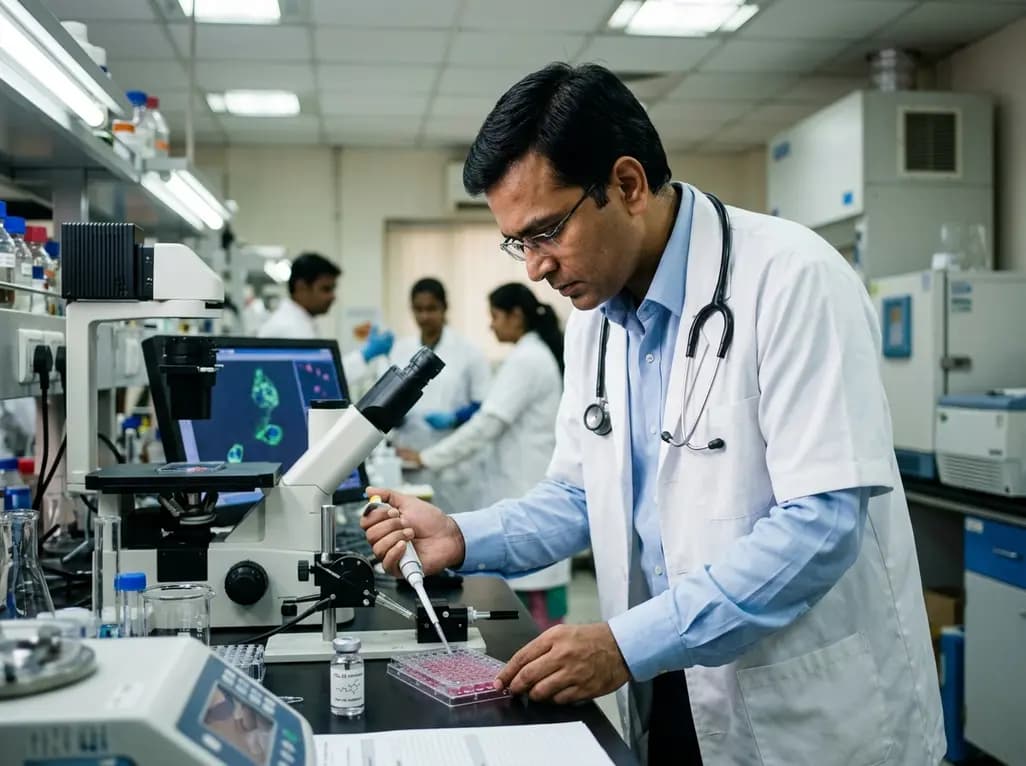
PZL-26, a selective small-molecule inhibitor of LONP1, reveals essential roles of this mitochondrial protease in complex I biogenesis, translation, and oxidative phosphorylation. These findings provide critical mechanistic insights for developing targeted anticancer therapies against proteostasis-dependent tumors.
Today

A nationwide South Korean study shows underreporting of smoking among women dropped from 57.4% to 36.4% between 2008 and 2021. However, over one-third of female smokers still conceal their habit, highlighting the critical role of cotinine verification in clinical risk assessment.
Today

PepsiCo and Monster Beverage have challenged the FSSAI ban on the energy drink label in Indian courts. While companies highlight supply disruptions, regulatory actions spotlight severe cardiovascular and metabolic risks associated with excessive caffeine, sugar, and taurine consumption, especially in adolescents.
Today
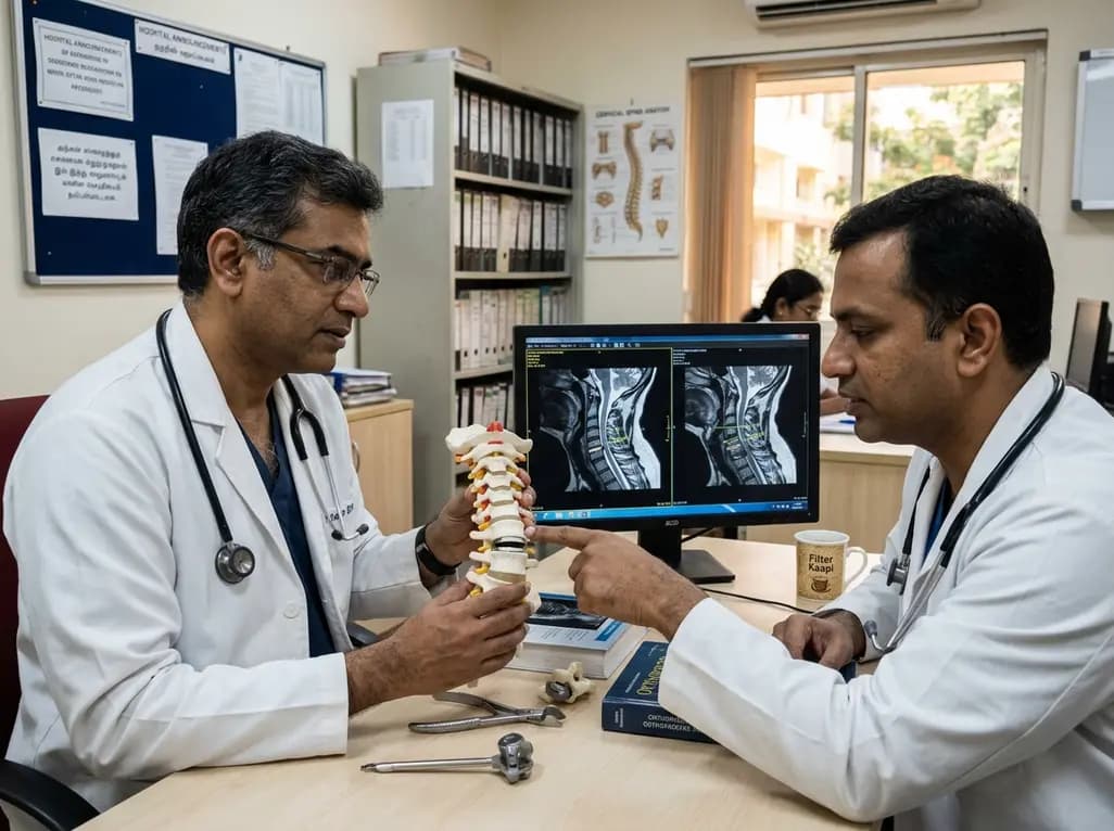
A recent investigation analyzes whether cervical disc arthroplasty effectively restores or merely preserves physiological range of motion, providing critical biomechanical insights for treating hypomobile cervical segments.
Today

A Bayesian multilevel meta-analysis reveals that aerobic training combined with moderate carbohydrate restriction modestly lowers HbA1c in type 2 diabetes. However, sparse data and very low certainty leave incremental benefits over exercise or diet alone unproven, highlighting the need for individualized care.
Yesterday
