.webp&w=384&q=75)

Loading, please wait...
Loading, please wait...
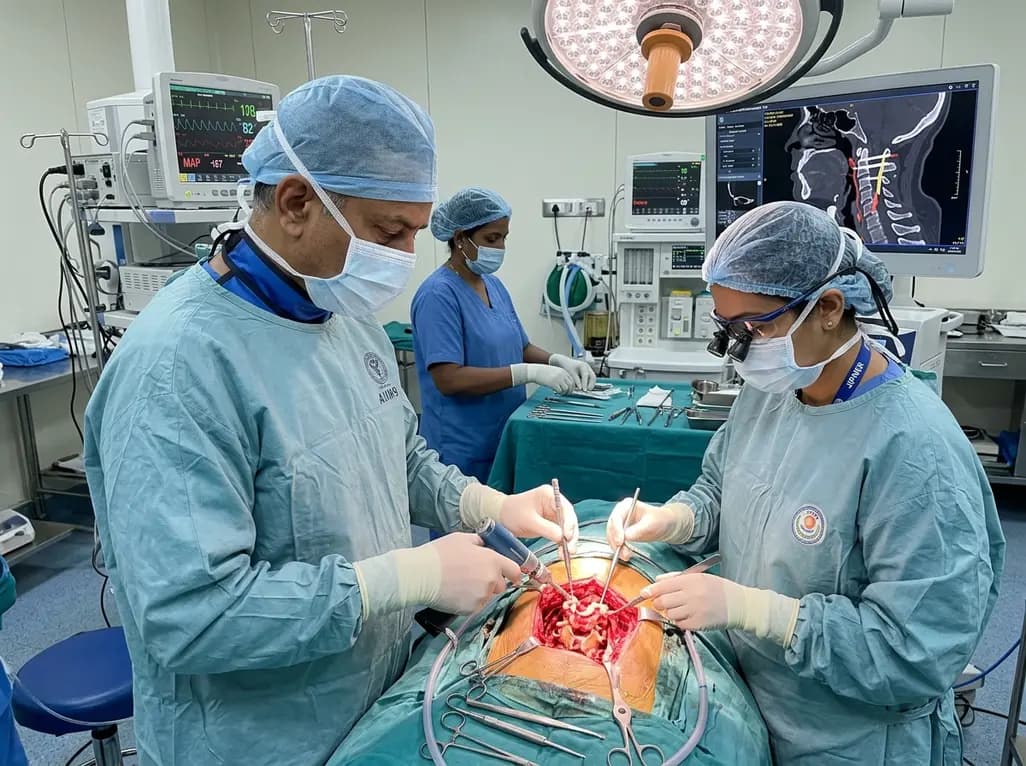
Atlantoaxial fixation remains a cornerstone intervention in complex craniovertebral junction surgery. However, managing challenging anatomy often creates significant surgical hurdles for spine specialists worldwide. The implementation of an individualized C2 screw placement strategy provides a structured blueprint to overcome anomalous neurovascular trajectories. Surgeons frequently face difficulties when patients present with high-riding vertebral arteries or narrow C2 pedicles. Therefore, adopting versatile fixation techniques combined with intraoperative vascular mobilization ensures maximum mechanical stability while prioritizing patient safety during delicate reconstructive procedures.
The anatomical architecture of the upper cervical spine exhibits substantial variation across patient populations. C2 pedicle screws represent the traditional gold standard for rigid posterior atlantoaxial stabilization. Nevertheless, anomalous bone structures or aberrant vascular courses can elevate operative risks considerably. Specifically, a high-riding vertebral artery significantly narrows the safe osseous corridor required for standard screw instrumentation. Inserting hardware without addressing these anatomical variations increases the risk of catastrophic arterial laceration or severe neurological compromise.
In addition, narrow C2 pedicles constrain the available bone stock, making conventional pedicle screw trajectory unfeasible. Consequently, alternative techniques such as pars, laminar, or transarticular screws have emerged as valuable rescue strategies. However, these alternatives may offer lower rigid construct stability compared to traditional pedicle fixation. Surgeons must therefore perform thorough preoperative imaging analyses to identify high-risk vascular variations early. By carefully evaluating three-dimensional computed tomography scans, clinical teams can anticipate technical challenges and tailor their surgical approach accordingly.
To address complex anatomical constraints, researchers recently evaluated a multi-strategy protocol combining customized screw selection with surgical vertebral artery mobilization. This advanced C2 screw placement strategy allows surgeons to choose optimal trajectory pathways based on individual patient anatomy. In cases with severe vascular encroachment, direct mobilization of the vertebral artery loop creates additional safe working space for screw entry.
Furthermore, integrating an atlantoaxial fusion cage significantly enhances overall structural support compared to traditional posterior bone grafting methods. Clinical data demonstrate that patients receiving fusion cage support achieved a 92.3% bone fusion rate at six months follow-up. In contrast, patients managed with interlaminar bone grafting alone reached a fusion rate of only 51.0%. This striking statistical difference highlights the clinical necessity of rigid interbody cage support in challenging reconstructions. Consequently, combining robust internal instrumentation with anterior cage placement offers superior biomechanical stabilization and superior bone healing outcomes.
Achieving precise hardware positioning is critical for preventing neurovascular injury during C2 instrumentation procedures. In a large retrospective cohort of 312 patients treated between 2017 and 2025, investigators evaluated screw placement accuracy using standard Gertzbein-Robbins criteria. Results confirmed that screw accuracy reached 100% clinically acceptable placement in patients with normal cervical anatomy.
Moreover, in complex cases requiring direct vertebral artery mobilization, surgeons maintained a clinically acceptable screw accuracy rate of 60%. Importantly, no patients experienced permanent neurovascular deficits, vertebral artery injury, or blood flow compromise throughout the operative process. This zero-percent vascular injury rate underscores the inherent safety of meticulous mobilization techniques. By exposing and retracting the artery under direct visualization, surgeons eliminate blind drilling risks through narrow pedicle corridors. As a result, this protocol successfully mitigates high-risk anatomical features while preserving essential cerebral blood flow.
Achieving early structural rigidity is paramount for long-term successful fusion across the atlantoaxial complex. Traditional posterior interlaminar bone grafts frequently suffer from mechanical displacement, resorption, or pseudarthrosis due to persistent rotational forces. Conversely, placing an atlantoaxial fusion cage directly into the C1-C2 joint space provides immediate axial load support and preserves vertical disc height.
Additionally, fusion cages offer a larger surface area for bone graft material, promoting rapid osteointegration across the joint space. Consequently, this construct minimizes micromotion and shields inserted C2 hardware from excessive fatigue stresses. The combined approach ensures that even patients with compromised bone density or narrow pedicle morphology achieve solid arthrodesis. Spine surgeons can therefore rely on fusion cages to maintain construct integrity, reduce hardware failure rates, and eliminate the need for prolonged external halo immobilization postoperatively.
Executing safe vertebral artery mobilization requires meticulous surgical technique and thorough anatomical exposure. First, surgeons dissect the posterior muscle layers to clearly identify the C2 lateral mass and transverse process. Next, specialized micro-instruments carefully remove the thin posterior wall of the vertebral artery groove. This step unroofs the arterial loop without causing mechanical trauma to the vessel wall.
Subsequently, the operating team gently mobilizes the artery inferiorly using a blunt dissector under direct microscopic visualization. Displacing the vessel creates direct line-of-sight exposure for pedicle or pars screw drilling. Furthermore, continuous intraoperative fluoroscopy or image guidance ensures high trajectory precision. Surgeons can then safely drill, tap, and place C2 screws while keeping the mobilized artery safely away from high-speed burs. Overall, this reproducible multi-strategy approach provides a safe, versatile option for complex craniovertebral junction surgeries.
A high-riding vertebral artery is an anatomical variant where the vessel loops abnormally high into the C2 pedicle or body. This anomaly significantly narrows the safe osseous corridor available for standard pedicle screw insertion. Inserting screws without modifying the technique carries a severe risk of arterial injury or stroke. Consequently, surgeons must utilize specialized techniques, such as arterial mobilization or alternative screw trajectories, to ensure safe fixation.
An atlantoaxial fusion cage provides immediate axial load support, maintains joint height, and creates a large surface area for bone fusion. Clinical studies demonstrate a 92.3% fusion rate with cages at six months, compared to just 51.0% for interlaminar grafting. Furthermore, cages reduce mechanical stress on posterior C2 screws, promoting rapid osteointegration and lowering the risk of hardware failure or nonunion in complex upper cervical spine reconstructions.
Yes, direct vertebral artery mobilization is a safe and highly effective surgical maneuver when executed under microscopic visualization. By unroofing the bony groove and gently retracting the vessel inferiorly, surgeons gain direct line-of-sight access to the C2 pedicle. Clinical trials report zero permanent neurovascular injuries or blood flow compromises during these procedures, making it a reliable solution for patients presenting with challenging, high-risk upper cervical anatomy.
Disclaimer: This content is for informational and educational purposes only and does not constitute medical advice, diagnosis, or treatment. Always seek the advice of a qualified healthcare provider with any questions you may have regarding a medical condition or surgical procedure. Refer to the latest local and national guidelines for clinical practice.
References

Read summarized clinical updates, watch expert medical content, and earn CME certifications right from your smartphone.
Open your smartphone camera to scan and install.





A novel multi-strategy C2 screw placement protocol integrating fusion cage support and vertebral artery mobilization achieves superior fusion rates and zero vascular complications in complex upper cervical spine surgery, offering a reliable solution for high-riding vertebral artery cases.
last month

The World Health Organization has launched a 10-year framework to address evidence gaps in traditional, complementary, and integrative medicine. Prioritizing conditions such as stroke, cancer, and depression, this global roadmap highlights rigorous clinical trials, pharmacovigilance, and safer patient care.
Today

A recent longitudinal study evaluated the link between prenatal loss of control eating and cardiovascular health using Life's Essential 8 frameworks. While direct associations across pregnancy were non-significant, the high prevalence of dysregulated eating highlights critical implications for obstetric practice.
Today

Explore modern perioperative management protocols for clinical xenotransplantation. Learn how costimulation blockade, anti-inflammatory perfusion, and coagulopathy rescue overcome interspecies hurdles.
Today
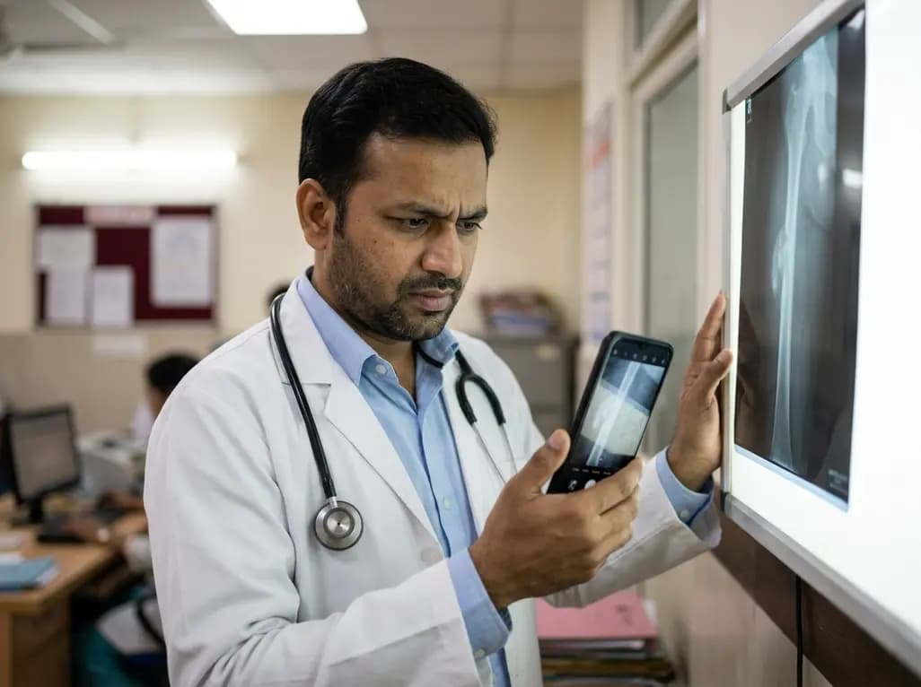
A retrospective study evaluating a locally deployed vision language model (Qwen3-VL) reveals that smartphone-captured radiographs and retrieval-augmented generation can paradoxically reduce diagnostic accuracy in bone tumor referrals due to optical noise and textual hallucinations.
Today

At Homa CME 2026 at Hyderabad's T-Hub, healthcare leaders emphasized moving beyond routine metrics through digital calculators and precision stratification. Discover why characterizing individual insulin secretion, resistance patterns, and vascular risks transforms chronic metabolic care in India.
Today
