.webp&w=384&q=75)

Loading, please wait...
Loading, please wait...

Bullous pemphigoid represents the most common autoimmune subepidermal blistering dermatosis encountered in elderly clinical practice. Clinicians frequently face significant diagnostic and therapeutic challenges when evaluating bullous pemphigoid disease severity. While circulating autoantibodies against BP180 and BP230 drive structural blister formation, cellular immunity plays an equally critical role. Recent clinical research highlights the biological contributions of eosinophils and immunoglobulin E (IgE) within this disease spectrum. Consequently, identifying precise histological and serological biomarkers helps dermatologists stratify clinical risk, predict progressive blistering, and optimize individualized therapeutic strategies for vulnerable elderly patients.
Bullous pemphigoid predominantly strikes older individuals, resulting in tense cutaneous bullae, intense pruritus, and severe systemic morbidity. Pathologists historically characterize this autoimmune condition by autoantibody deposition against hemidesmosomal antigens along the basement membrane zone. Consequently, immune complexes trigger complement activation, recruit inflammatory cells, and produce subepidermal separation. Although clinicians traditionally focus on immunoglobulin G antibodies, both eosinophils and immunoglobulin E direct local tissue destruction.
In particular, eosinophils synthesize cytotoxic granule proteins, including major basic protein and eosinophil cationic protein. These aggressive enzymes digest junctional proteins and accelerate detachment of the dermal-epidermal junction. Furthermore, eosinophils generate numerous pro-inflammatory cytokines such as interleukin-5 and leukotrienes. These chemical mediators recruit additional inflammatory cells to the dermis, intensifying local erythema and blister expansion. Therefore, bullous pemphigoid disease severity reflects not only humoral autoantibody activity but also profound cellular infiltration. Recognizing how cellular mediators drive dermal injury allows dermatologists to anticipate severe exacerbations earlier in the therapeutic journey. In addition, persistent eosinophil activation amplifies intractable pruritus, which severely degrades sleep quality and functional independence in older adults.
A prospective observational study from South India evaluated thirty-five patients with confirmed bullous pemphigoid to determine inflammatory activity markers. The investigators assessed clinical extent using the Bullous Pemphigoid Disease Area Index (BPDAI). Interestingly, higher disease activity, defined as a BPDAI score of at least 20, was present in seventy-seven percent of the cohort.
Histopathological evaluation revealed that eosinophil-predominant dermal infiltrates strongly correlated with severe disease manifestations. Specifically, patients exhibiting prominent tissue eosinophilia demonstrated an odds ratio of 9.638 for elevated disease activity compared to those with sparse dermal infiltration. This statistically significant association underscores the direct destructive role of tissue eosinophils at lesion sites. While circulating eosinophils circulate transiently, dermal eosinophils localize directly where blistering occurs. Consequently, local degranulation causes mechanical shearing and microvesicle coalescence. Therefore, examining the intensity of tissue eosinophilia on diagnostic skin biopsy specimens offers valuable prognostic insight. Pathologists and dermatologists should routinely quantify dermal eosinophils to identify individuals requiring intensified therapeutic intervention. Furthermore, this quantifiable tissue infiltration provides objective confirmation of disease severity before secondary infections obscure the histological picture.
Peripheral blood eosinophilia occurs frequently in patients with bullous pemphigoid, often prompting clinicians to suspect extensive skin disease. However, the South Indian prospective investigation demonstrated no statistically significant association between circulating eosinophil counts above 500 cells per microliter and higher BPDAI scores.
This notable finding highlights a critical clinical distinction between systemic circulation and end-organ tissue involvement. Although circulating eosinophils reflect systemic immunological priming, they do not necessarily reflect the magnitude of cutaneous blister formation at any single moment. For instance, eosinophils rapidly migrate from the vascular compartment into inflamed skin tissues under the chemotactic guidance of eotaxin and other chemokines. As a result, peripheral counts may fluctuate depending on transit time, bone marrow release, and concurrent corticosteroid administration. Therefore, clinicians must exercise caution when relying solely on complete blood counts to gauge skin disease severity. Instead, combining serological parameters with rigorous histological examination ensures a more accurate assessment. Dermal eosinophil density remains a far more reliable indicator of active cutaneous destruction than routine peripheral leukocyte differentials alone. Consequently, dermatologists should avoid making aggressive management decisions based exclusively on isolated peripheral blood eosinophil counts.
Immunoglobulin E antibodies play an emerging role in the immunopathogenesis of autoimmune blistering conditions. In bullous pemphigoid, IgE antibodies bind to the NC16A domain of BP180, activating mast cells and eosinophils via high-affinity Fc receptors. However, the South Indian prospective study observed no statistically significant correlation between elevated total serum IgE levels exceeding 500 IU per milliliter and high BPDAI scores.
Similarly, direct immunofluorescence detection of IgE deposition along the basement membrane zone did not independently correlate with higher disease scores in this cohort. Nevertheless, researchers observed that IgE-mediated severity becomes more evident when investigators concurrently detect IgE within affected tissues alongside prominent eosinophils. Small sample sizes, biopsy site variability, and specialized technical nuances of immunofluorescence likely explain why peripheral IgE levels diverge from clinical presentation. Furthermore, serum IgE levels often remain elevated due to non-specific atopic tendencies or age-related immune dysregulation. Therefore, circulating IgE alone serves poorly as an isolated marker of immediate disease severity. Instead, clinicians should interpret total serum IgE in conjunction with tissue-based immunopathological findings. Ultimately, local tissue interactions between IgE and resident immune cells dictate clinical blistering far more than free circulating antibodies.
These prospective findings carry vital practical implications for dermatologists and general physicians managing bullous pemphigoid. Because tissue eosinophilia correlates strongly with severe disease activity, clinicians should carefully review skin biopsy reports for dermal eosinophil density. Patients with marked tissue eosinophil infiltration warrant proactive treatment escalation, such as potent topical corticosteroids, systemic steroids, or steroid-sparing immunosuppressive agents.
Furthermore, the clear pathogenic role of eosinophils supports the therapeutic rationale for targeted biologic agents. Therapies targeting interleukin-5 pathways, including mepolizumab and benralizumab, directly deplete eosinophils and reduce inflammatory burden. Similarly, dupilumab, which inhibits interleukin-4 and interleukin-13 signaling, effectively suppresses the Th2 axis driving both eosinophilia and IgE synthesis. By neutralizing these pathways, clinicians can achieve disease control while minimizing the adverse effects of prolonged high-dose corticosteroid therapy in frail elderly patients. Moreover, clinicians should track clinical scores using the BPDAI alongside histological evaluation to ensure comprehensive monitoring. Adopting this integrated, multi-modal diagnostic approach improves therapeutic precision, limits treatment-related toxicity, and ultimately improves quality of life for patients battling severe autoimmune blistering diseases. Consequently, personalized biomarker-guided strategies will redefine the standard of care for bullous pemphigoid in contemporary dermatological practice.
Tissue eosinophils directly participate in dermal-epidermal separation by releasing destructive enzymes, including major basic protein and eosinophil cationic protein, directly at the basement membrane. Conversely, peripheral blood eosinophil counts merely reflect circulating leukocytes prior to chemotactic tissue recruitment. Furthermore, circulating levels fluctuate rapidly with diurnal cycles or prior steroid exposure. Therefore, histological tissue infiltration represents true localized immunopathology, correlating far more reliably with clinical blistering and objective disease activity scores.
The Bullous Pemphigoid Disease Area Index, known as BPDAI, provides a standardized, validated scoring system that objectively evaluates cutaneous blisters, urticarial erythema, and mucosal erosion. Moreover, it separates objective anatomical lesions from subjective pruritus severity. Clinicians use established BPDAI thresholds, such as a score of twenty, to differentiate moderate-to-severe disease from mild cases. Consequently, serial BPDAI assessments allow physicians to gauge treatment response accurately, adjust corticosteroid dosing, and identify impending clinical relapses.
Targeted biologics provide steroid-sparing alternatives for patients with recalcitrant disease or contraindications to systemic corticosteroids. Specifically, anti-interleukin-5 monoclonal antibodies like mepolizumab directly suppress eosinophil maturation and cutaneous recruitment. In addition, dupilumab blocks interleukin-4 and interleukin-13 receptor signaling, dampening Th2-mediated inflammation and reducing both tissue eosinophilia and total IgE production. Therefore, incorporating these targeted biologics mitigates systemic corticosteroid toxicity while successfully achieving disease remission in vulnerable elderly patients.
Disclaimer: This content is for informational and educational purposes only... Refer to the latest local and national guidelines for clinical practice.
References

Read summarized clinical updates, watch expert medical content, and earn CME certifications right from your smartphone.
Open your smartphone camera to scan and install.





A prospective study evaluates the correlation of serum and tissue eosinophils and IgE with bullous pemphigoid disease severity. While circulating markers showed variable associations, prominent tissue eosinophilia strongly correlated with elevated disease activity (OR = 9.638), guiding targeted care.
Today
A comprehensive analysis of serum bile acid variability establishes robust reference ranges and reveals how sex, age, BMI, and smoking alter circulating profiles.
Today
A retrospective cost-minimization study of 16,695 Medicare patients reveals that pelvic floor muscle therapy and pessaries incur less than half the 1-year costs of primary sling surgery, supporting conservative management as initial therapy.
Today
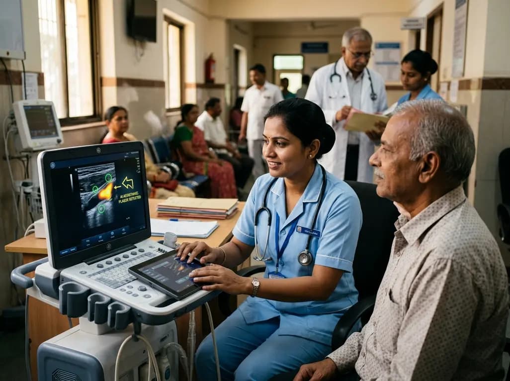
A user-centered study demonstrates that AI-assisted carotid ultrasound enables nonexpert primary care staff to detect subclinical atherosclerosis. With real-time guidance and workflow optimization, this technology supports early cardiovascular risk communication and task-shifting in routine clinical practice.
Today

A hyaluronic acid-modified metal-polyphenol nanocomposite successfully eliminates reactive oxygen species in chondrocytes, halts cartilage breakdown, and promotes tissue anabolism, presenting a novel disease-modifying strategy for early osteoarthritis.
Today

A pilot randomized trial demonstrates that digital wellness applications and medically tailored meals significantly attenuate rapid weight regain following GLP-1 receptor agonist discontinuation, providing valuable transitional support for long-term obesity management.
Today
